Pipkin IV Femoral Head Fracture with Acetabular Rim Involvement.
Score and Comment on this Case
Clinical Details
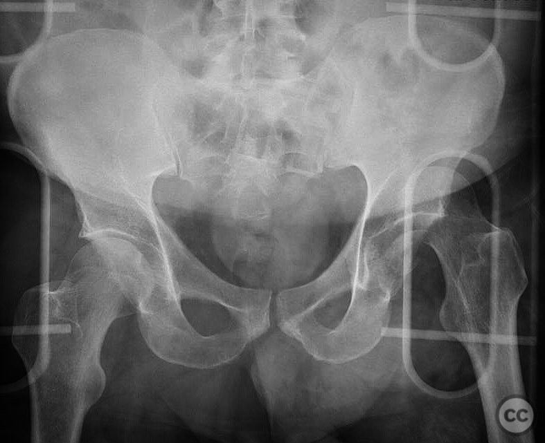
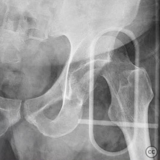
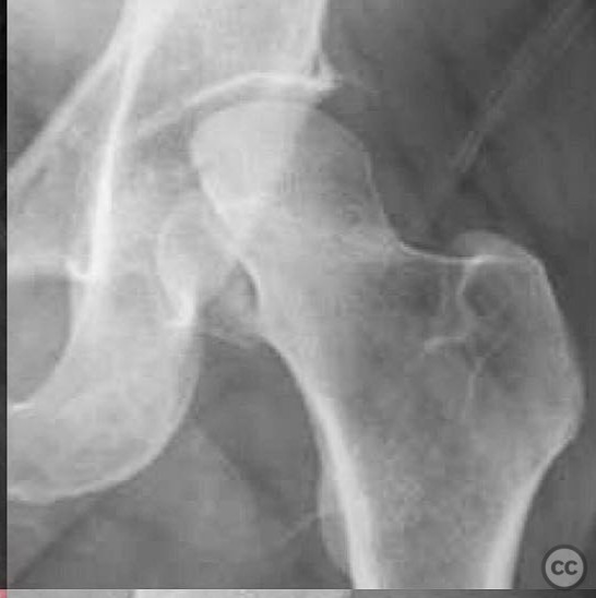
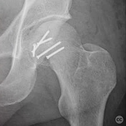
Clinical and radiological findings: A 40-year-old male skier sustained a high-energy injury after skiing off a cliff in early season conditions, resulting in a Pipkin IV femoral head fracture. The injury is characterized by a Pipkin II fracture with an additional cranial peripheral acetabular fracture. Initial radiographs confirmed the fracture pattern, and a closed reduction was performed in the emergency room. The neurovascular examination was unremarkable.
Preoperative Plan
Planning remarks: The preoperative plan involved an open reduction and internal fixation of the femoral head fracture through a modified anterior approach. The plan included the use of buried lag screws for fixation. An examination under anesthesia (EUA) was planned post-fixation to assess the stability of the hip and determine the necessity of acetabular rim fixation.
Surgical Discussion
Patient positioning: Supine position on a radiolucent table to facilitate fluoroscopic imaging and access to the anterior aspect of the hip.
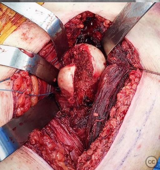
Anatomical surgical approach: Modified anterior approach to the hip, involving an incision over the anterior superior iliac spine extending distally along the sartorius muscle. Dissection proceeded through the interval between the sartorius and tensor fascia lata, with careful protection of the lateral femoral cutaneous nerve. Capsulotomy was performed to expose the femoral head for fracture reduction and fixation.
Operative remarks:Following successful reduction and fixation of the femoral head using 2.0 mm and 2.4 mm lag screws, an EUA was conducted to evaluate hip stability. The hip demonstrated stability, indicating that fixation of the acetabular rim fracture was not required. The decision to forego acetabular fixation was based on intraoperative stability findings.
Postoperative protocol: Postoperative rehabilitation included protected weight-bearing with crutches for 6 weeks, followed by gradual progression to full weight-bearing as tolerated. Range of motion exercises were initiated early to prevent stiffness, with emphasis on avoiding excessive flexion and rotation during the initial healing phase.
Follow up: Not specified.
Orthopaedic implants used: 2.0 mm and 2.4 mm lag screws.
Search for Related Literature

orthopaedic_trauma
- United States , Seattle
- Area of Specialty - General Trauma
- Position - Specialist Consultant

Industry Sponsership
contact us for advertising opportunities

Article viewed 199 times
05 Aug 2025
Add to Bookmarks
Full Citation
Cite this article:
Surname, Initial. (2025). Pipkin IV Femoral Head Fracture with Acetabular Rim Involvement.. Journal of Orthopaedic Surgery and Traumatology. Case Report 13650913 Published Online Aug 05 2025.