Proximal Tibiofibular Joint Dislocation with Tibial Shaft Fracture.
Score and Comment on this Case
Clinical Details
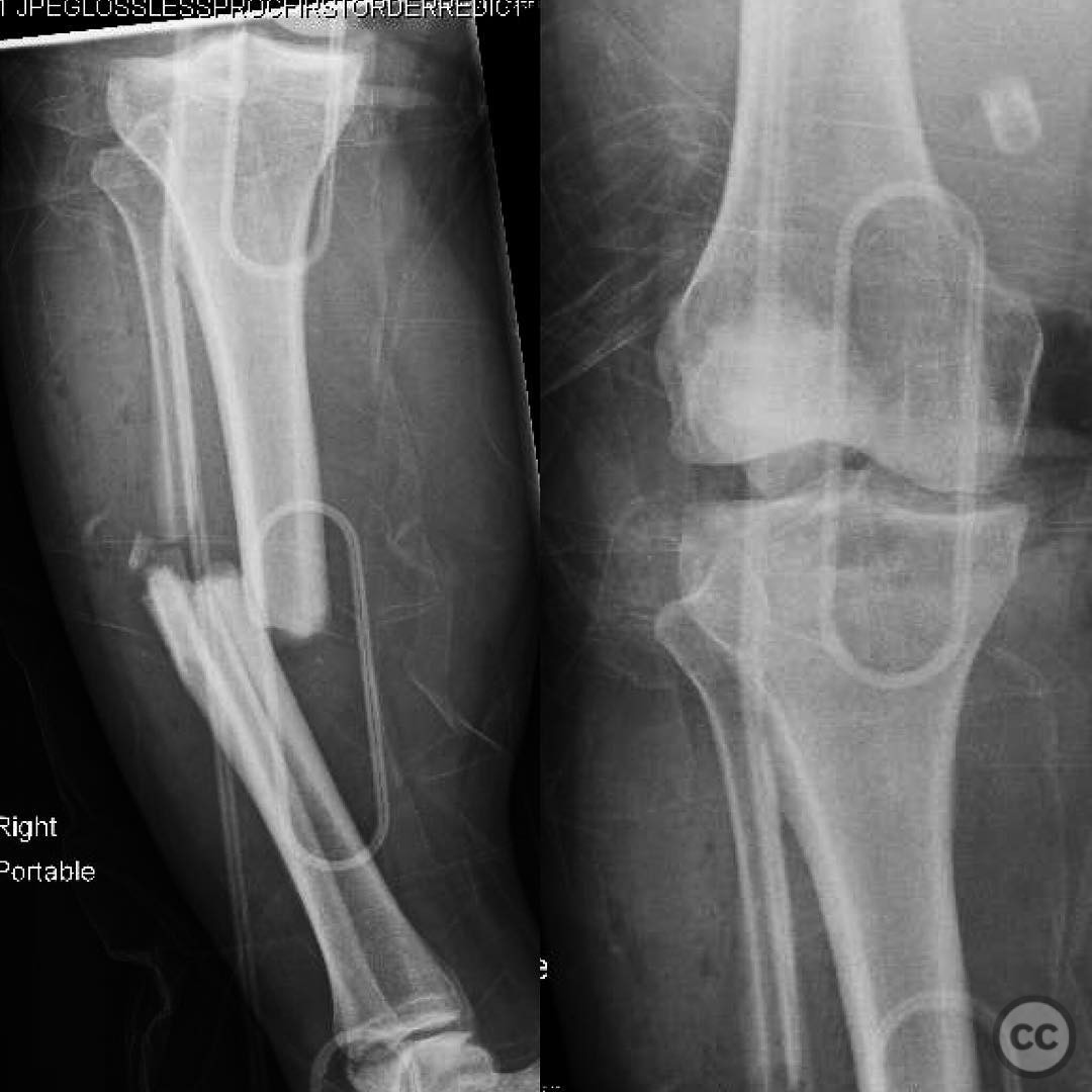
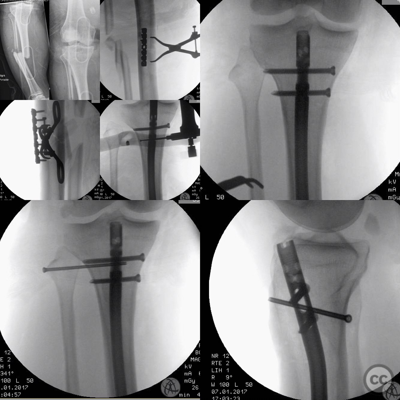
Clinical and radiological findings: A 33-year-old morbidly obese male was involved in a motorcycle collision with a tree, resulting in a complex injury to the lower extremity. Initial clinical examination revealed absent distal pulses and peroneal nerve palsy. Vascular assessment via CTA was normal, suggesting arterial spasm, and dopplerable pulses returned without vascular intervention. Radiographic evaluation identified an open tibial shaft fracture with proximal tibiofibular joint dislocation. The patient also sustained injuries to the PCL and ACL.
Preoperative Plan
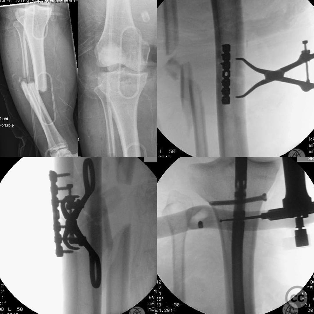
Planning remarks: The preoperative plan included debridement and irrigation of the open fracture, followed by provisional stabilization with clamping and plating, and definitive fixation with intramedullary nailing. Attention was directed towards addressing the proximal tibiofibular joint dislocation.
Surgical Discussion
Patient positioning: Supine positioning on a radiolucent table to facilitate intraoperative imaging and access to the lower extremity.
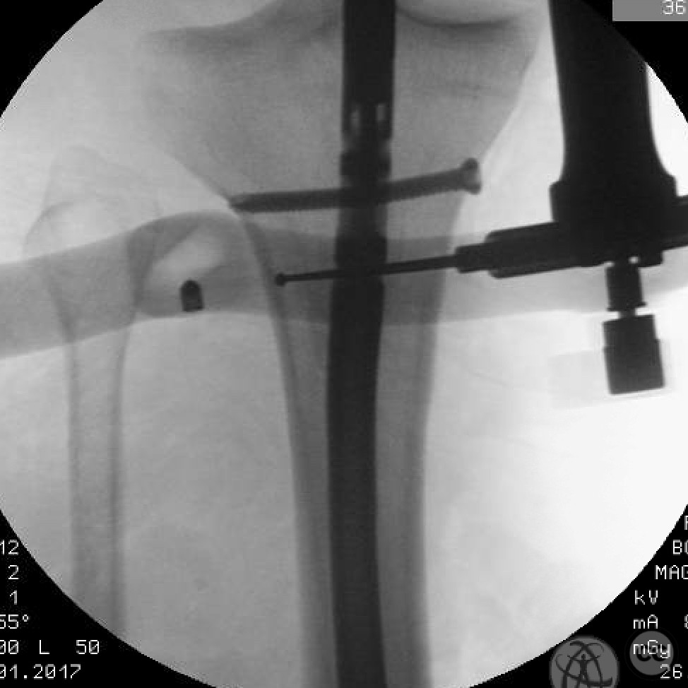
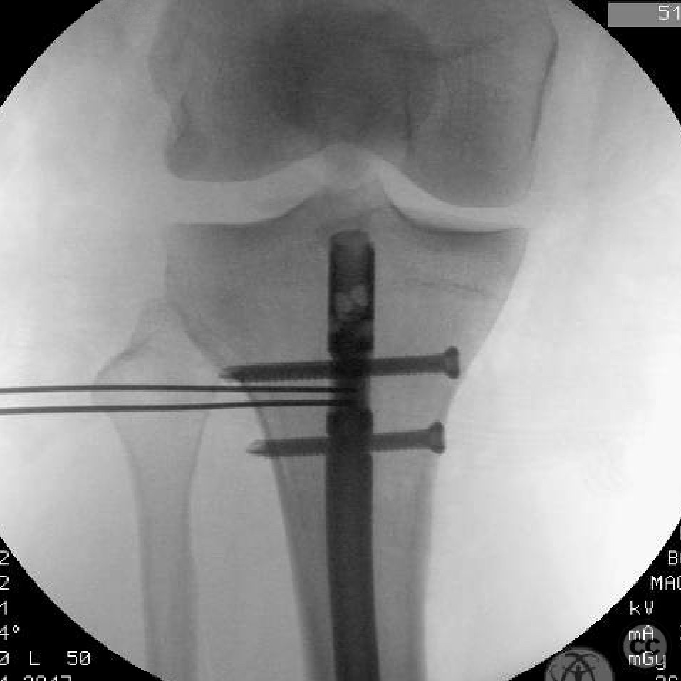
Anatomical surgical approach: A longitudinal incision was made over the anteromedial aspect of the tibia for debridement and irrigation of the open fracture site. The proximal tibiofibular joint was approached laterally, with care taken to avoid iatrogenic injury to the common peroneal nerve. The tibial shaft was stabilized using an intramedullary nail inserted through a standard entry point at the proximal tibia.
Operative remarks:The proximal tibiofibular joint dislocation was identified intraoperatively, requiring careful reduction to prevent long-term instability and dysfunction. The reduction was confirmed under fluoroscopy, ensuring proper alignment and stability of the joint. Given the complexity of the injury, a multidisciplinary approach involving both orthopaedic trauma and sports medicine specialists was considered for potential ligamentous reconstruction at a later stage.
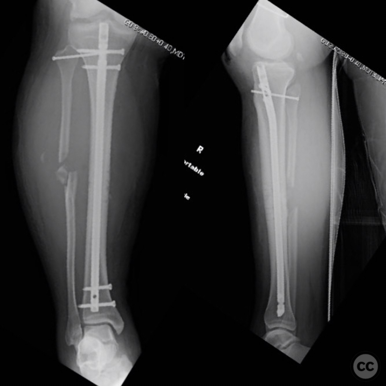
Postoperative protocol: Postoperative rehabilitation included non-weight bearing on the affected limb for 6 weeks, with gradual progression to partial weight bearing as tolerated. Early range of motion exercises were initiated to prevent joint stiffness, with close monitoring of neurovascular status.
Follow up: Not specified.
Orthopaedic implants used: Intramedullary nail, provisional plate fixation (specific implant details not provided).
Search for Related Literature

orthopaedic_trauma
- United States , Seattle
- Area of Specialty - General Trauma
- Position - Specialist Consultant

Industry Sponsership
contact us for advertising opportunities
Article viewed 228 times
26 Jul 2025
Add to Bookmarks
Full Citation
Cite this article:
Surname, Initial. (2025). Proximal Tibiofibular Joint Dislocation with Tibial Shaft Fracture.. Journal of Orthopaedic Surgery and Traumatology. Case Report 8175953 Published Online Jul 26 2025.