. Smokes a bit_ horribly contr(.jpg)
Lisfranc Fracture Dislocation with ORIF and K-wire Fixation.
Score and Comment on this Case
Clinical Details
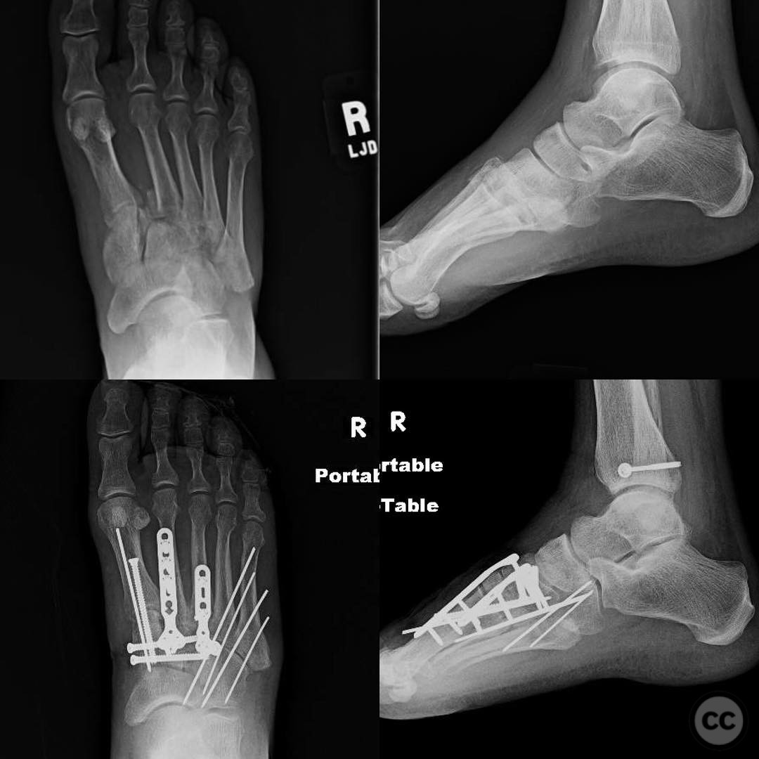
Clinical and radiological findings: A female patient in her 40s, with poorly controlled diabetes (HbA1c ~12) and a history of frequent falls, presented with a Lisfranc fracture dislocation two weeks post-injury. The patient has a history of a similar injury on the contralateral side, which was treated operatively with satisfactory healing. The current injury was characterized by significant midfoot instability and malalignment. Initial radiographs confirmed the presence of a Lisfranc injury with displacement of the tarsometatarsal joints.
Preoperative Plan
Planning remarks: The preoperative plan involved open reduction and internal fixation (ORIF) of the Lisfranc joint complex. The approach was planned to address the medial, middle, and lateral columns of the foot. Fixation strategy included the use of plates and screws for stabilization of the second metatarsal to the middle cuneiform and the third metatarsal to the lateral cuneiform, as well as a cannulated screw from the first metatarsal to the medial cuneiform.
Surgical Discussion
Patient positioning: Supine position with a bump under the ipsilateral hip to allow for easier access to the dorsal aspect of the foot.
Anatomical surgical approach: A dorsal longitudinal incision was made over the tarsometatarsal joints. Subcutaneous tissues were carefully dissected to expose the extensor tendons, which were retracted to provide access to the Lisfranc joint complex. The joint capsule was incised longitudinally, and careful reduction of the dislocated joints was achieved under direct visualization.
Operative remarks:Due to the patient's status as a smoker and brittle diabetic, a more conservative fixation approach was chosen, avoiding extensive soft tissue dissection. K-wires were used for temporary stabilization, and plates were applied to reinforce the reduction. The decision not to perform a Castro slide was based on concerns regarding wound healing and potential complications associated with her comorbidities.
Postoperative protocol: Postoperatively, the patient was placed in a non-weight-bearing cast for 8 weeks. Transition to partial weight-bearing in a controlled ankle motion (CAM) boot was planned after removal of K-wires, with gradual progression to full weight-bearing as tolerated.
Follow up: Not specified.
Orthopaedic implants used: K-wires, plates for second metatarsal to middle cuneiform and third metatarsal to lateral cuneiform, cannulated screw from first metatarsal to medial cuneiform.
Search for Related Literature

orthopaedic_trauma
- United States , Seattle
- Area of Specialty - General Trauma
- Position - Specialist Consultant

Industry Sponsership
contact us for advertising opportunities
Article viewed 594 times
28 Jul 2025
Add to Bookmarks
Full Citation
Cite this article:
Surname, Initial. (2025). Lisfranc Fracture Dislocation with ORIF and K-wire Fixation.. Journal of Orthopaedic Surgery and Traumatology. Case Report 6955469 Published Online Jul 28 2025.