 posterior elbow dislocation. She presented with a banged up but perfused (mostly) hand._1.jpg)
 posterior elbow dislocation. She presented with a banged up but perfused (mostly) hand._2.jpg)
 posterior elbow dislocation. She presented with a banged up but perfused (mostly) hand._3.jpg)
 posterior elbow dislocation. She presented with a banged up but perfused (mostly) hand._4.jpg)
 posterior elbow dislocation. She presented with a banged up but perfused (mostly) hand._5.jpg)
 posterior elbow dislocation. She presented with a banged up but perfused (mostly) hand._8.jpg)
 posterior elbow dislocation. She presented with a banged up but perfused (mostly) hand._6.jpg)
 posterior elbow dislocation. She presented with a banged up but perfused (mostly) hand._7.jpg)
 posterior elbow dislocation. She presented with a banged up but perfused (mostly) hand._9.jpg)
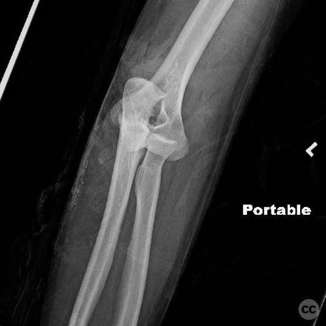
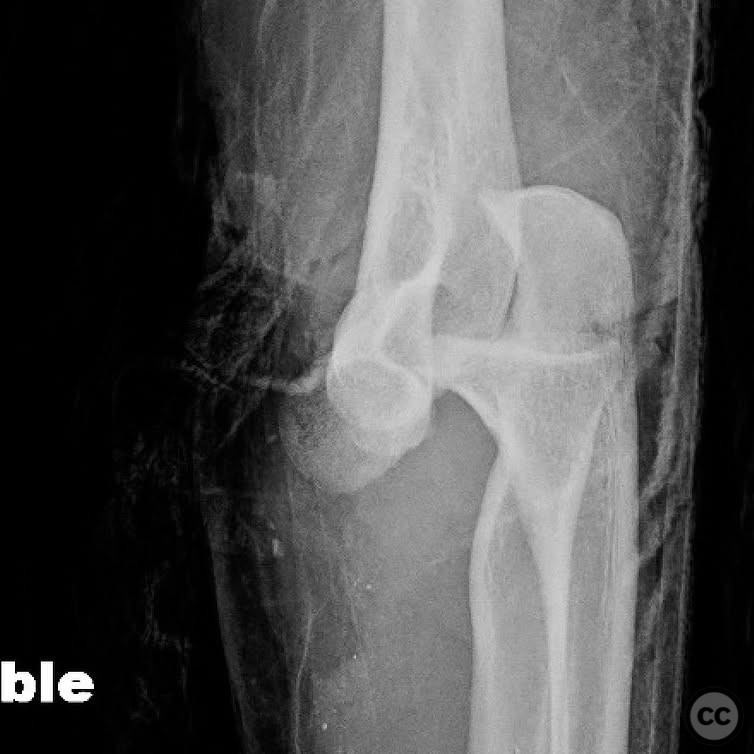
Open Posterior Elbow Dislocation with Internal Fixation
Score and Comment on this Case
Clinical Details
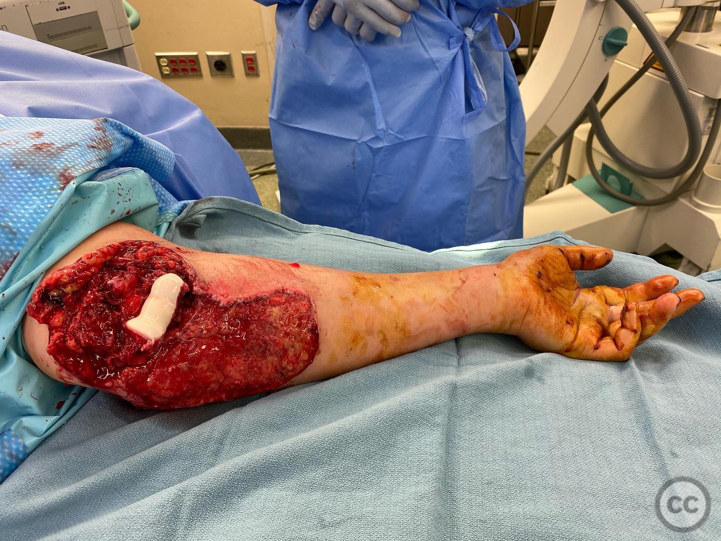
Clinical and radiological findings: The patient presented with an open posterior elbow dislocation and a compromised, yet mostly perfused hand. The neurovascular examination was challenging upon presentation. Urgent surgical intervention was performed for debridement, irrigation, and exploration. All major nerves around the elbow were found to be in continuity, with no arterial injury noted. The wound was massively contaminated.
Preoperative Plan
Planning remarks: The preoperative plan involved a second-look procedure 48 hours post-initial debridement. The approach was planned via a Kocher incision to address the lateral structures, given the inability to repair medial structures due to extensive soft tissue damage.
Surgical Discussion
Patient positioning: The patient was positioned supine with the arm placed on an arm table to allow optimal access to the lateral aspect of the elbow.
Anatomical surgical approach: A Kocher approach was utilized, involving an incision over the lateral aspect of the elbow. Dissection proceeded through the interval between the extensor carpi ulnaris and anconeus muscles to expose the lateral epicondyle. The lateral ulnar collateral ligament (LUCL) and common extensor origin (CEO) were identified and repaired.
Operative remarks:The surgeon noted that the brachialis and joint capsule were severely damaged and not repairable, while the triceps remained intact. Given the global instability and inability to repair medial structures, a decision was made to perform a reduction, repair the LUCL and CEO, and apply an internal external fixator for augmented stability and motion. Primary closure of the soft tissue injury was achieved with a split-thickness skin graft (STSG).
Postoperative protocol: Postoperative rehabilitation included early motion as permitted by soft tissue healing, with removal of the internal fixator planned between 3 to 6 months post-surgery.
Follow up: Not specified.
Orthopaedic implants used: Internal external fixator system, split-thickness skin graft (STSG).
Search for Related Literature

orthopaedic_trauma
- United States , Seattle
- Area of Specialty - General Trauma
- Position - Specialist Consultant

Industry Sponsership
contact us for advertising opportunities
Article viewed 221 times
13 Jul 2025
Add to Bookmarks
Full Citation
Cite this article:
Surname, Initial. (2025). Open Posterior Elbow Dislocation with Internal Fixation. Journal of Orthopaedic Surgery and Traumatology. Case Report 6798430 Published Online Jul 13 2025.