


Challenging Elbow Trauma: Low Distal Humerus Articular Fracture with Olecranon Dislocation in a Type II Open Injury
Score and Comment on this Case
Clinical Details
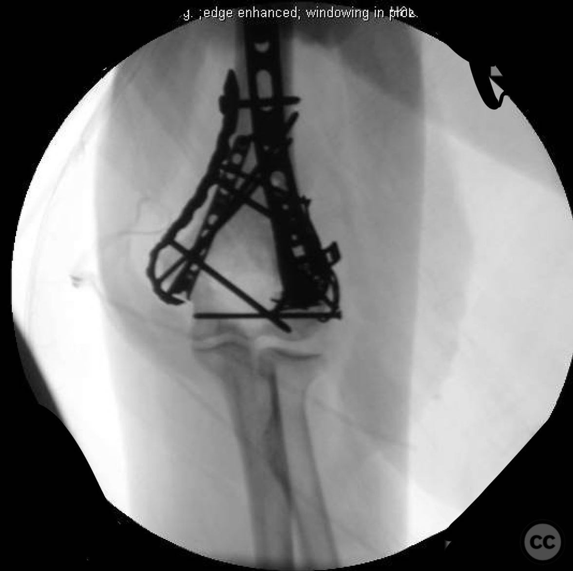
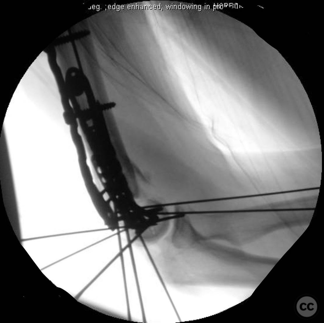
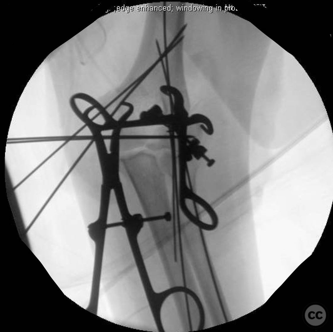
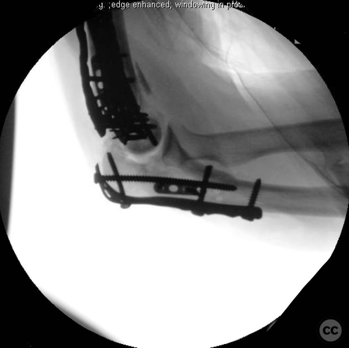
Clinical and radiological findings: A 56-year-old female presented following a high-speed motorcycle collision, sustaining a type 2 open fracture of the distal humerus with an associated olecranon fracture dislocation. The distal humerus fracture was classified as AO/OTA 13C3, characterized by a multifragmentary articular involvement with the capitellum in three pieces and the lateral epicondyle significantly displaced. The trochlea was split, with part integrated into the medial column fragment. The olecranon fracture was associated with a dislocation, complicating the elbow injury.
Preoperative Plan
Planning remarks: The preoperative plan involved utilizing the olecranon fracture as an access point to address the distal humerus fracture. Anatomic reduction of the articular fragments was prioritized, with consideration for stable fixation using dual plating of the medial and lateral columns. Given the complexity and open nature of the fracture, a backup plan for conversion to total elbow arthroplasty (TEA) was prepared if fixation proved unfeasible.
Surgical Discussion
Patient positioning: The patient was positioned supine on the operating table with the arm placed across the chest on a padded support to allow for optimal access to the posterior aspect of the elbow.
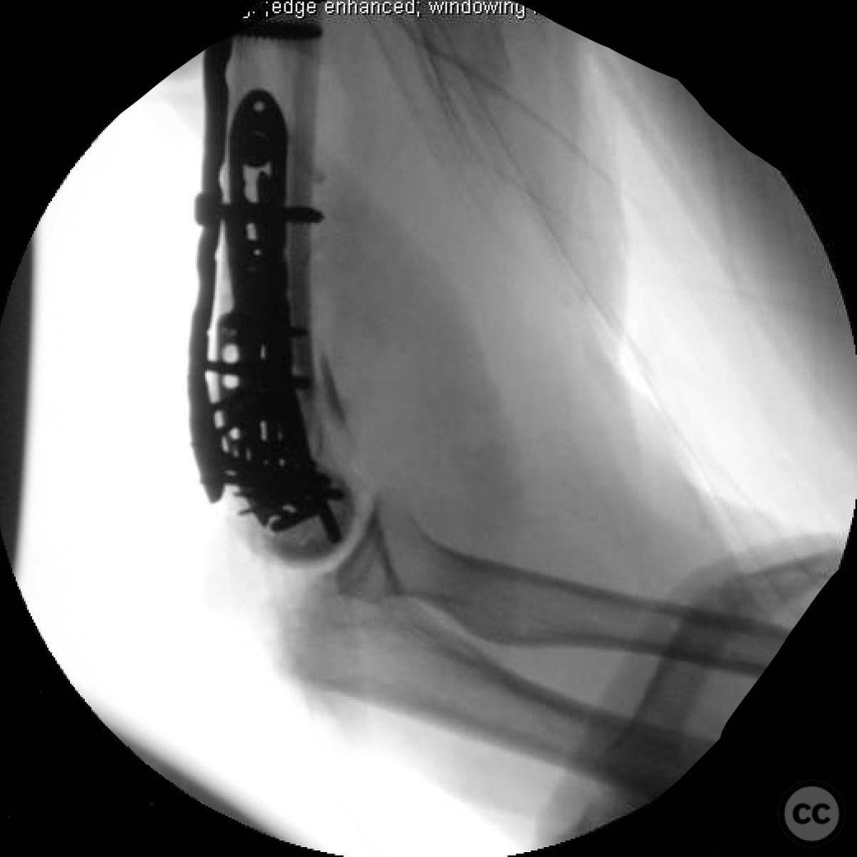
Anatomical surgical approach: A posterior approach was utilized, taking advantage of the existing olecranon fracture to access the distal humerus. The fracture allowed direct entry to the joint, facilitating visualization and manipulation of the articular fragments. Reconstruction focused on anatomically reducing the comminuted capitellum, split trochlea, and restoring column integrity, while assessing the feasibility of stable fixation throughout the procedure.
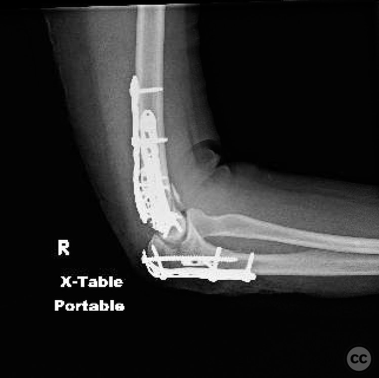
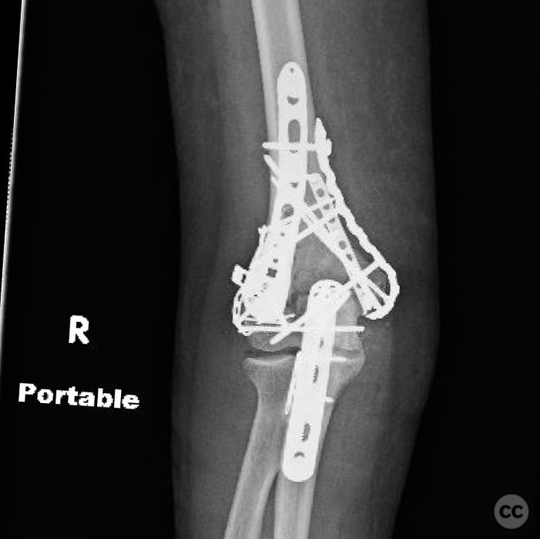
Operative remarks:Intraoperatively, achieving stable fixation was challenging due to limited bone stock and multiple small fragments. The decision was made to proceed with limited open reduction and internal fixation (ORIF) of the medial and lateral columns and olecranon. The capitellum was deemed non-viable for fixation and excised. A cement spacer was considered for temporary stabilization, with plans for delayed conversion to TEA if necessary.
Postoperative protocol: Postoperatively, the patient was immobilized in a posterior splint with the elbow in 90 degrees of flexion for two weeks. Early passive range of motion exercises were initiated at two weeks post-surgery, progressing to active-assisted exercises by six weeks, contingent on radiographic evidence of healing.
Follow up: Not specified.
Orthopaedic implants used: Dual plating system for medial and lateral column fixation, olecranon plate, potential use of a cement spacer.
Search for Related Literature

orthopaedic_trauma
- United States , Seattle
- Area of Specialty - General Trauma
- Position - Specialist Consultant

Industry Sponsership
contact us for advertising opportunities
Article viewed 302 times
19 Jul 2025
Add to Bookmarks
Full Citation
Cite this article:
Surname, Initial. (2025). Challenging Elbow Trauma: Low Distal Humerus Articular Fracture with Olecranon Dislocation in a Type II Open Injury. Journal of Orthopaedic Surgery and Traumatology. Case Report 6593618 Published Online Jul 19 2025.