Sequential Reduction and Grafting of Comminuted Acetabular Fracture
Score and Comment on this Case
Clinical Details
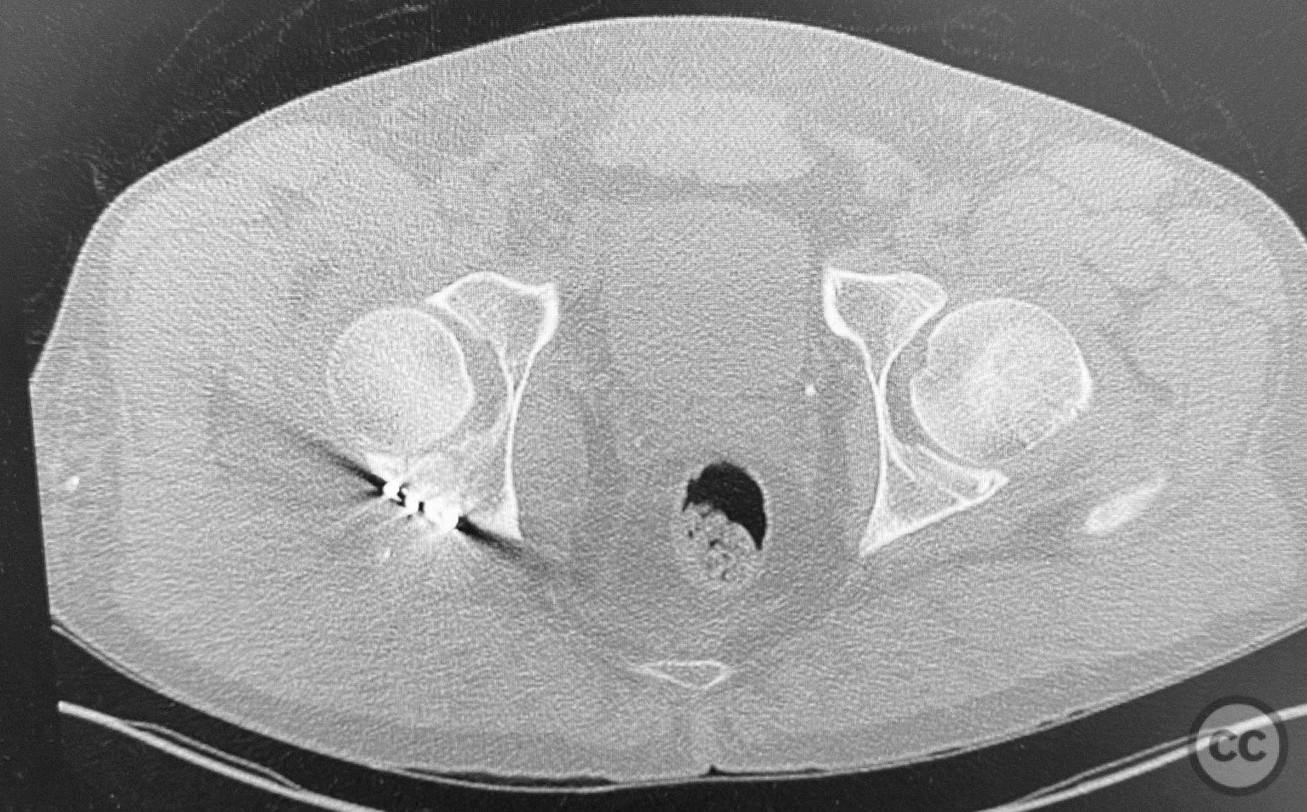
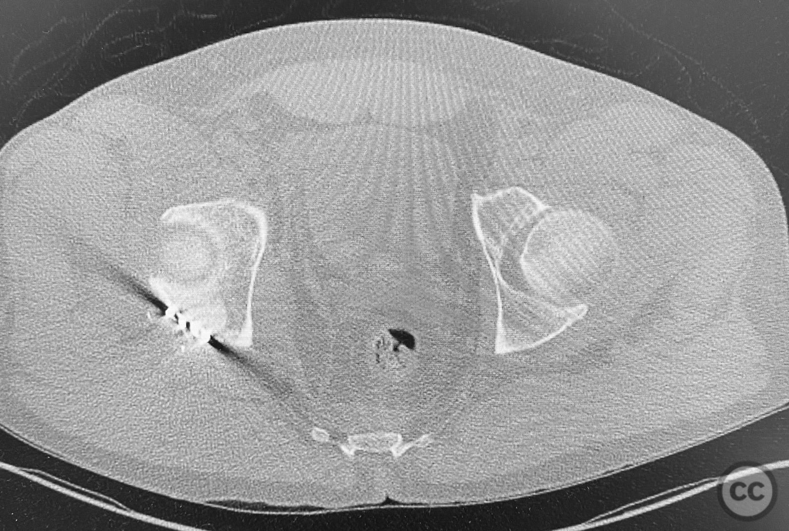
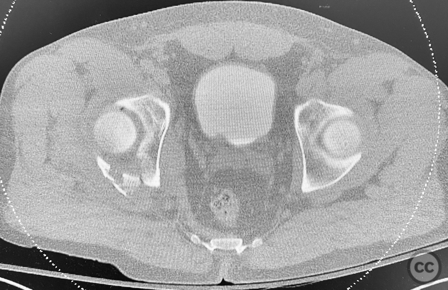
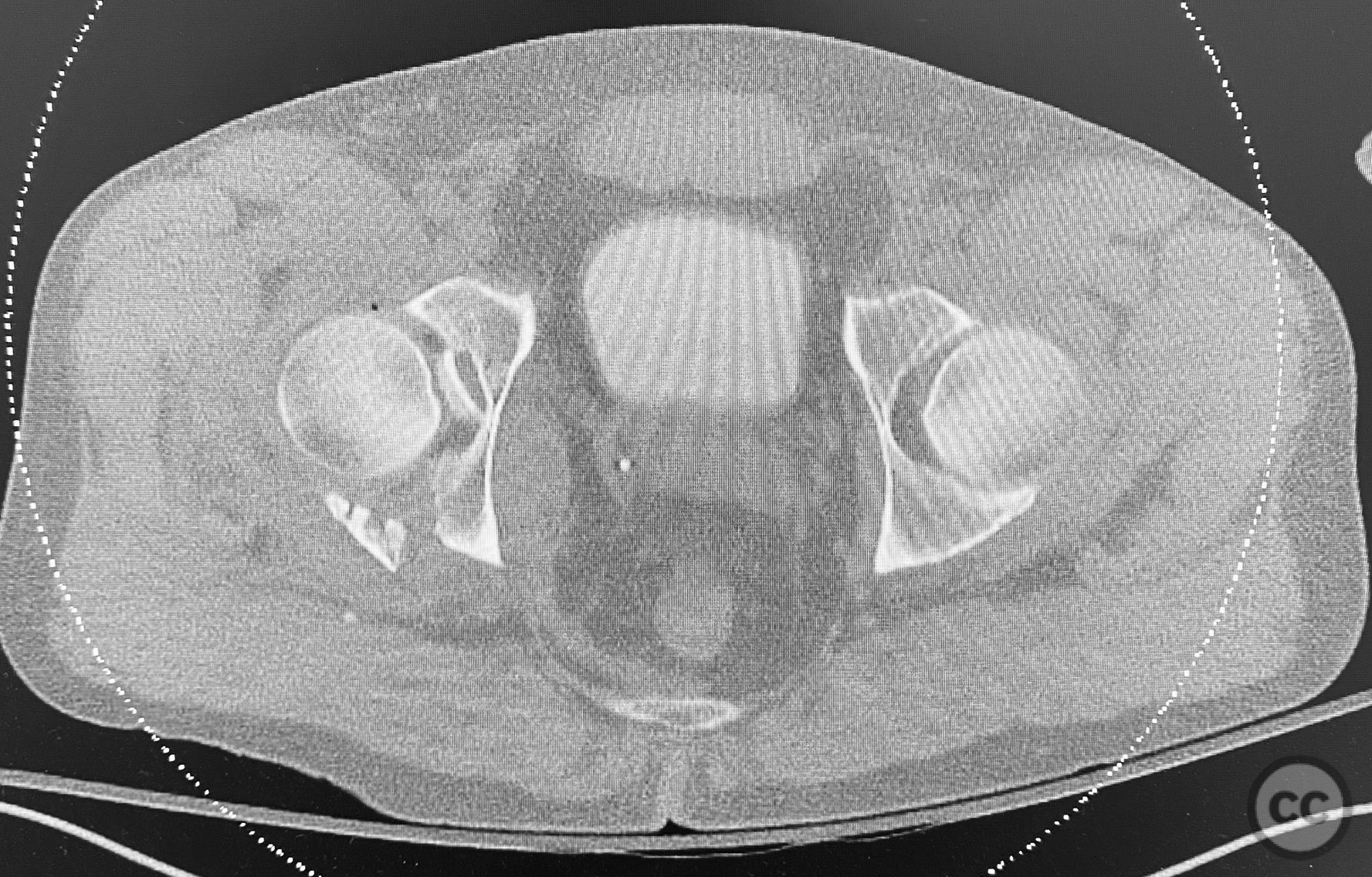
Clinical and radiological findings: The patient sustained a comminuted acetabular fracture with multiple chondrocancellous articular fragments. The injury pattern is consistent with an AO/OTA 62-B3 (both column) acetabular fracture. Initial radiographs and computed tomography demonstrated displaced intra-articular fragments with associated segmental bone loss and wall involvement. No neurovascular compromise was documented.
Preoperative Plan
Planning remarks: The preoperative plan involved an extensile approach to the acetabulum, with the intention to carefully extract all chondrocancellous fragments for ex vivo cleansing, followed by dense packing of allograft bone grit into metaphyseal defects to support articular reconstruction. Sequential reduction of articular fragments was planned, followed by reduction and fixation of the acetabular wall using plate osteosynthesis.
Surgical Discussion
Operative remarks:All chondrocancellous fragments were meticulously removed from the wound and cleansed of hematoma and debris. Dense allograft bone grit was packed into metaphyseal defects to provide subchondral support. Each articular fragment was anatomically reduced and provisionally stabilized. The posterior wall was then reduced and definitive fixation achieved using contoured reconstruction plates. Attention was paid to restoring the congruity of the acetabular dome and maintaining anatomic reduction throughout the sequence.
Postoperative protocol: Postoperatively, toe-touch weight bearing was instituted for 8 weeks, with passive and active-assisted range of motion exercises initiated on postoperative day one. Progression to partial weight bearing at 8 weeks, with full weight bearing as tolerated after radiographic evidence of healing.
Follow up: Not specified
Orthopaedic implants used: Contoured reconstruction plate(s), allograft bone grit
Search for Related Literature


Industry Sponsership
contact us for advertising opportunities
Article viewed 417 times
12 Sep 2025
Add to Bookmarks
Full Citation
Cite this article:
Routt, ML. (2025). Sequential Reduction and Grafting of Comminuted Acetabular Fracture. Journal of Orthopaedic Surgery and Traumatology. Case Report 49086970 Published Online Sep 12 2025.