Pathological Pilon Fracture with Benign Cystic Lesion
Score and Comment on this Case
Clinical Details
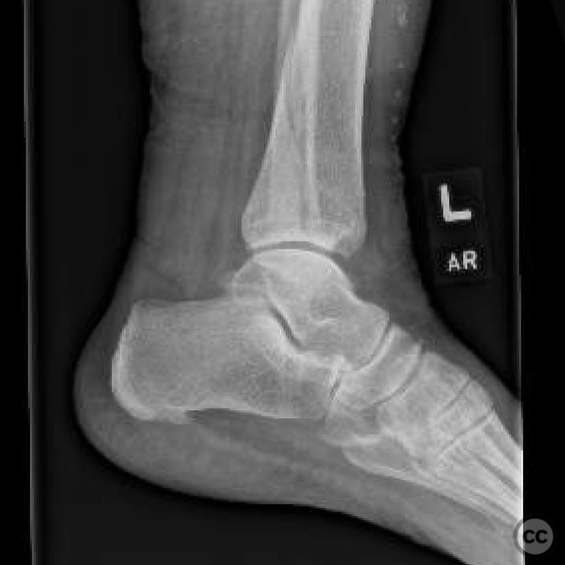
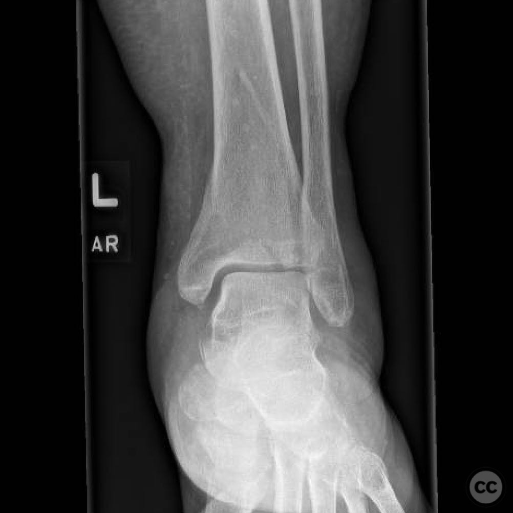
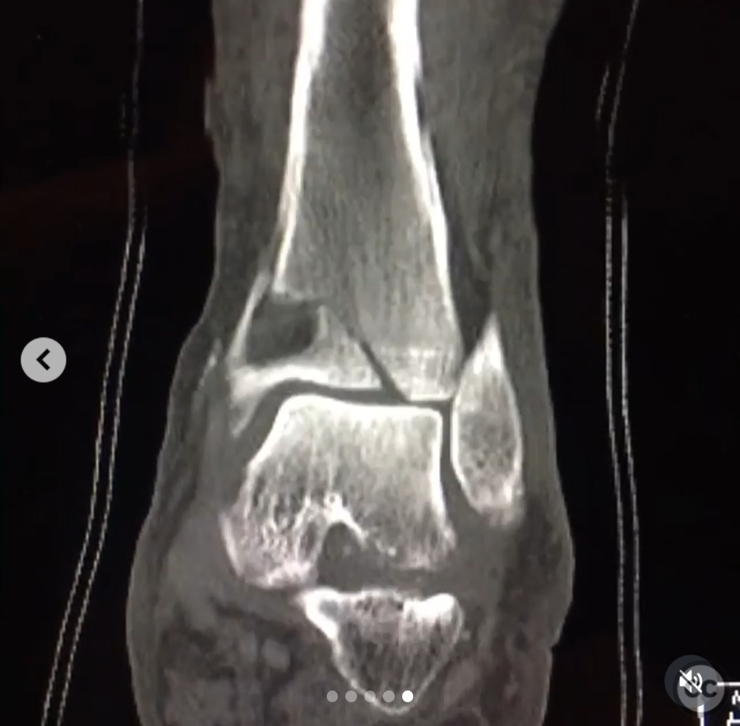
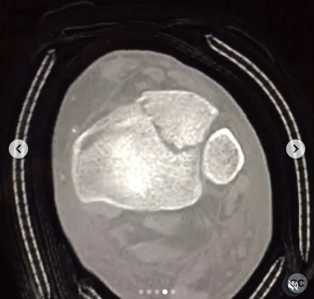
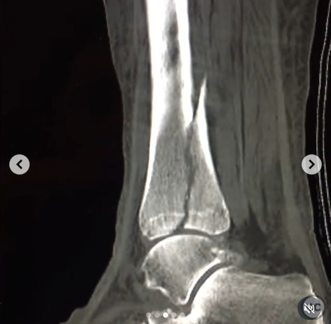
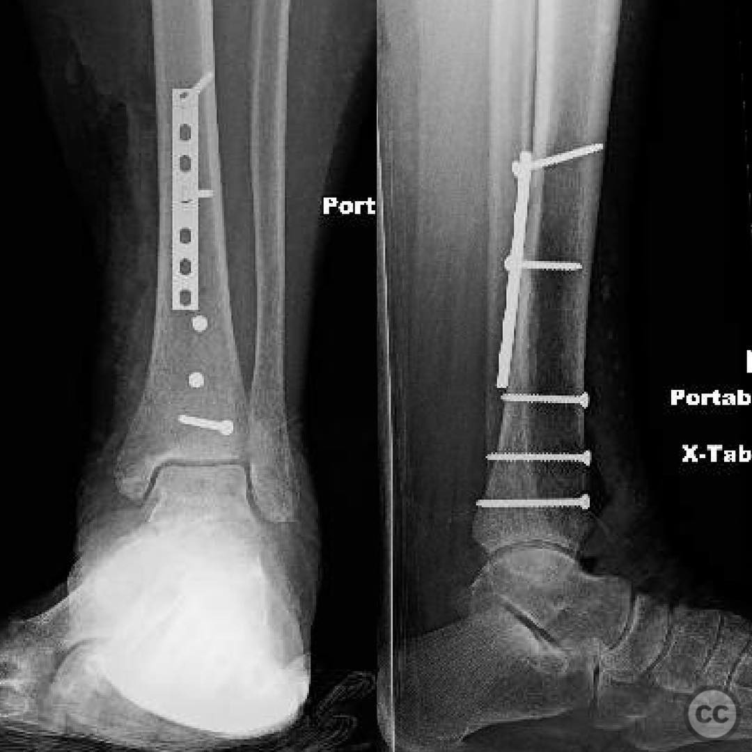
Clinical and radiological findings: A 56-year-old male presented following a low-energy fall from a curb while attempting to retrieve his dog. The patient had no prior history of ankle pain. Radiographic evaluation revealed a low-energy pilon fracture with an anteromedial meta/epiphyseal cystic lesion. The lesion was eccentrically positioned, well-marginated, non-expansile, and without cortical destruction or soft tissue involvement, suggestive of a benign process. However, given the patient's demographic, metastatic disease was considered a differential diagnosis.
Preoperative Plan
Planning remarks: The preoperative plan included obtaining a biopsy with intra-operative frozen section analysis to rule out primary bone malignancy and ensure adequate tissue for final pathological diagnosis. Upon confirmation of non-specific inflammation and absence of malignancy, surgical fixation was planned. The fracture pattern was classified as AO/OTA 43-B type, indicating a partial articular fracture.
Surgical Discussion
Patient positioning: The patient was positioned supine on the operating table with the affected limb prepared for a posteromedial approach.
Anatomical surgical approach: A posteromedial approach was utilized, involving an incision along the medial aspect of the distal tibia. Subperiosteal dissection was performed to expose the fracture site and underlying cystic lesion.
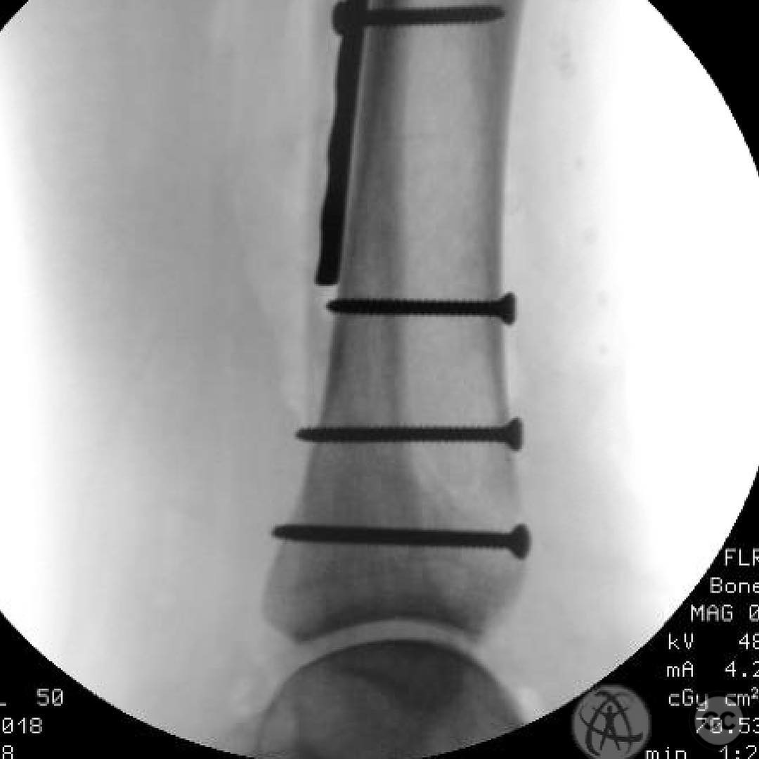
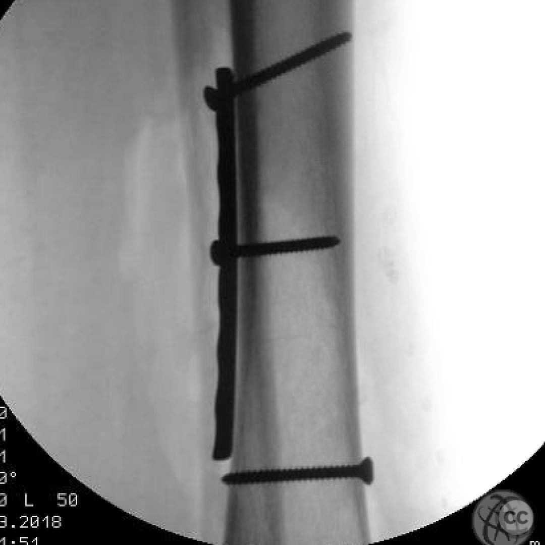
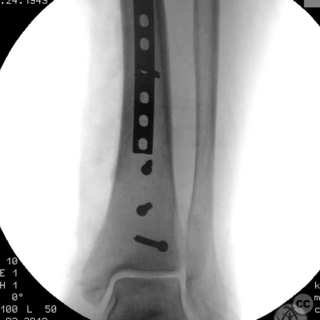
Operative remarks:Intraoperative frozen section analysis revealed non-specific inflammation with no evidence of malignancy, allowing for fracture fixation to proceed. Direct anatomic reduction of the articular surface was achieved at the fracture apex. An anti-glide buttress plate was applied for stabilization, supplemented by interfragmentary compression using lag screws.
Postoperative protocol: Postoperatively, the patient was advised to maintain non-weight bearing status for 6 weeks, followed by progressive weight bearing as tolerated. Range of motion exercises were initiated early to prevent joint stiffness.
Follow up: Not specified.
Orthopaedic implants used: Anti-glide buttress plate, interfragmentary lag screws.
Search for Related Literature

orthopaedic_trauma
- United States , Seattle
- Area of Specialty - General Trauma
- Position - Specialist Consultant

Industry Sponsership
contact us for advertising opportunities
Article viewed 301 times
23 Jul 2025
Add to Bookmarks
Full Citation
Cite this article:
Surname, Initial. (2025). Pathological Pilon Fracture with Benign Cystic Lesion. Journal of Orthopaedic Surgery and Traumatology. Case Report 47767805 Published Online Jul 23 2025.