._._._._This is a simple set _5.jpg)
._._._._This is a simple set o(.jpg)
._._._._This is a simple set _2.jpg)
._._._._This is a simple set _1.jpg)
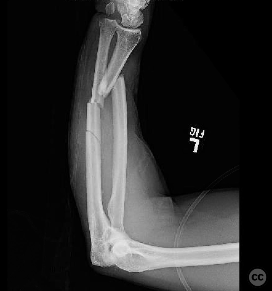
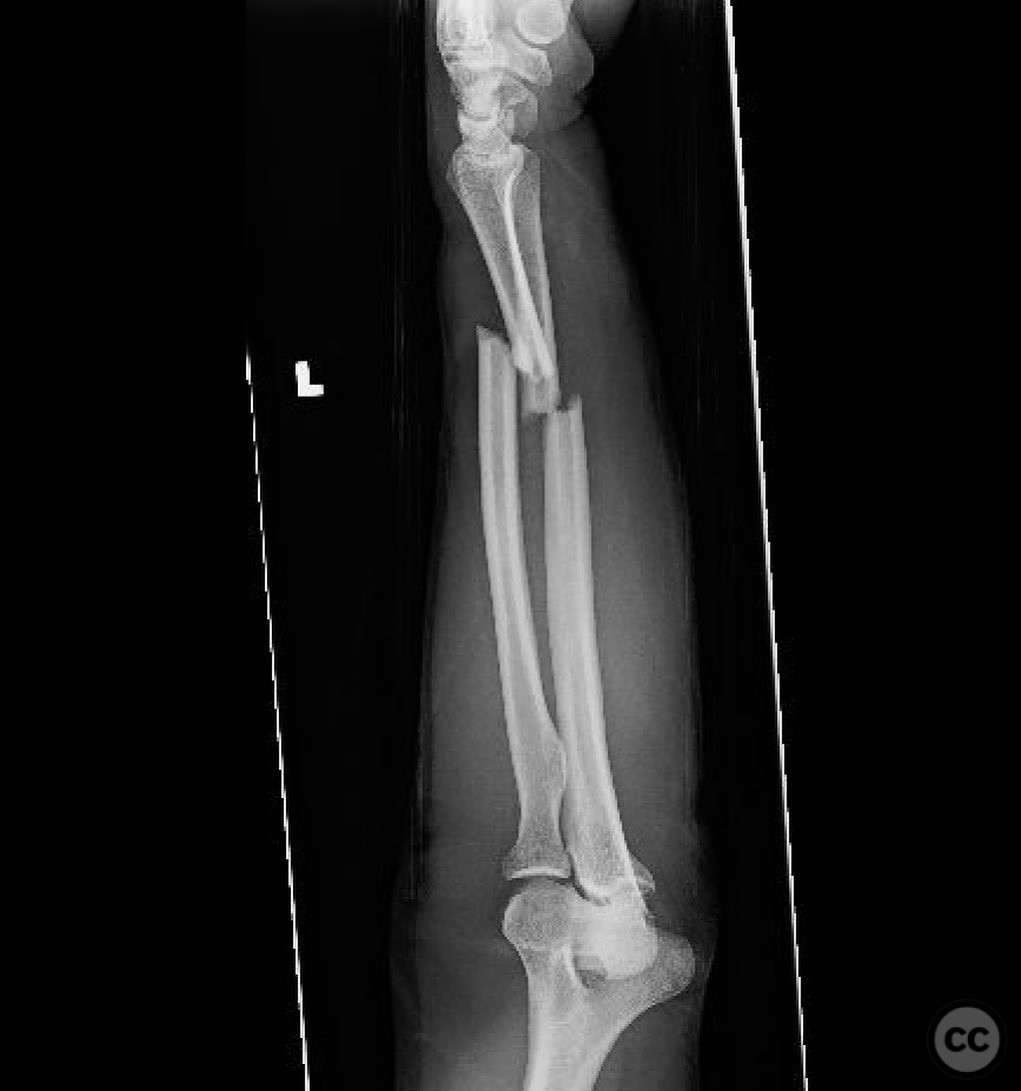
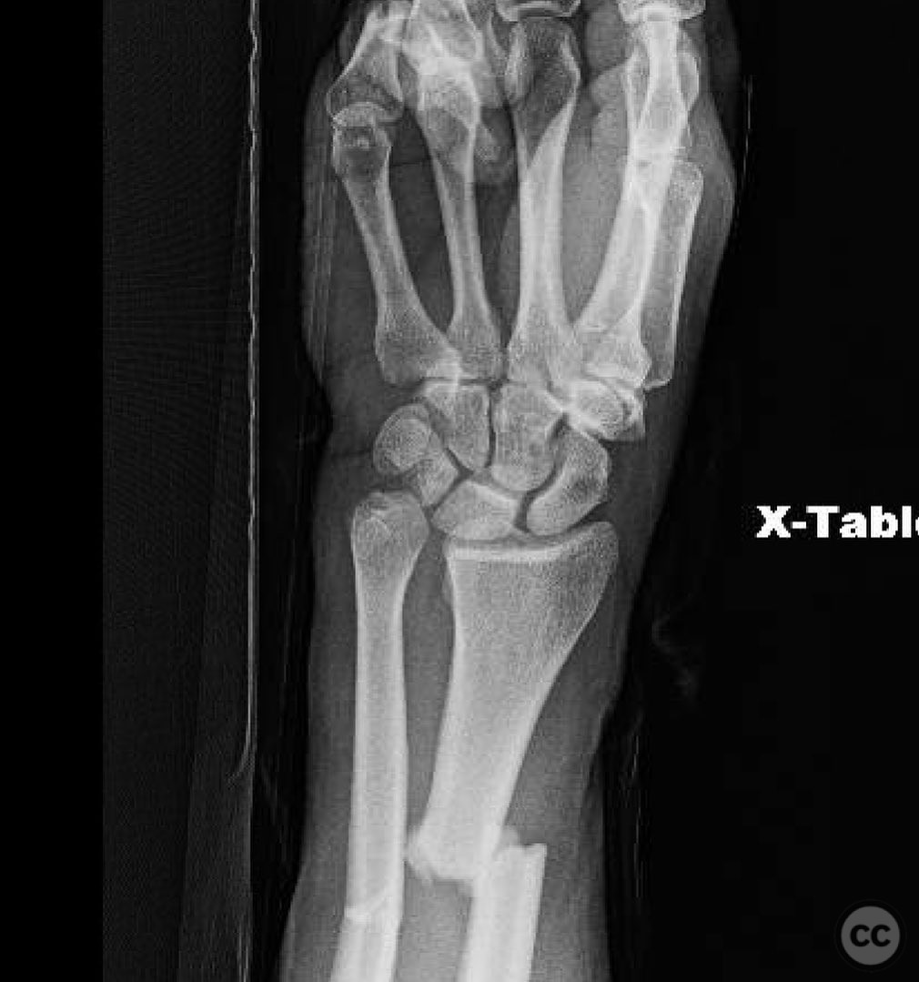
Galeazzi-like Both Bones Forearm Fracture with DRUJ Dislocation.
Score and Comment on this Case
Clinical Details
Clinical and radiological findings: The patient presented with a both bones forearm fracture, characterized by a simple fracture pattern in the radius and a segmental fracture in the ulna, accompanied by a distal radioulnar joint (DRUJ) dislocation. The radial fracture required restoration of normal length and alignment, particularly the radial bow, to ensure proper function and stability of the DRUJ.
Preoperative Plan
Planning remarks: The preoperative plan involved a volar Henry approach for the radius to achieve direct anatomic reduction and interfragmentary compression using compression plating. For the ulna, a subcutaneous approach was planned with the plate placed on the dorsal surface under the extensor carpi ulnaris (ECU), utilizing a lag screw for the oblique fracture and a compression plate for the transverse fracture.
Surgical Discussion
Patient positioning: Supine positioning with the arm on an arm board to allow for optimal access to both the volar and dorsal aspects of the forearm.
Anatomical surgical approach: A volar Henry approach was employed for the radius, involving an incision along the flexor carpi radialis tendon, with careful retraction of the radial artery and flexor tendons to expose the fracture site. For the ulna, a subcutaneous dorsal approach was used, with exposure of the dorsal surface under the ECU for plate placement.
Operative remarks:The surgeon emphasized the importance of achieving absolute stability through compression plating for both fractures. For the DRUJ, examination in pronation, neutral, and supination was conducted to assess stability. Due to concerns about patient compliance, cross pinning with 2.0mm pins was performed in a neutral position to maintain DRUJ stability.
Postoperative protocol: Postoperative rehabilitation included immobilization in a splint with restricted forearm rotation for 6 weeks, followed by gradual range of motion exercises and strengthening as tolerated.
Follow up: Not specified.
Orthopaedic implants used: 3.5mm dynamic compression plates (DCP), 2.0mm cross pins.
Search for Related Literature

orthopaedic_trauma
- United States , Seattle
- Area of Specialty - General Trauma
- Position - Specialist Consultant

Industry Sponsership
contact us for advertising opportunities
Article viewed 241 times
19 Jul 2025
Add to Bookmarks
Full Citation
Cite this article:
Surname, Initial. (2025). Galeazzi-like Both Bones Forearm Fracture with DRUJ Dislocation.. Journal of Orthopaedic Surgery and Traumatology. Case Report 47452430 Published Online Jul 19 2025.