


Complex Pilon Fracture with Segmental Bone Loss and Syndesmotic Disruption
Score and Comment on this Case
Clinical Details
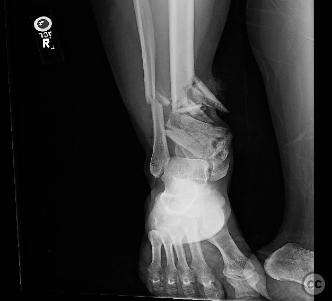
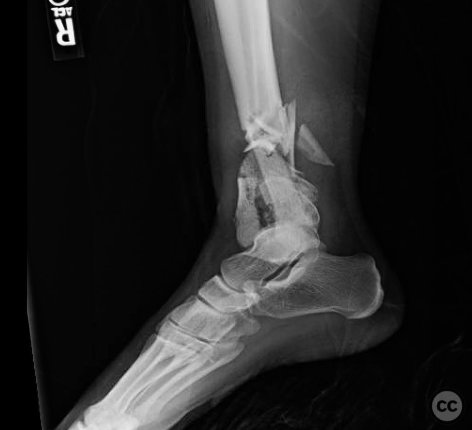
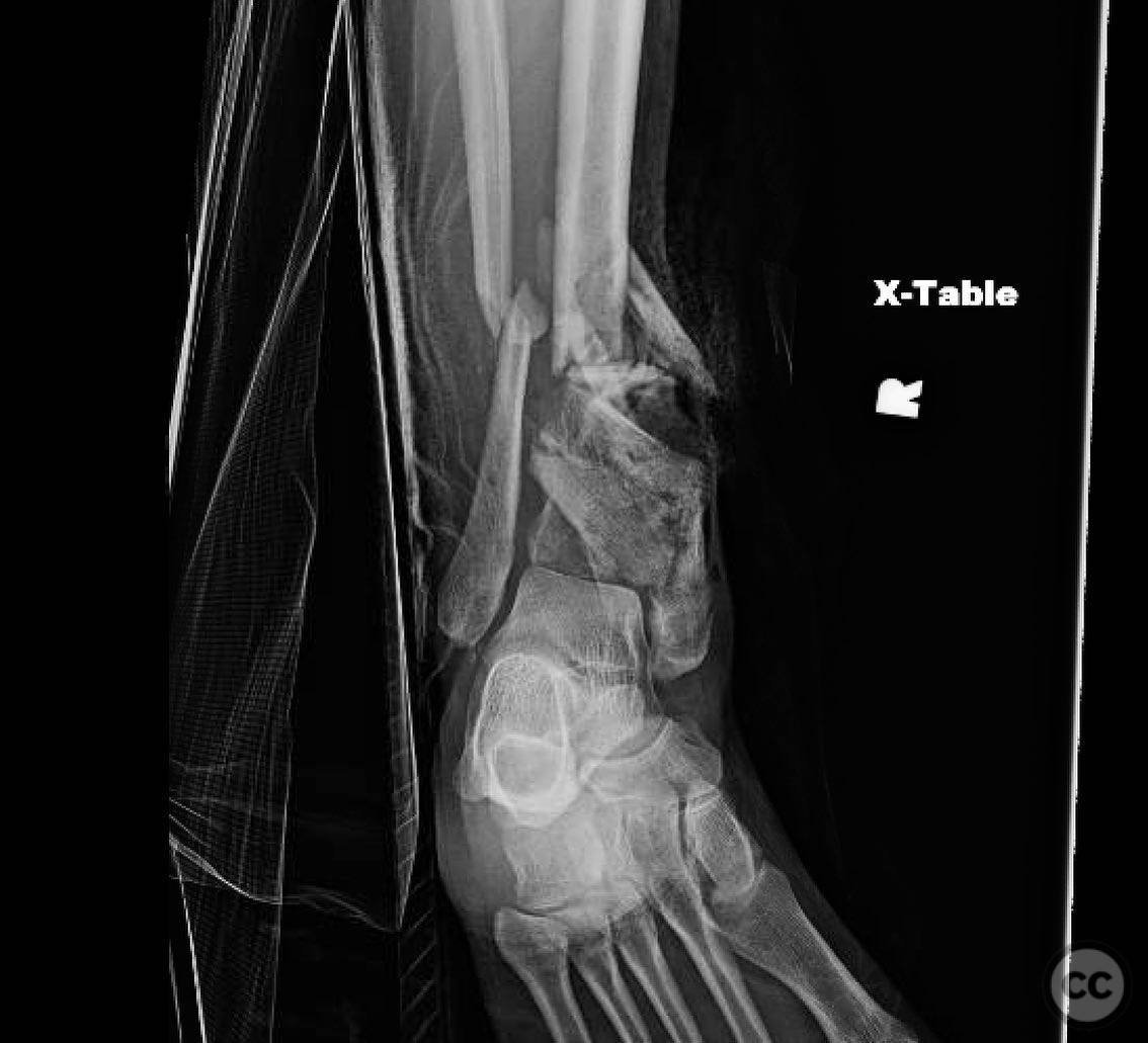
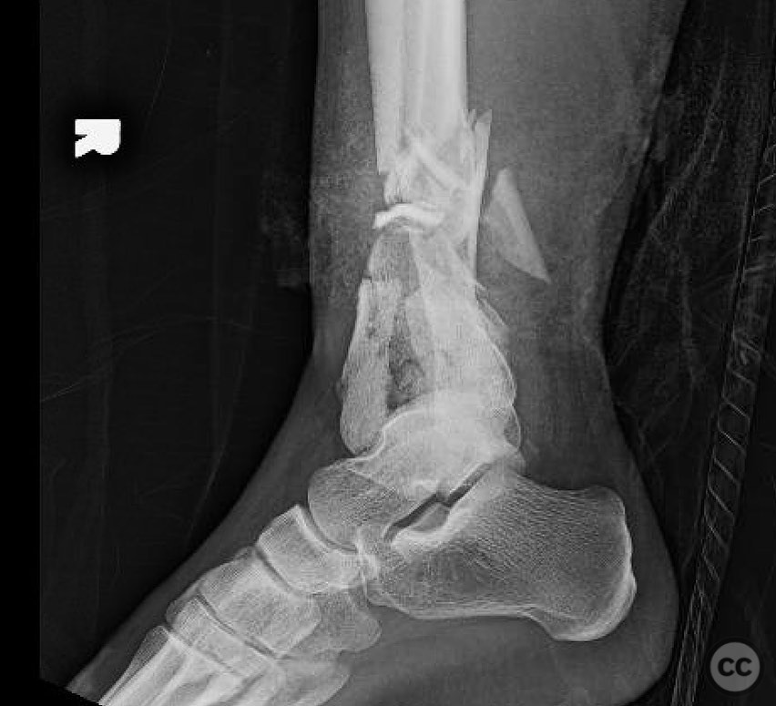
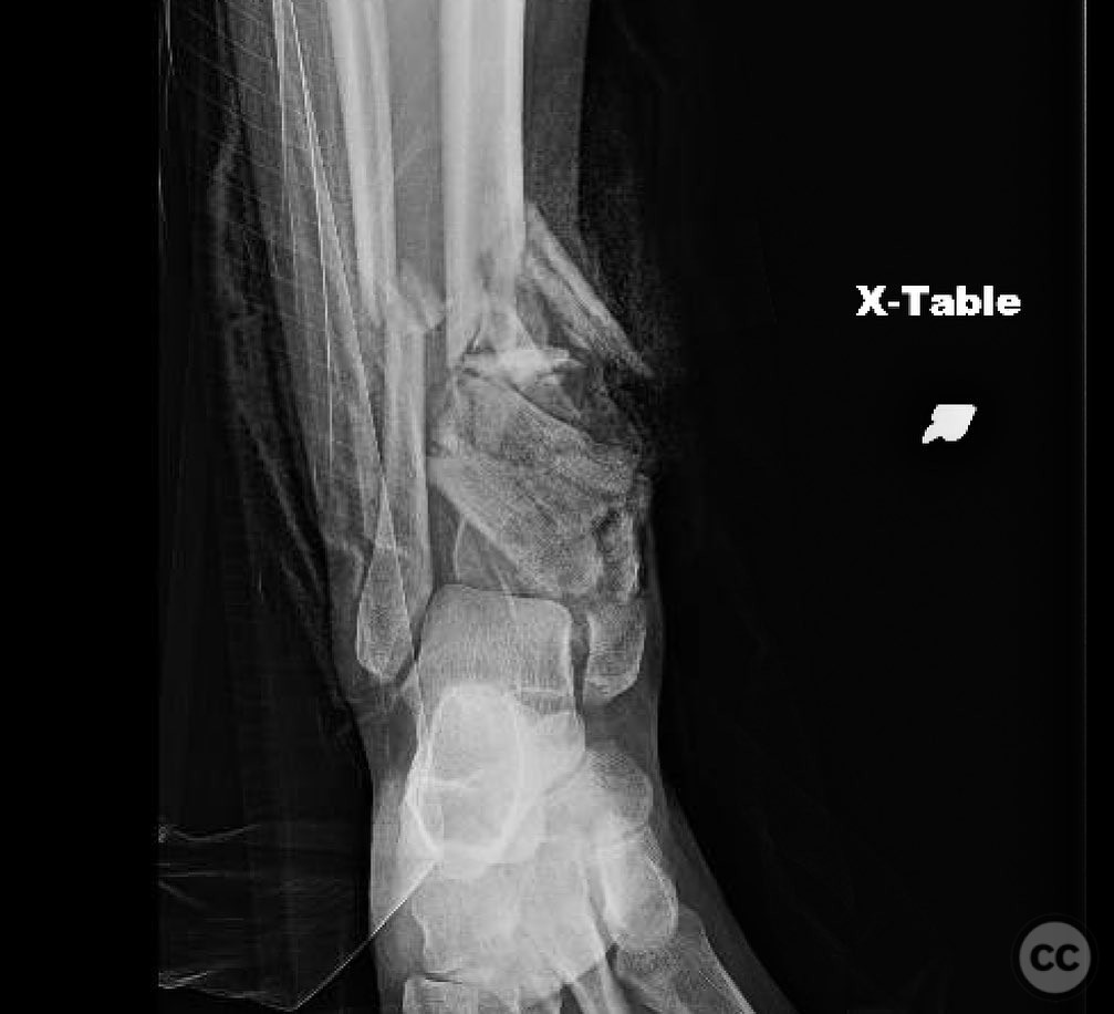
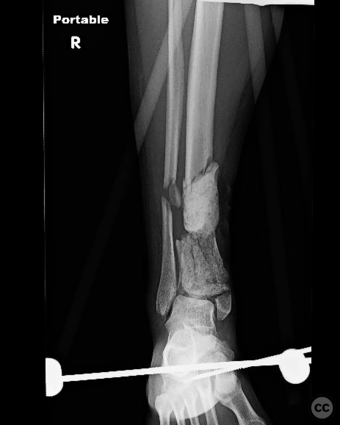
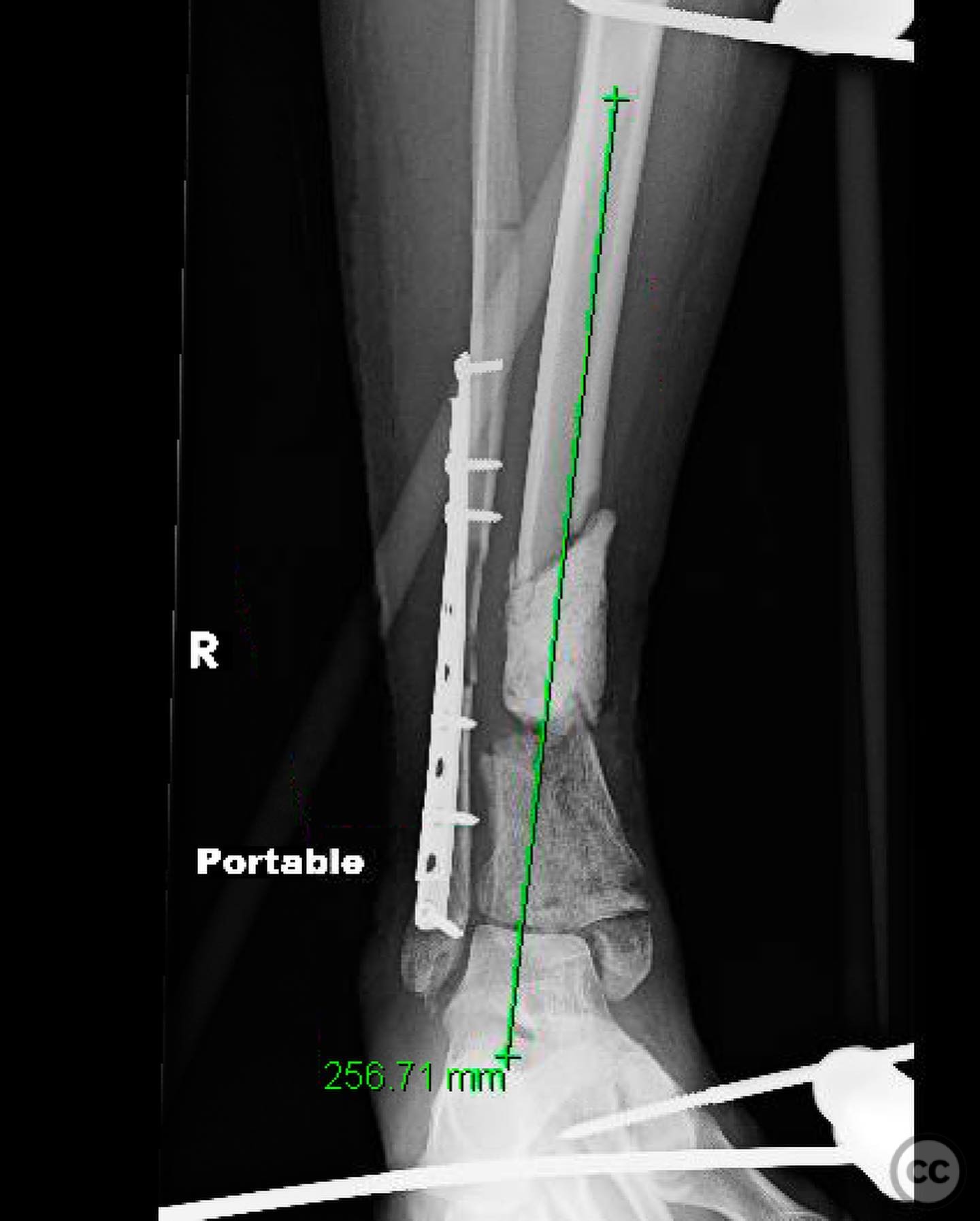
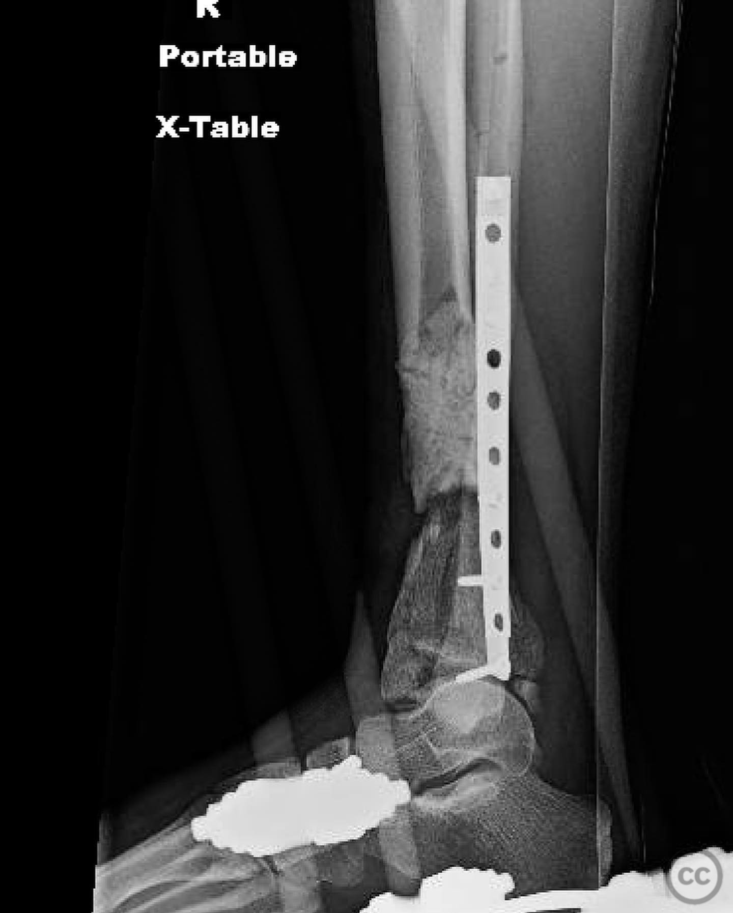
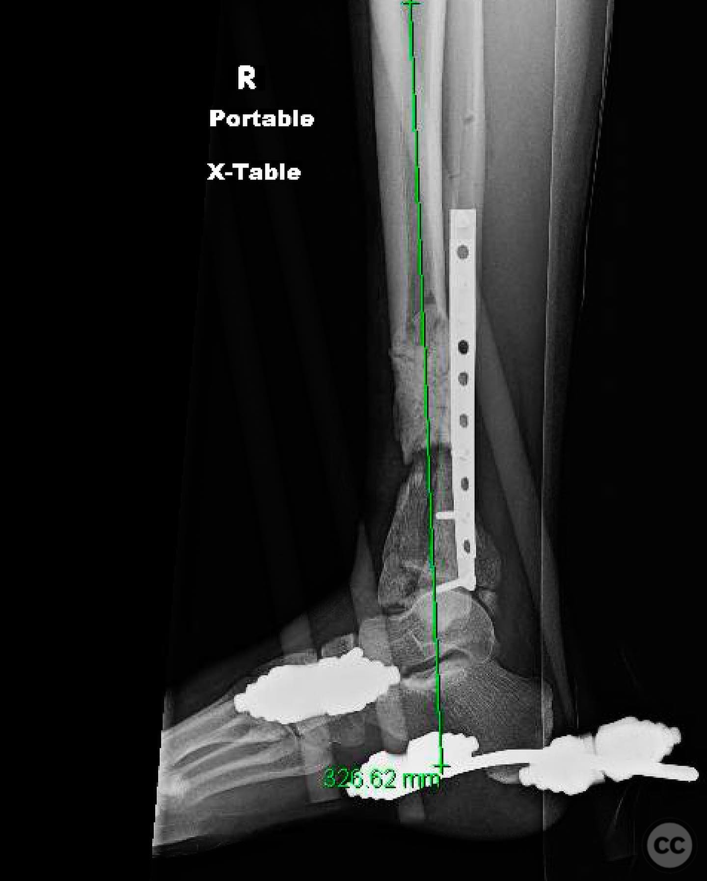
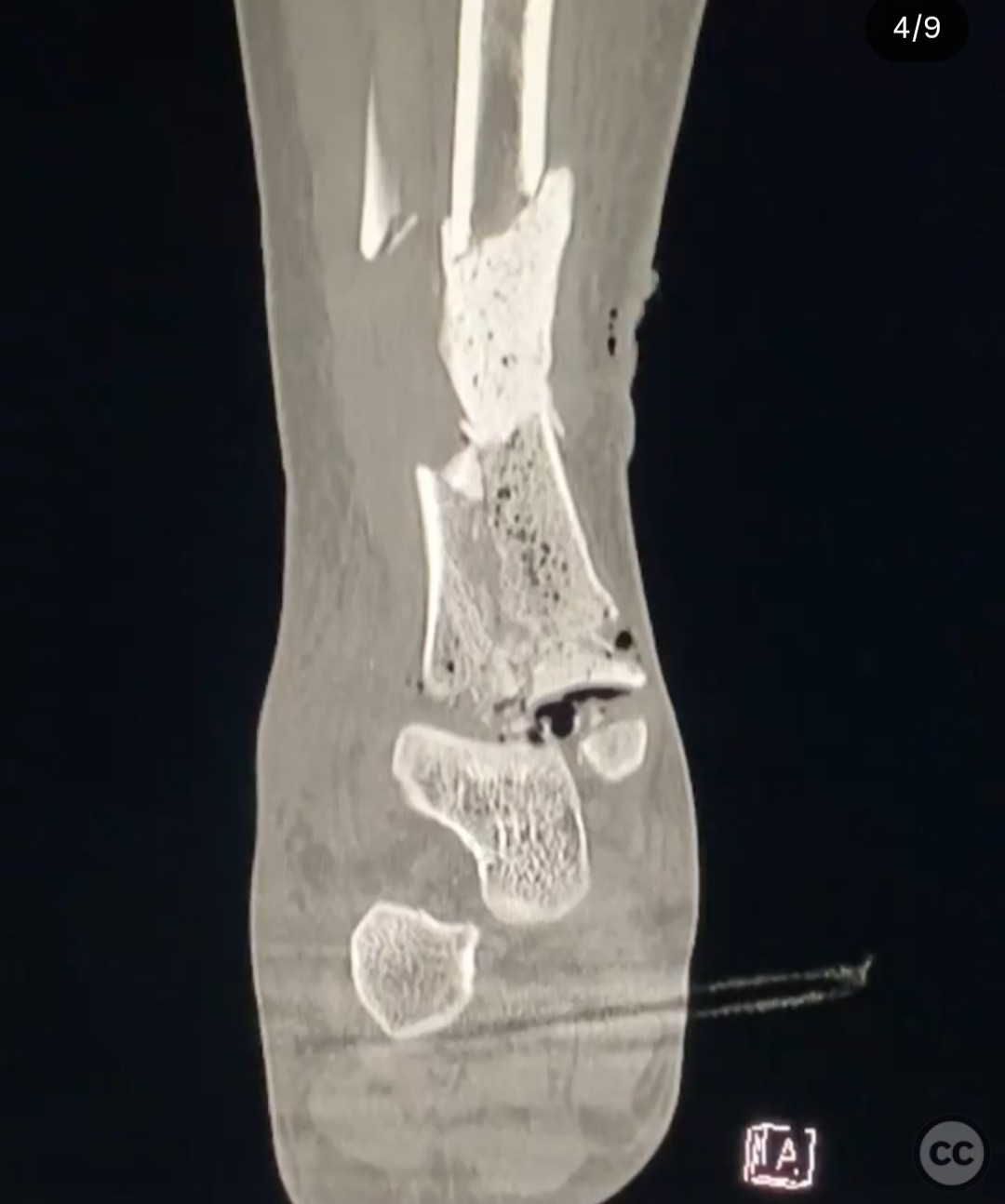
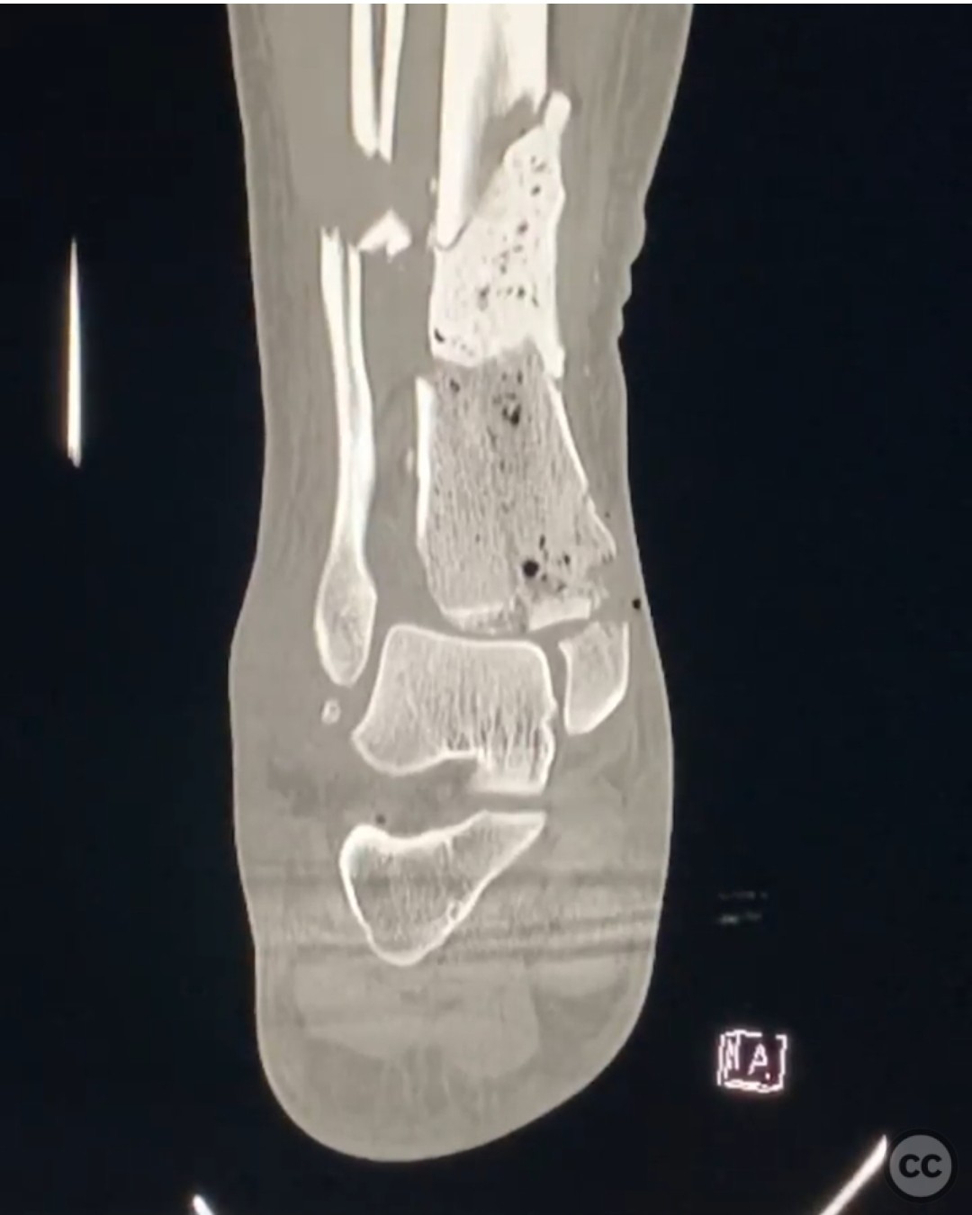
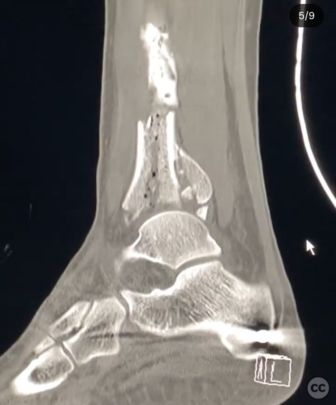
Clinical and radiological findings: A 23-year-old male sustained a fall from a height of 60 feet, resulting in a complex open fracture of the distal tibia (pilon fracture) with segmental bone loss and complete syndesmotic injury. The open wound was located medially, measuring 4 cm, with oblique orientation at the meta-diaphyseal junction. The underlying skin was blanched, and the wound was contaminated with dirt. The limb was well perfused with no vascular or neurological deficits, and compartment pressures were normal. An ipsilateral closed femur fracture was also present. Initial imaging and clinical examination confirmed the extent of the injury, classified as a 3A open fracture according to the Gustilo-Anderson classification.
Preoperative Plan
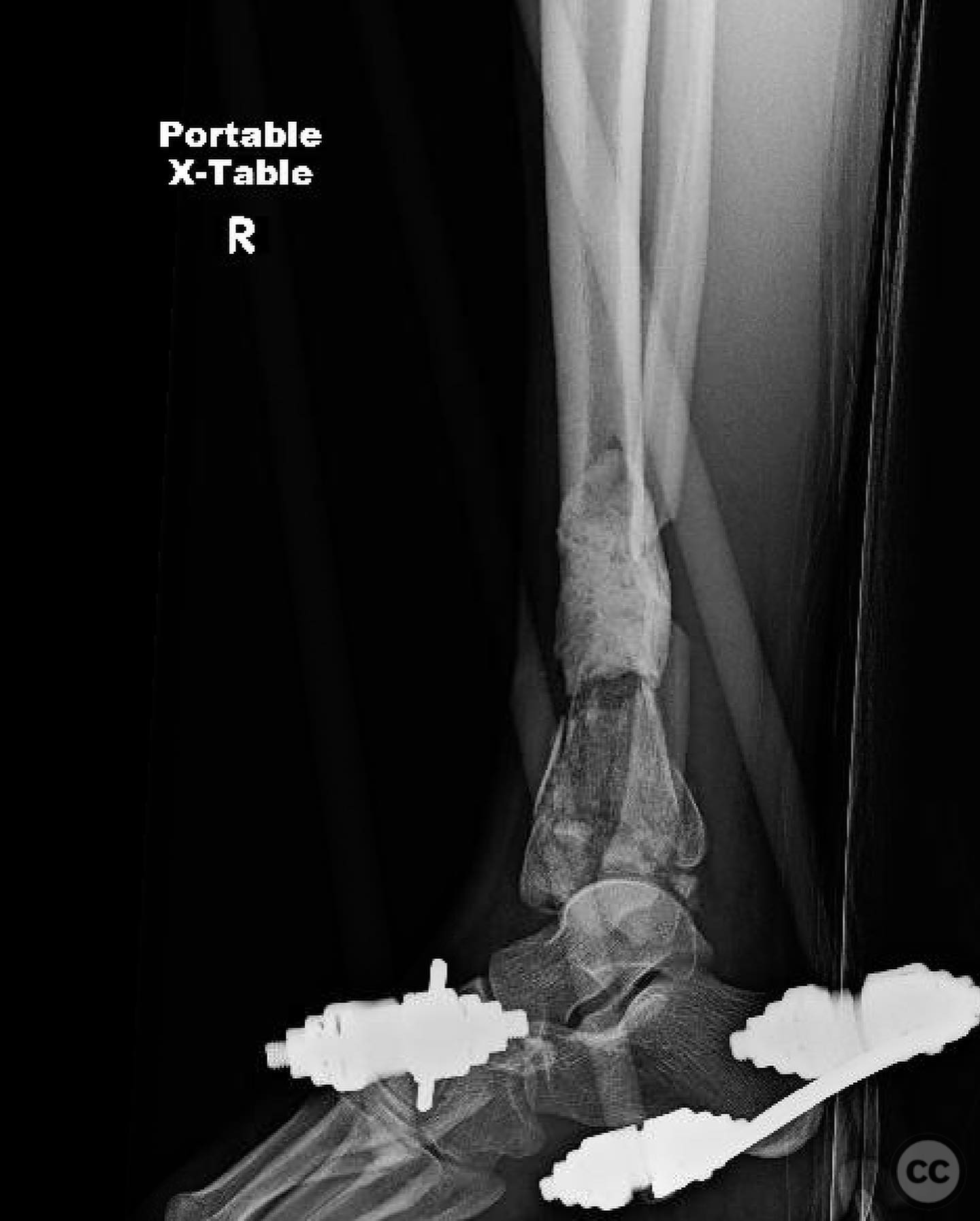
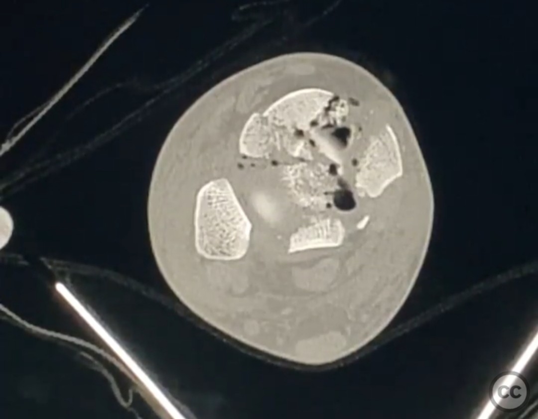
Planning remarks: The initial surgical plan involved urgent debridement and irrigation of the open wound, followed by reduction and application of an external fixator to stabilize the fracture temporarily. A CT scan was obtained post-initial stabilization to assess the fracture pattern and plan for definitive fixation. The fibula was addressed with open reduction and internal fixation on the following day.
Surgical Discussion
Patient positioning: The patient was positioned supine on the operating table for both the initial debridement and subsequent fibular fixation.
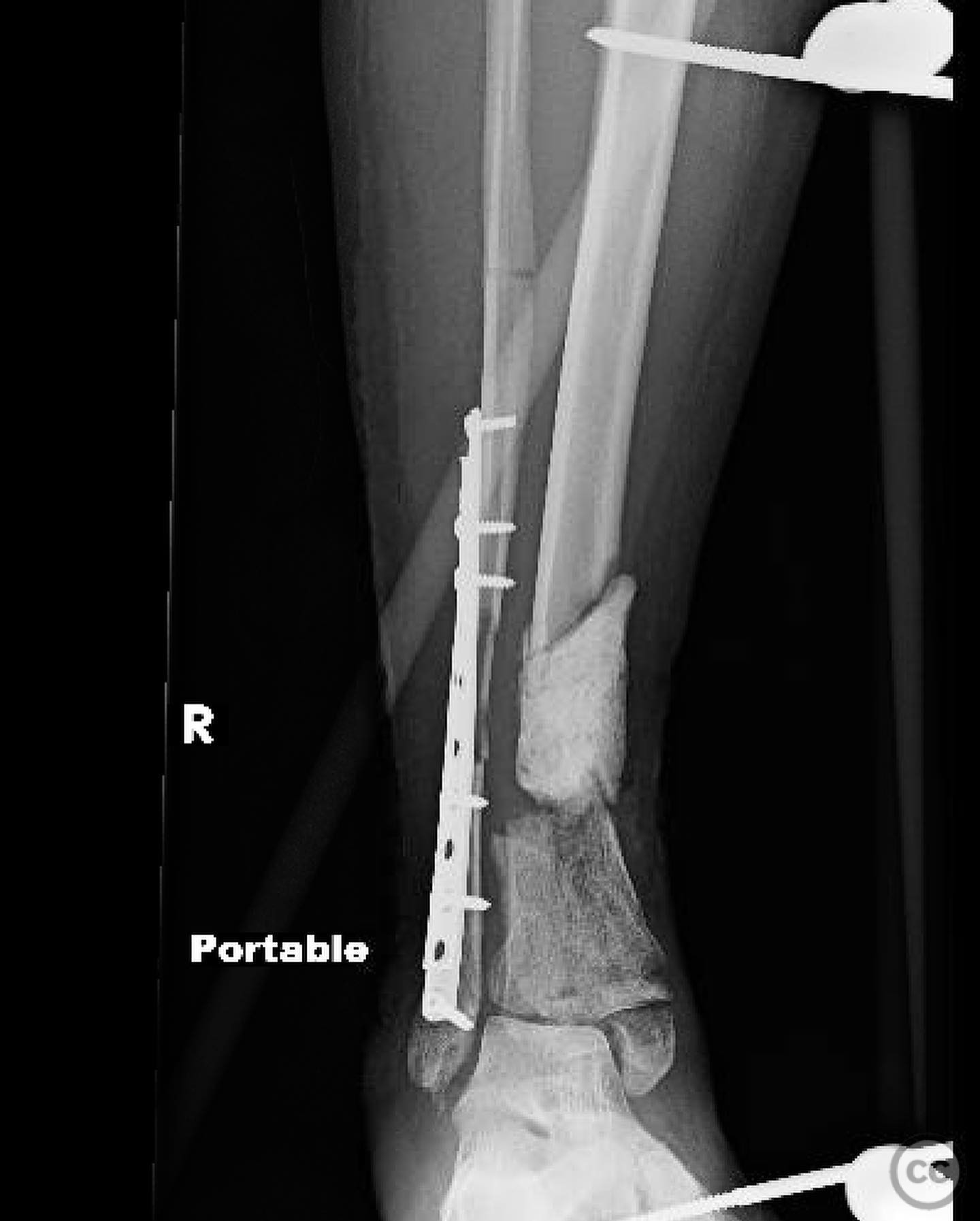
Anatomical surgical approach: A medial approach was avoided due to the location of the traumatic wound. Instead, a lateral approach was utilized for fibular fixation, involving an incision over the fibula with careful dissection to expose and reduce the fracture site.
Operative remarks:The surgical team encountered significant challenges due to the absence of ligamentotaxis and reference fragments for reconstruction. The articular block was initially reconstructed too distally relative to the fibula, necessitating intraoperative adjustment. A suture anchor was used to recreate the anterior inferior tibiofibular ligament (AITFL), and a syndesmosis screw was placed into cement to stabilize the syndesmosis due to lack of intact tibial bone stock.
Postoperative protocol: Postoperatively, the patient was placed in a non-weight-bearing status with a focus on maintaining range of motion exercises for adjacent joints. Weight-bearing status would be reassessed following definitive bone grafting and stabilization.
Follow up: Not specified
Orthopaedic implants used: External fixator, suture anchor for AITFL reconstruction, syndesmosis screw into cement, internal fixation for fibula (specific implant not detailed).
Search for Related Literature

orthopaedic_trauma
- United States , Seattle
- Area of Specialty - General Trauma
- Position - Specialist Consultant

Industry Sponsership
contact us for advertising opportunities
Article viewed 324 times
16 Jul 2025
Add to Bookmarks
Full Citation
Cite this article:
Surname, Initial. (2025). Complex Pilon Fracture with Segmental Bone Loss and Syndesmotic Disruption. Journal of Orthopaedic Surgery and Traumatology. Case Report 46777828 Published Online Jul 16 2025.