 Pay attention to the subtle details on the injury and traction views. Go back to the previous post- you can s_1.jpg)
 Pay attention to the subtle details on the injury and traction views. Go back to the previous post- you can s_2.jpg)
 Pay attention to the subtle details on the injury and traction views. Go back to the previous post- you can s_3.jpg)
 Pay attention to the subtle details on the injury and traction views. Go back to the previous post- you can s_4.jpg)
 Pay attention to the subtle details on the injury and traction views. Go back to the previous post- you can s_5.jpg)
 Pay attention to the subtle details on the injury and traction views. Go back to the previous post- you can s_7.jpg)
 Pay attention to the subtle details on the injury and traction views. Go back to the previous post- you can s_6.jpg)
 Pay attention to the subtle details on the injury and traction views. Go back to the previous post- you can s_8.jpg)
 Pay attention to the subtle details on the injury and traction views. Go back to the previous post- you can s_9.jpg)
 Pay attention to the subtle details on the injury and traction views. Go back to the previous post- you can se(.jpg)
Comminuted Capitellum Fracture with Lateral Epicondyle Involvement in a Hockey Player
Score and Comment on this Case
Clinical Details
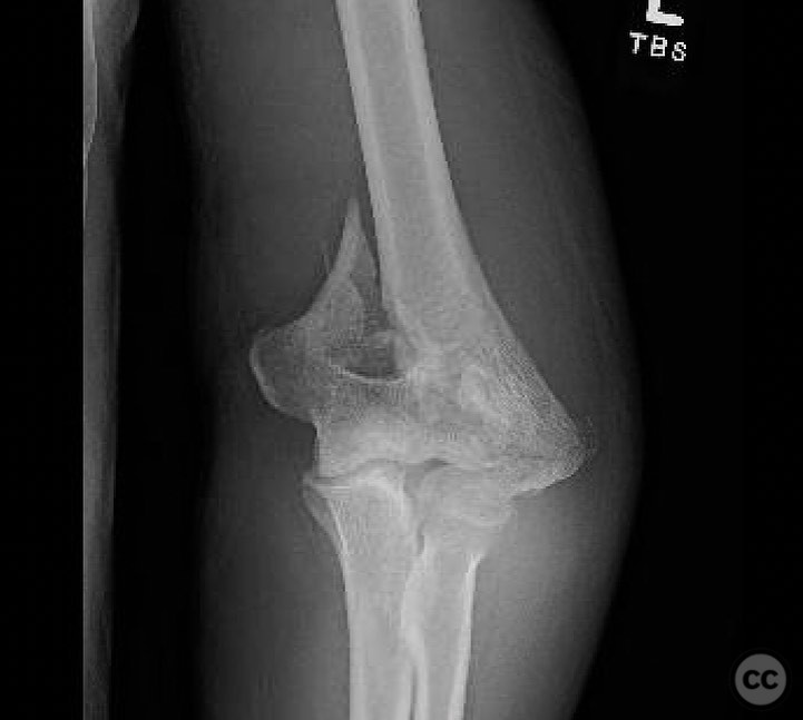
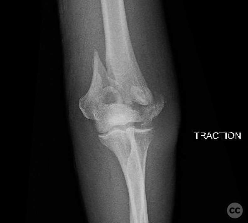
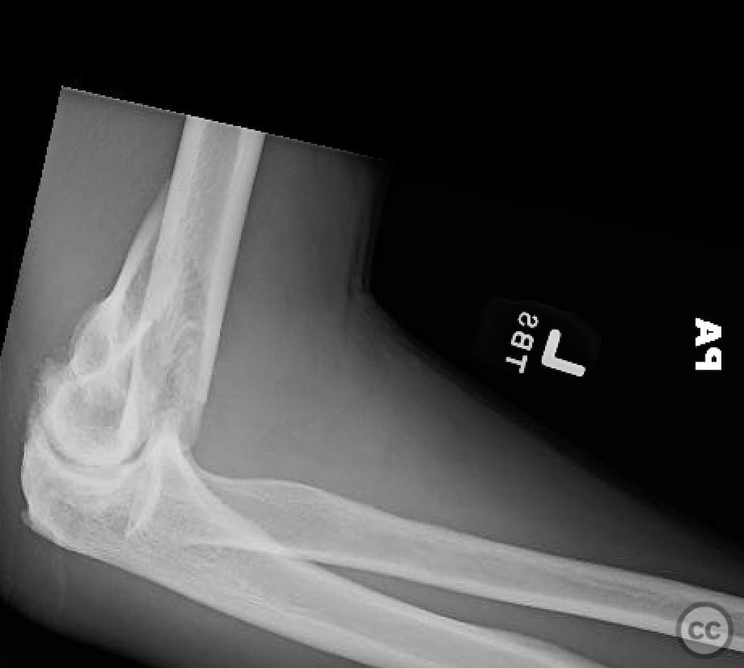
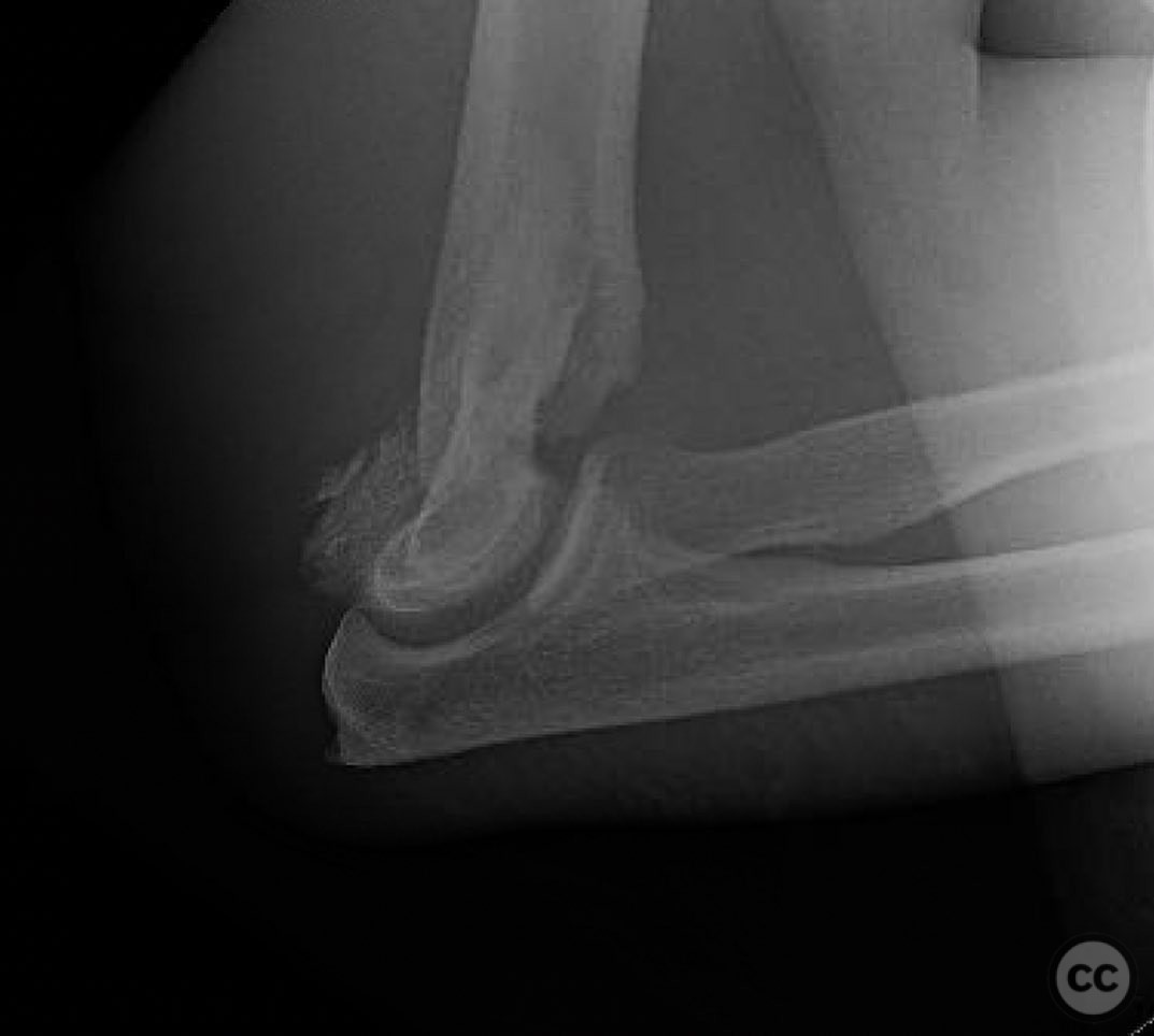
Clinical and radiological findings: A 31-year-old male hockey player sustained a closed, isolated injury to the elbow following a fall. Initial radiographs were challenging to interpret due to the complexity of the distal humerus anatomy. A traction view was utilized, revealing a comminuted fracture of the capitellum with multiple suspicious densities on the lateral side. The lateral epicondyle was also involved, but the trochlea appeared uninjured.
Preoperative Plan
Planning remarks: The preoperative plan involved exploiting the lateral epicondyle fracture to gain access to the capitellum. An olecranon osteotomy was deemed unnecessary as the fracture pattern was radial to the trochlea. The surgical approach aimed to move the lateral epicondyle, LUCL, and muscular origins out of the way to visualize and address the capitellum fracture.
Surgical Discussion
Patient positioning: The patient was positioned supine with the arm supported on a hand table, allowing for optimal access to the lateral aspect of the elbow.
Anatomical surgical approach: A lateral approach was employed, utilizing the existing fracture line of the lateral epicondyle for osteotomy. The LUCL and muscular origins were carefully retracted to expose the anterior aspect of the capitellum for direct visualization and reduction.
Operative remarks:The surgeon noted that the fracture pattern required meticulous reconstruction akin to solving a puzzle. Although an olecranon osteotomy is traditionally used to visualize the trochlea, it was not helpful in this case since the pathology was primarily radial to the trochlea. Instead, access to the comminuted capitellum was achieved via a lateral epicondyle osteotomy (or by utilizing the existing lateral epicondyle fracture), which allowed direct visualization and reduction. Intraoperatively, the “double bubble” sign was observed, indicating partial fixation of the capitellum. Subsequent lateral views confirmed successful reduction and fixation, with complete resolution of the double bubble sign.
Postoperative protocol: Postoperative rehabilitation included immobilization in a posterior splint for two weeks, followed by gradual range of motion exercises. Strengthening exercises were introduced at six weeks post-surgery.
Follow up: Not specified.
Orthopaedic implants used: Orthopaedic implants used included screws for fixation of the capitellum and lateral epicondyle.
Search for Related Literature

orthopaedic_trauma
- United States , Seattle
- Area of Specialty - General Trauma
- Position - Specialist Consultant

Industry Sponsership
contact us for advertising opportunities
Article viewed 313 times
16 Jul 2025
Add to Bookmarks
Full Citation
Cite this article:
Surname, Initial. (2025). Comminuted Capitellum Fracture with Lateral Epicondyle Involvement in a Hockey Player. Journal of Orthopaedic Surgery and Traumatology. Case Report 46301516 Published Online Jul 16 2025.