


Pertrochanteric Malunion with Osteotomy and Revision Fixation
Score and Comment on this Case
Clinical Details
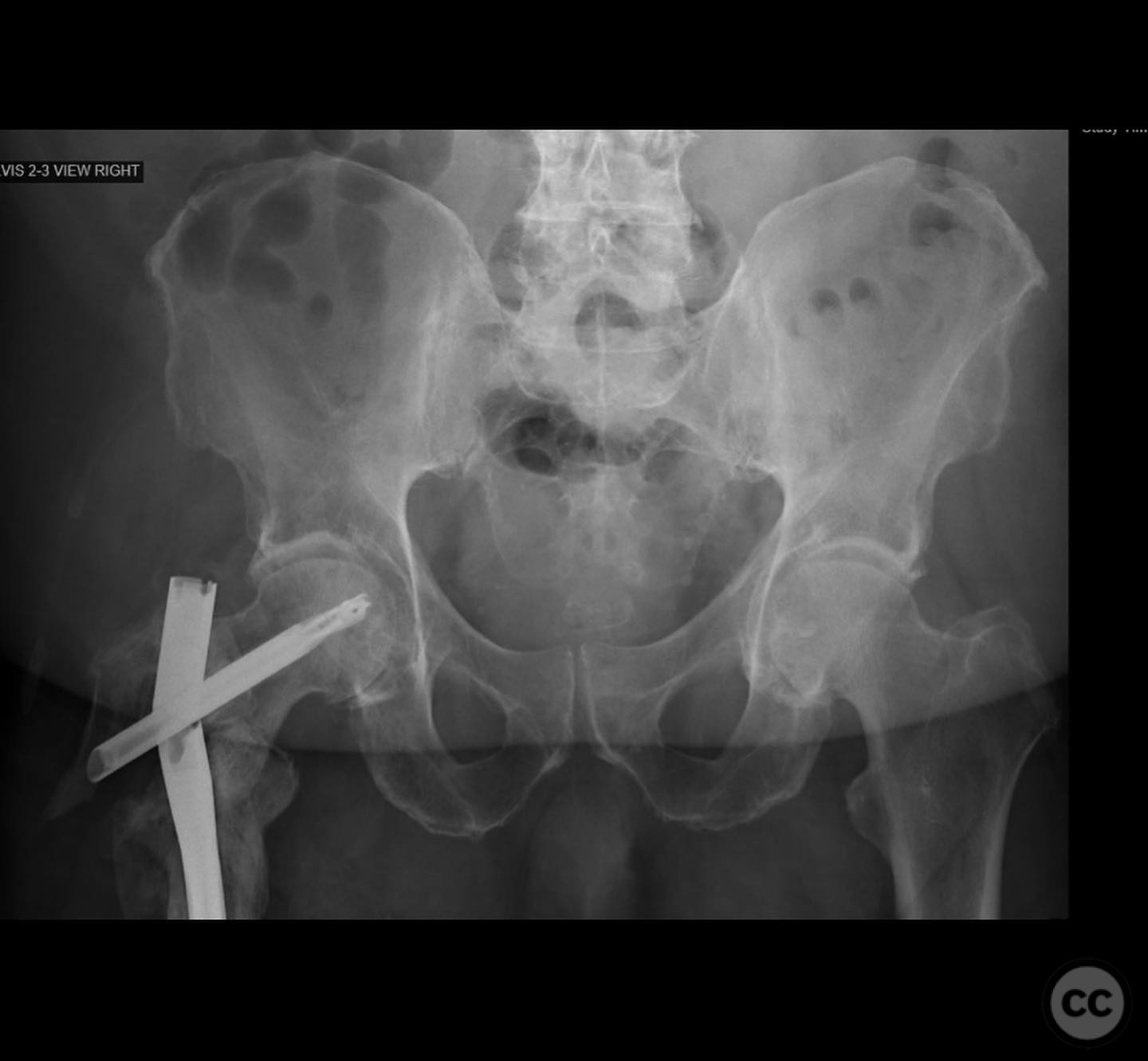
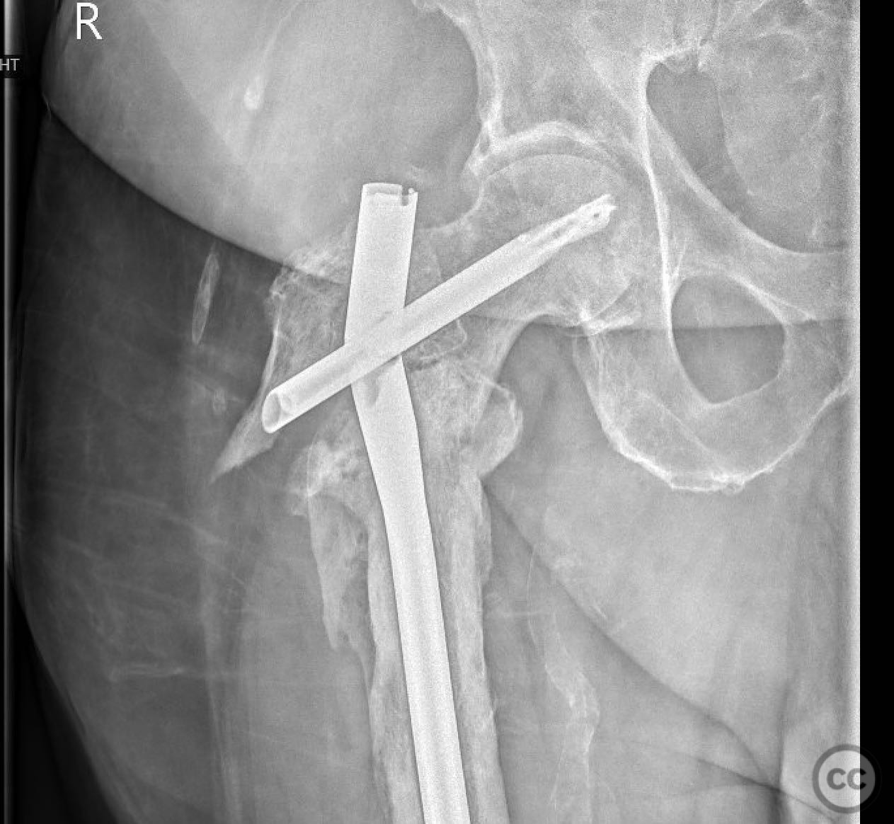
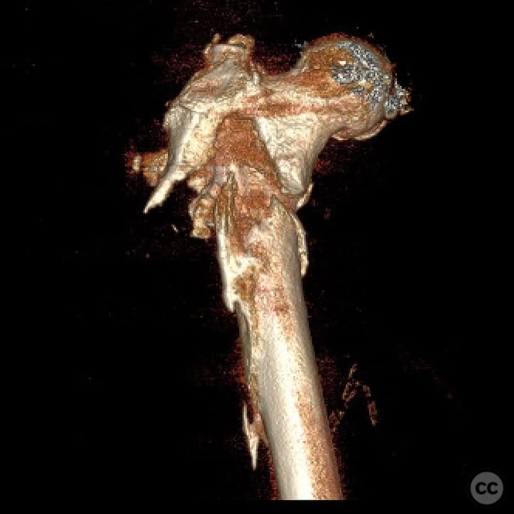
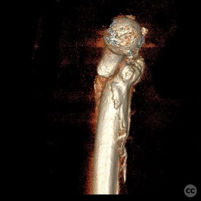
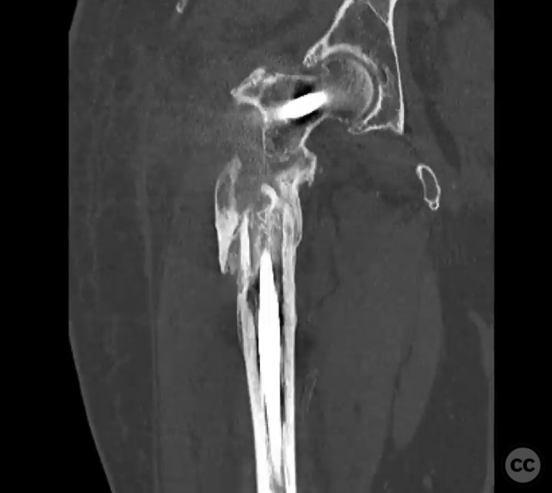
Clinical and radiological findings: The patient presented with a pertrochanteric malunion involving both the greater and lesser trochanters. Radiological assessment revealed significant malunion deformities, with the lesser trochanter malunion impeding sagittal plane correction. The greater trochanter malunion was also noted but was considered less complex to address.
Preoperative Plan
Planning remarks: The preoperative plan included performing osteotomies of both the greater and lesser trochanters to facilitate deformity correction. A revision intramedullary nailing combined with compression plating was planned to achieve stable fixation and correct alignment.
Surgical Discussion
Patient positioning: The patient was positioned supine on a fracture table to allow for optimal access and manipulation of the proximal femur.
Anatomical surgical approach: A lateral approach to the proximal femur was utilized. This involved a longitudinal incision over the lateral aspect of the hip, followed by subperiosteal dissection to expose the greater and lesser trochanters. Osteotomies were performed on both trochanters to correct the malunions.
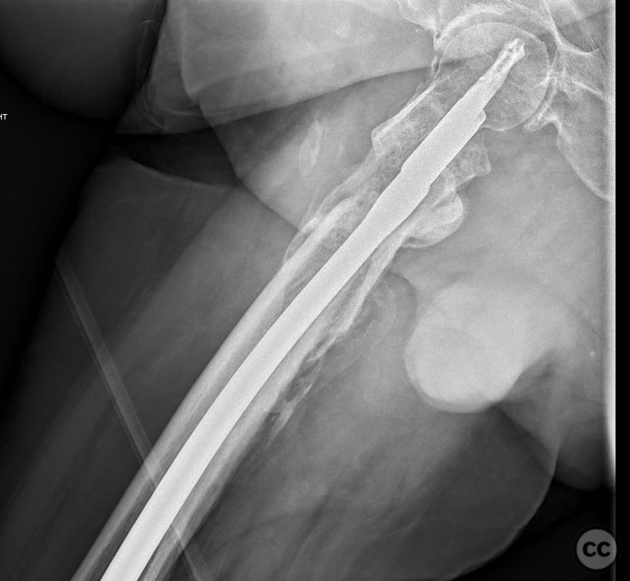
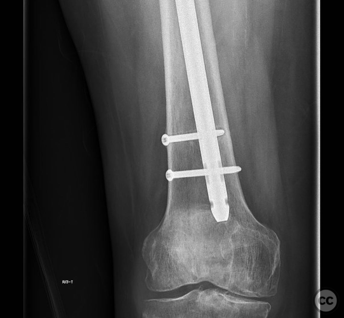
Operative remarks:The lesser trochanter malunion posed significant challenges in achieving sagittal plane correction, necessitating an osteotomy for successful realignment. The greater trochanter malunion was corrected with a straightforward osteotomy. A 4.5 narrow combi plate was employed for compression and stabilization, while a trapezoidal-shaped intramedullary nail was selected for its proximal press-fit capability and dual screw design, providing robust proximal fixation. Biological augmentation was achieved using a combination of cytokines, bone morphogenetic proteins (BMPs), and allograft.
Postoperative protocol: Postoperative rehabilitation included protected weight-bearing with gradual progression as tolerated, focusing on maintaining range of motion and strengthening exercises.
Follow up: Not specified.
Orthopaedic implants used: 4.5 narrow combi plate, trapezoidal intramedullary nail, dual proximal screws, allograft, cytokines, BMPs.
Search for Related Literature

orthopaedic_trauma
- United States , Seattle
- Area of Specialty - General Trauma
- Position - Specialist Consultant

Industry Sponsership
contact us for advertising opportunities
Article viewed 263 times
12 Jul 2025
Add to Bookmarks
Full Citation
Cite this article:
Surname, Initial. (2025). Pertrochanteric Malunion with Osteotomy and Revision Fixation. Journal of Orthopaedic Surgery and Traumatology. Case Report 46148654 Published Online Jul 12 2025.