Open Supracondylar/Intercondylar Distal Femur Fracture with Nail/Plate Fixation
Score and Comment on this Case
Clinical Details
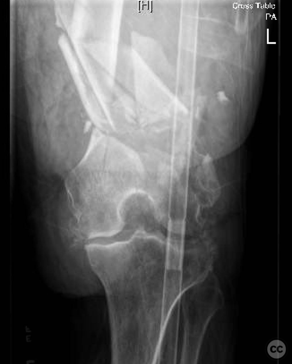
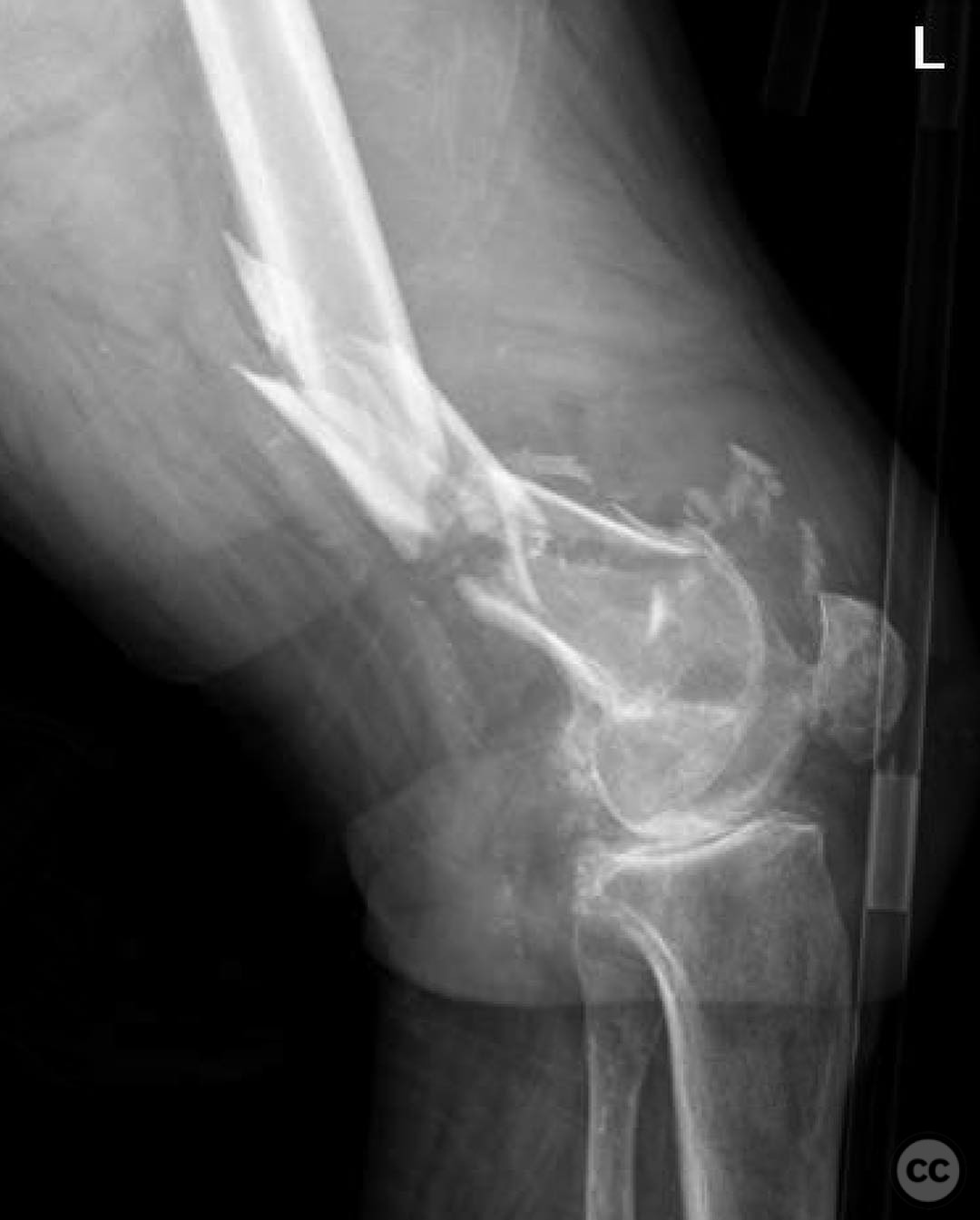
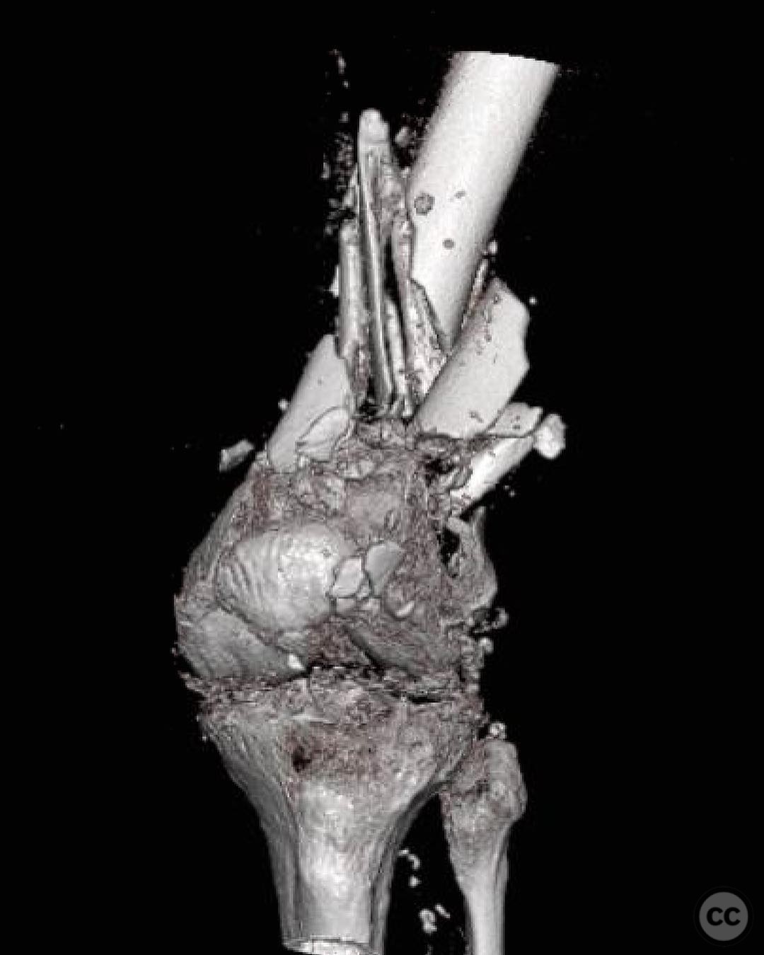
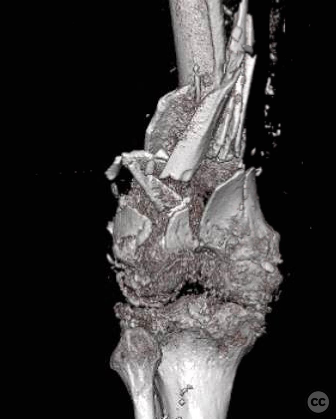
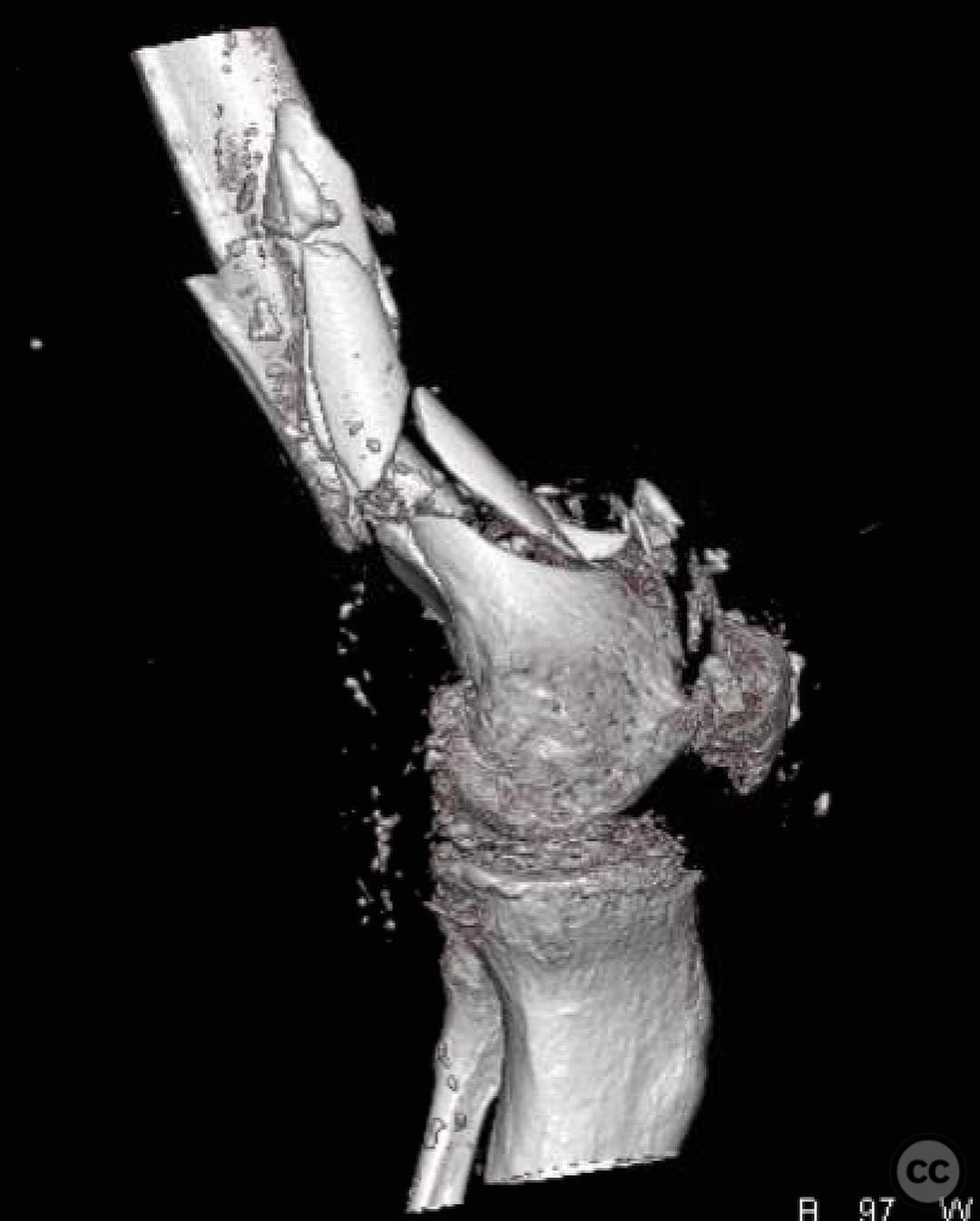
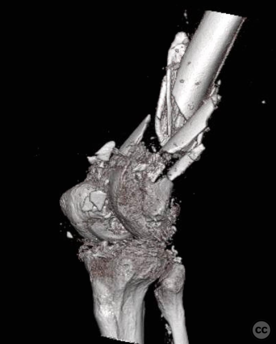
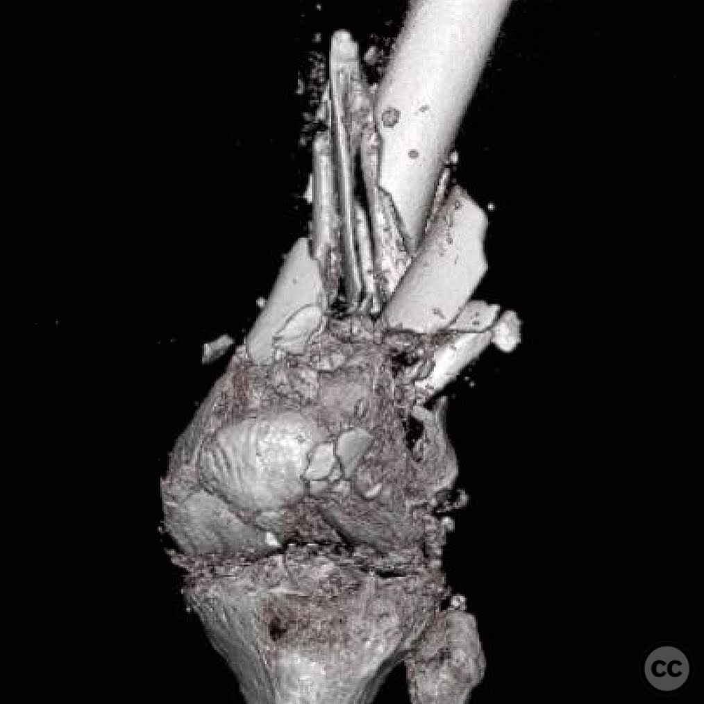
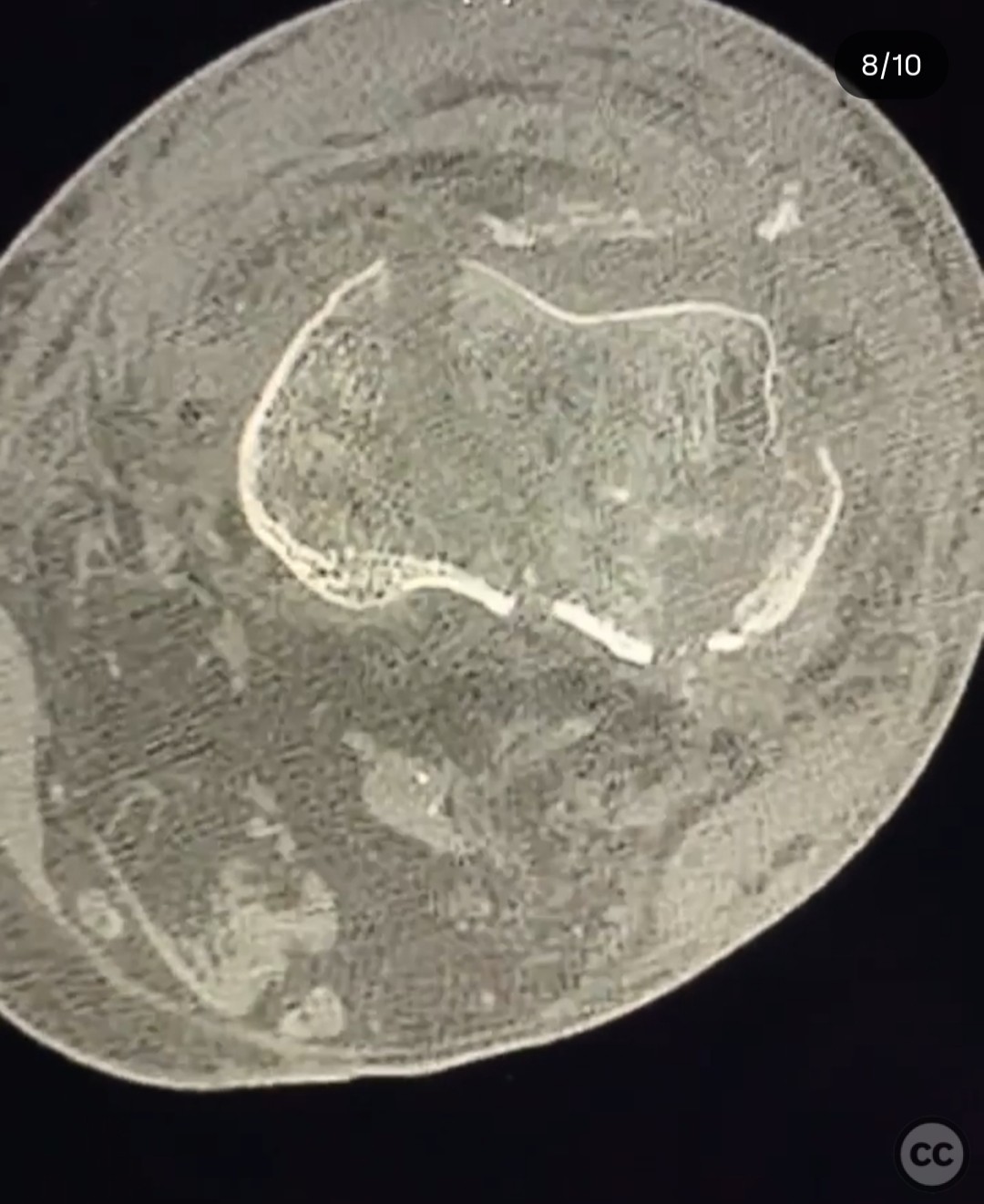
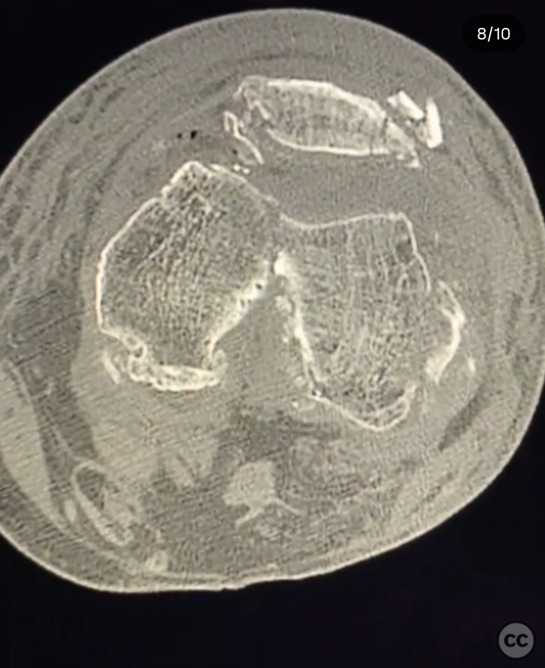
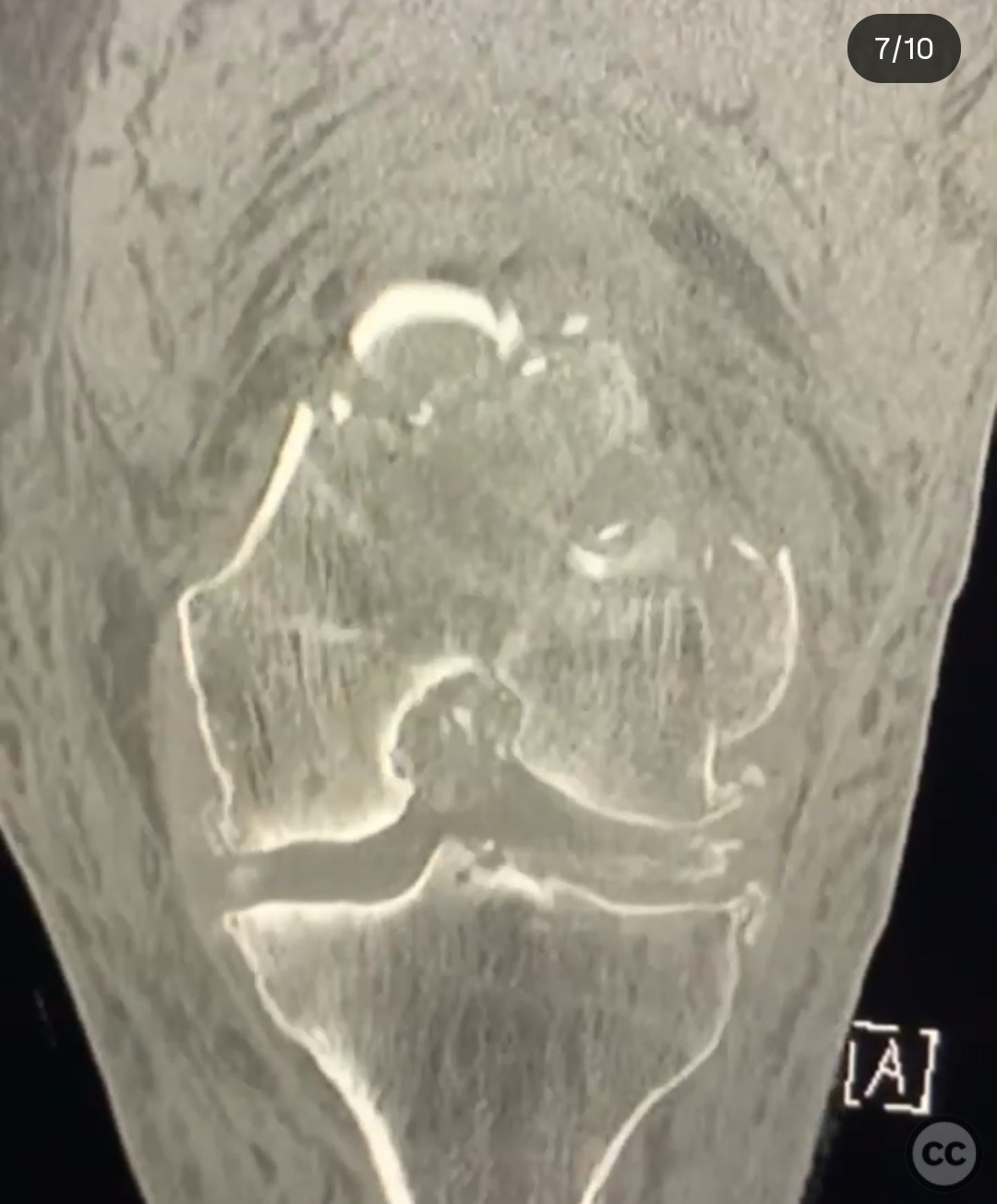
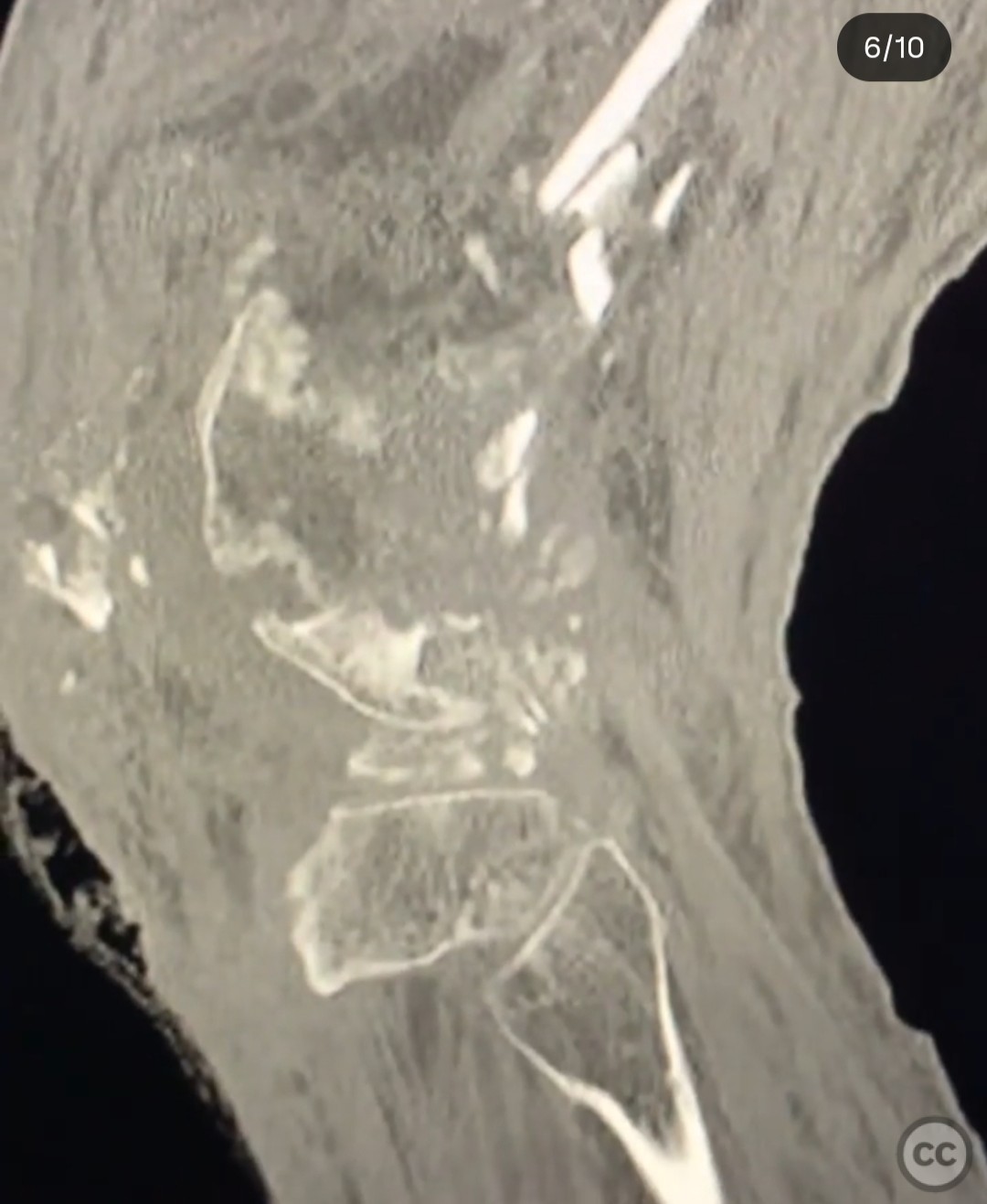
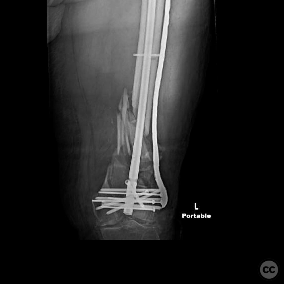
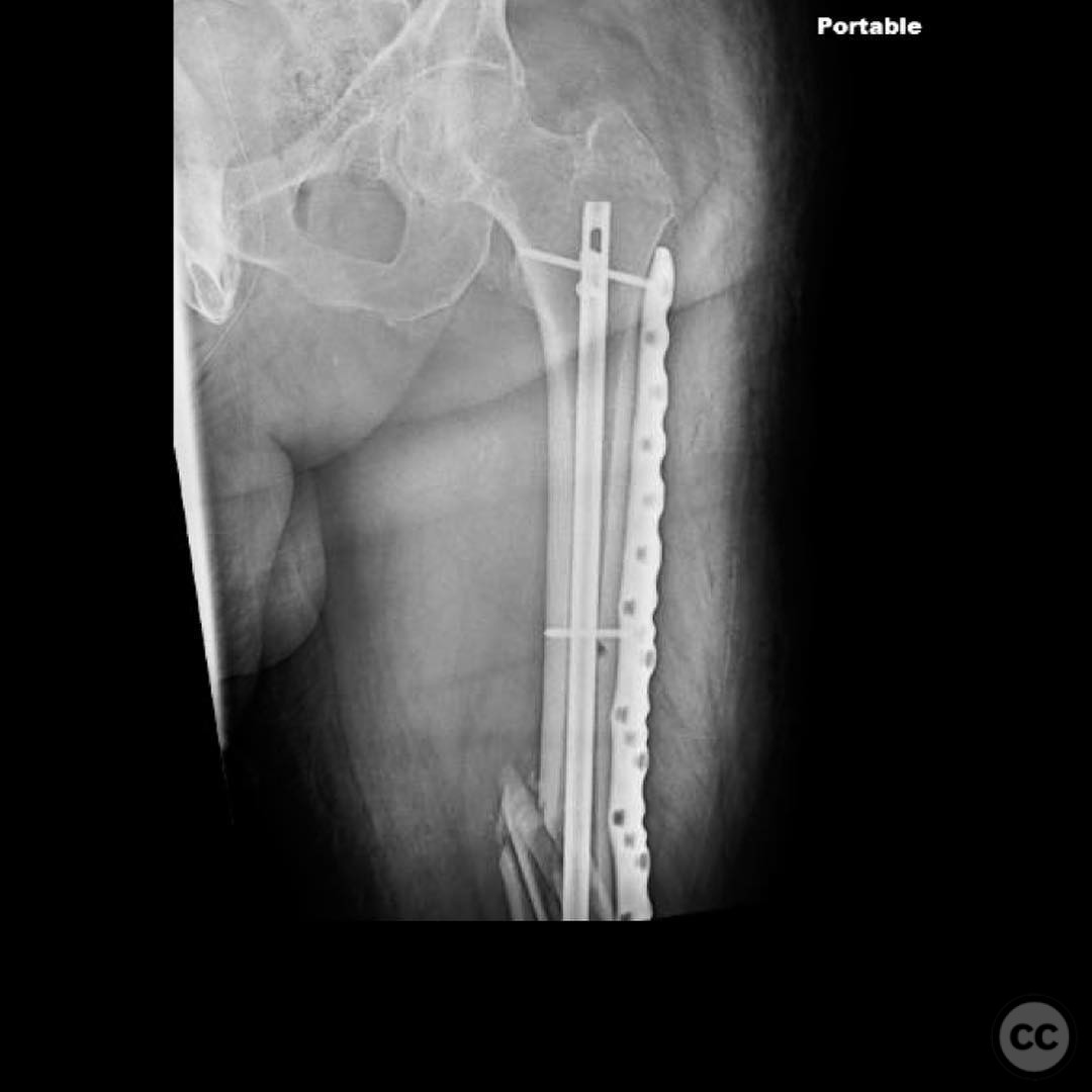
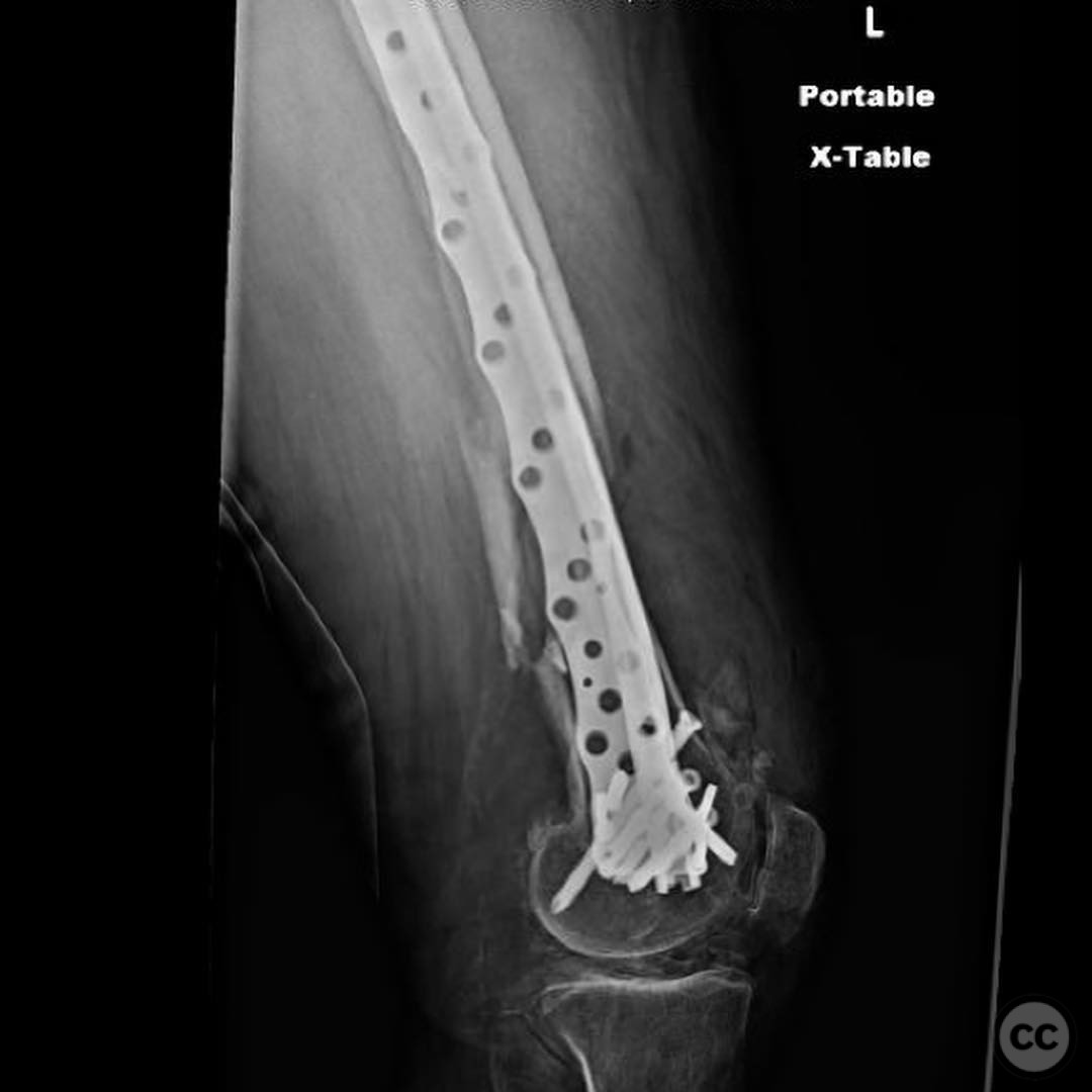
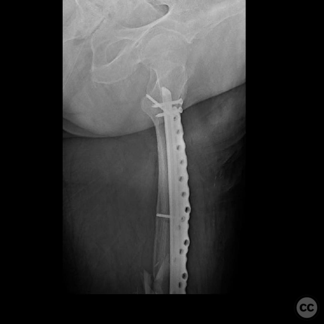
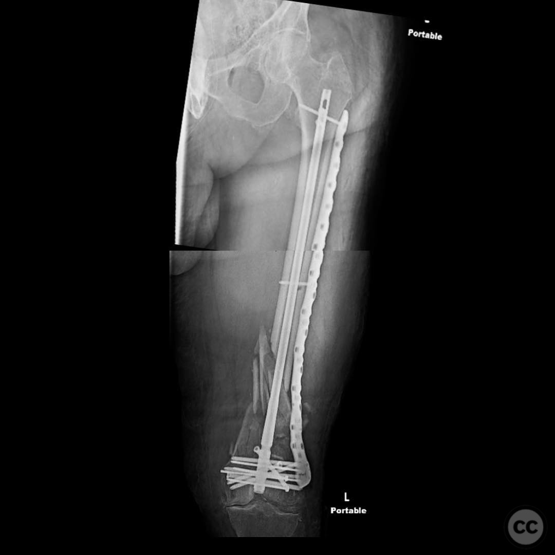
Clinical and radiological findings: A 72-year-old male involved in an auto vs tree accident presented with a 3a open supracondylar/intercondylar distal femur fracture. The patient was otherwise healthy and active, with no significant knee pain prior to the injury. The open fracture was characterized by a 3-4 cm skin laceration, which was not grossly contaminated. Radiological assessment revealed a relatively simple joint injury pattern, classified as AO/OTA 33C fracture.
Preoperative Plan
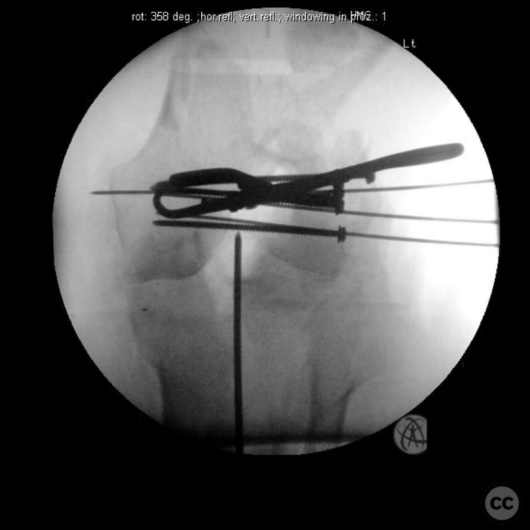
Planning remarks: The preoperative plan included achieving anatomic reduction, interfragmentary compression, and absolute stability of the joint using lag screws. For the metaphyseal component, the goal was to restore length, alignment, and rotation, providing relative stability for secondary bone healing. A nail/plate combination was chosen to ensure adequate stability for the short distal segment and to modulate construct stiffness for early weight bearing.
Surgical Discussion
Patient positioning: Supine position on a radiolucent table, with the affected limb prepared for intraoperative imaging.
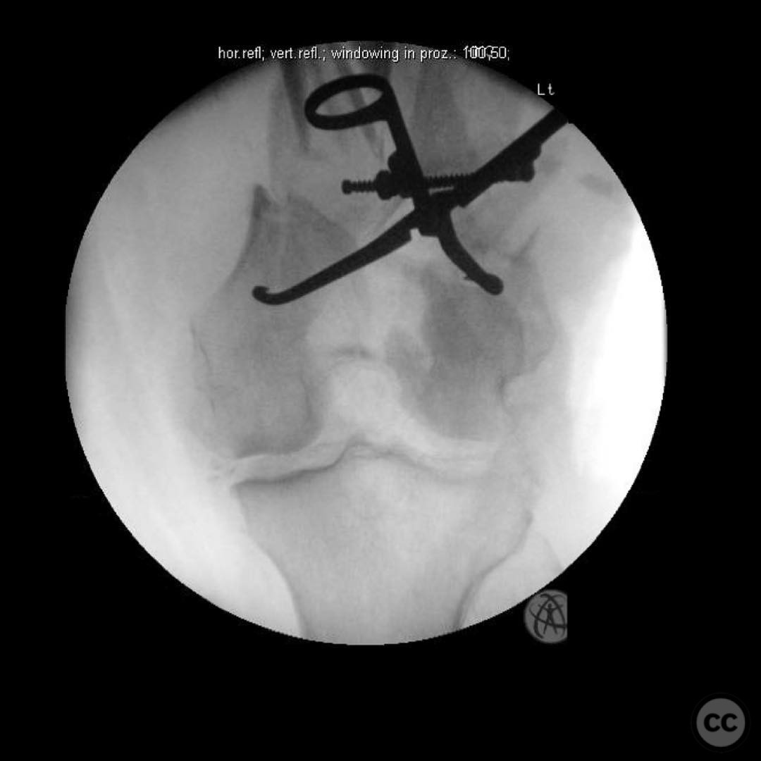
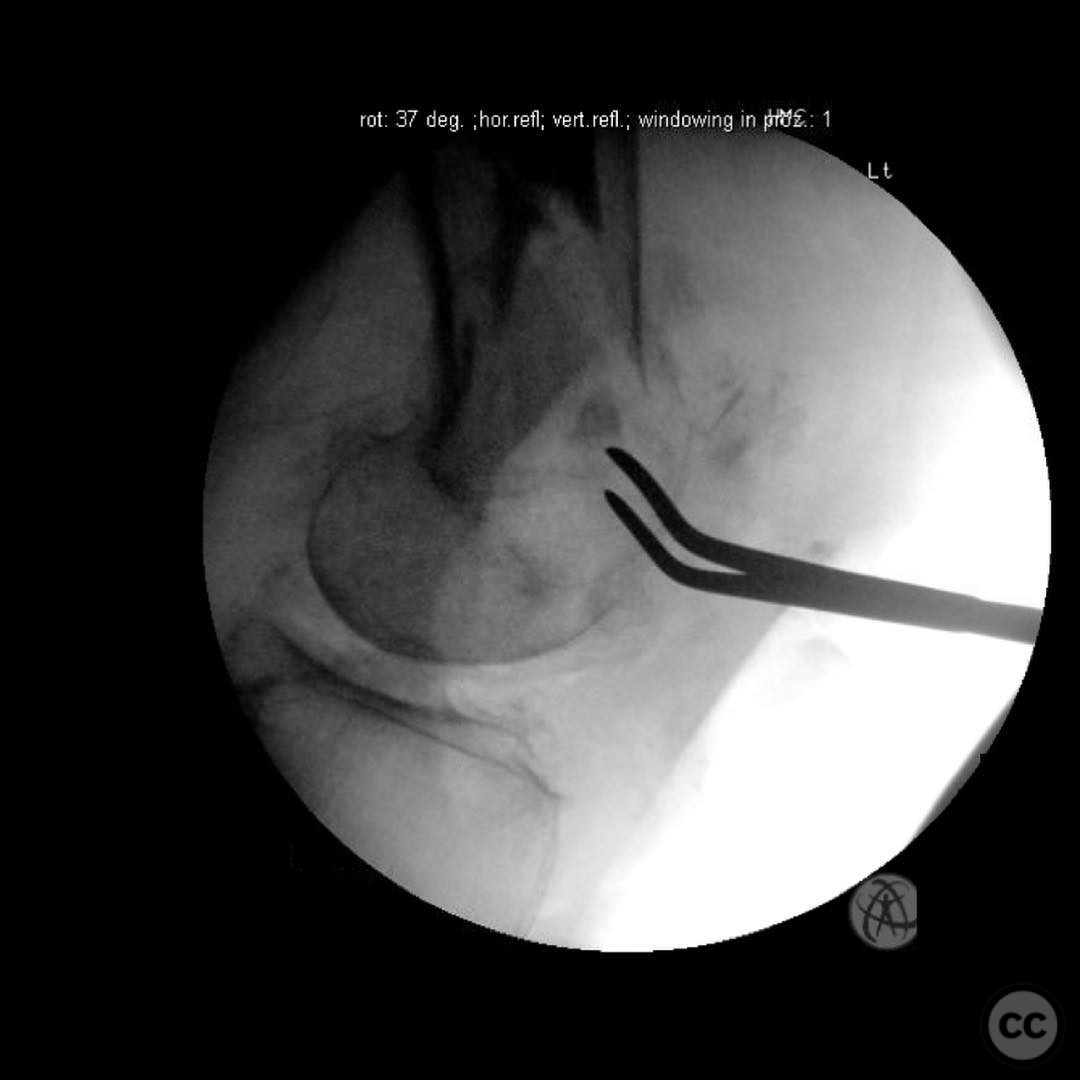
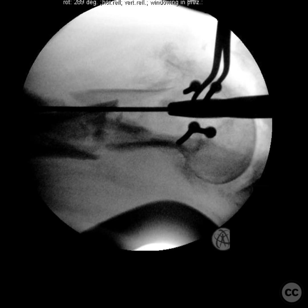
Anatomical surgical approach: A lateral approach to the distal femur was performed. The incision extended along the lateral aspect of the thigh, allowing access to the distal femur. Subperiosteal dissection was carried out to expose the fracture site. The joint was addressed first with lag screws for interfragmentary compression, followed by fixation of the metaphyseal component using a nail/plate combination.
Operative remarks:The surgeon emphasized the importance of achieving absolute stability in the joint with lag screws to allow early weight bearing. The nail/plate combination provided distal fixation and medial column support, offering a long working length and low screw density. This approach was deemed optimal for this geriatric 33C fracture, balancing stability and flexibility to prevent early failure under load.
Postoperative protocol: Early mobilization with weight-bearing as tolerated was encouraged postoperatively. The rehabilitation protocol focused on restoring range of motion and strength while monitoring for signs of infection due to the open nature of the fracture.
Follow up: Not specified.
Orthopaedic implants used: Intramedullary nail, locking plate, lag screws.
Search for Related Literature

orthopaedic_trauma
- United States , Seattle
- Area of Specialty - General Trauma
- Position - Specialist Consultant

Industry Sponsership
contact us for advertising opportunities
Article viewed 172 times
21 Jul 2025
Add to Bookmarks
Full Citation
Cite this article:
Surname, Initial. (2025). Open Supracondylar/Intercondylar Distal Femur Fracture with Nail/Plate Fixation. Journal of Orthopaedic Surgery and Traumatology. Case Report 44623988 Published Online Jul 21 2025.