_0.jpg)
_1.jpg)
_2.jpg)
vascular injury vs compartment syndrome and 2) prox tib fib joint dislocations._._._._1.jpg)
vascular injury vs compartment syndrome and 2) prox tib fib joint dislocations._._._._2.jpg)
vascular injury vs compartment syndrome and 2) prox tib fib joint dislocations._._._._7.jpg)
vascular injury vs compartment syndrome and 2) prox tib fib joint dislocations._._._._3.jpg)
vascular injury vs compartment syndrome and 2) prox tib fib joint dislocations._._._._4.jpg)
vascular injury vs compartment syndrome and 2) prox tib fib joint dislocations._._._._6.jpg)
vascular injury vs compartment syndrome and 2) prox tib fib joint dislocations._._._._5.jpg)
vascular injury vs compartment syndrome and 2) prox tib fib joint dislocations._._._._8.jpg)
vascular injury vs compartment syndrome and 2) prox tib fib joint dislocations._._._._9.jpg)
vascular injury vs compartment syndrome and 2) prox tib fib joint dislocations._._._._(.jpg)
Looks Like Compartment… But It’s Not: The Hidden Vascular Blowout + A Wobbly PTFJ
Score and Comment on this Case
Clinical Details
Clinical and radiological findings: A 22-year-old male was involved in a high-energy motor vehicle accident, sustaining an open tibial shaft fracture. The patient presented to the emergency department 30 minutes post-injury with a 1 cm wound located at the anteromedial aspect of the tibia, consistent with the site of fracture. The leg was massively swollen. Vascular examination revealed normal Ankle-Brachial Index (ABI) above 0.9, with palpable dorsalis pedis and posterior tibial pulses. Despite moderate pain, there was no exacerbation with passive toe stretch, raising suspicion for vascular injury rather than compartment syndrome.
Preoperative Plan
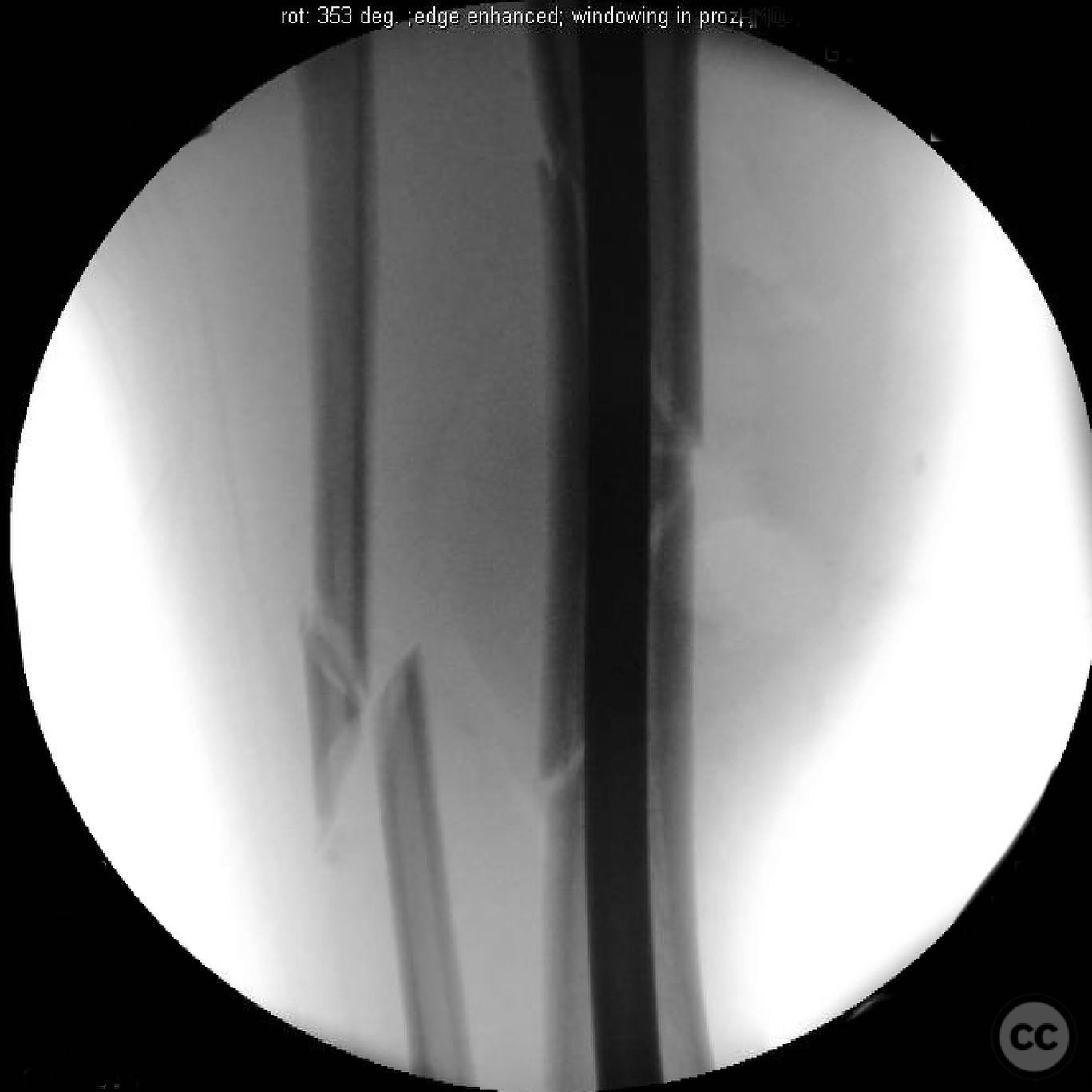
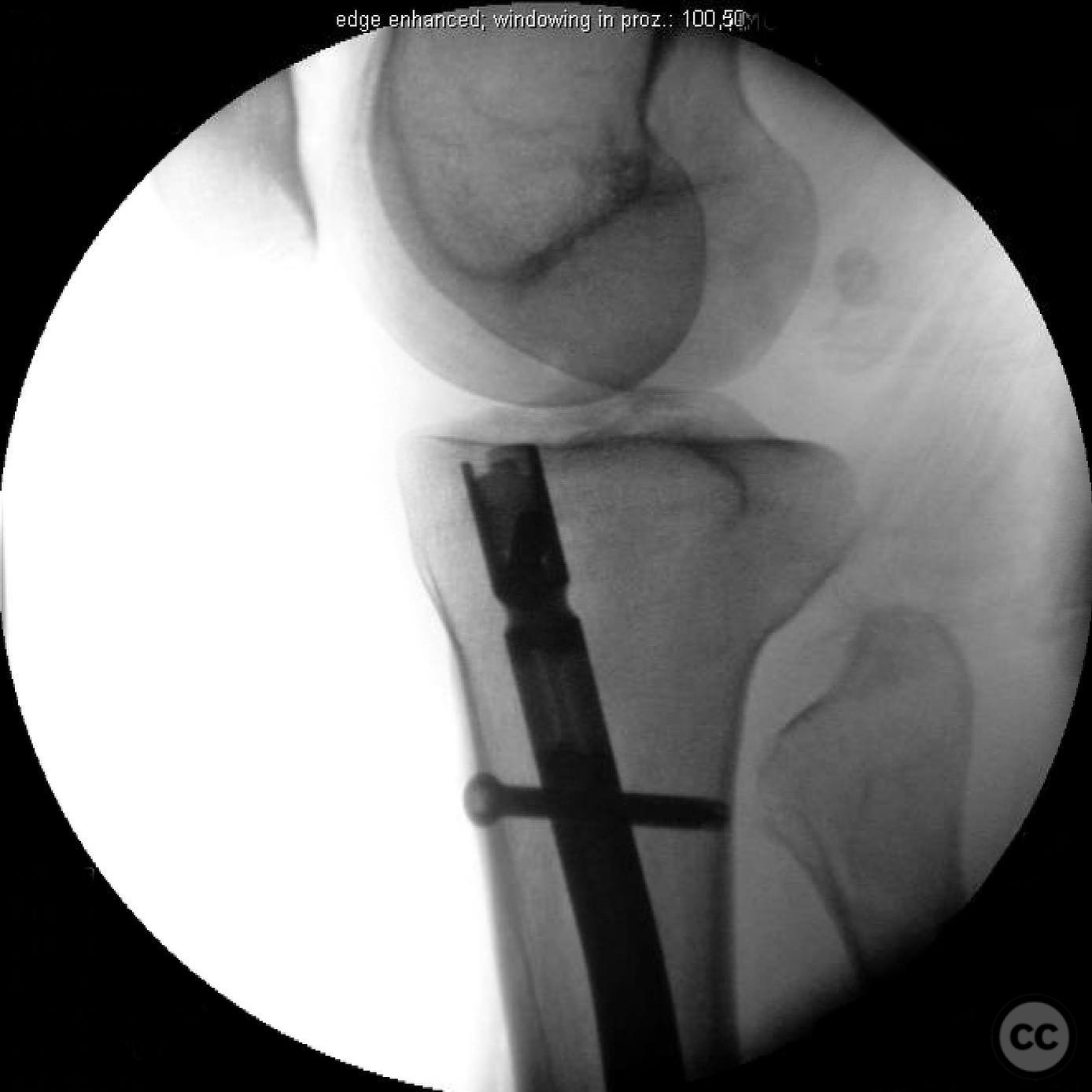
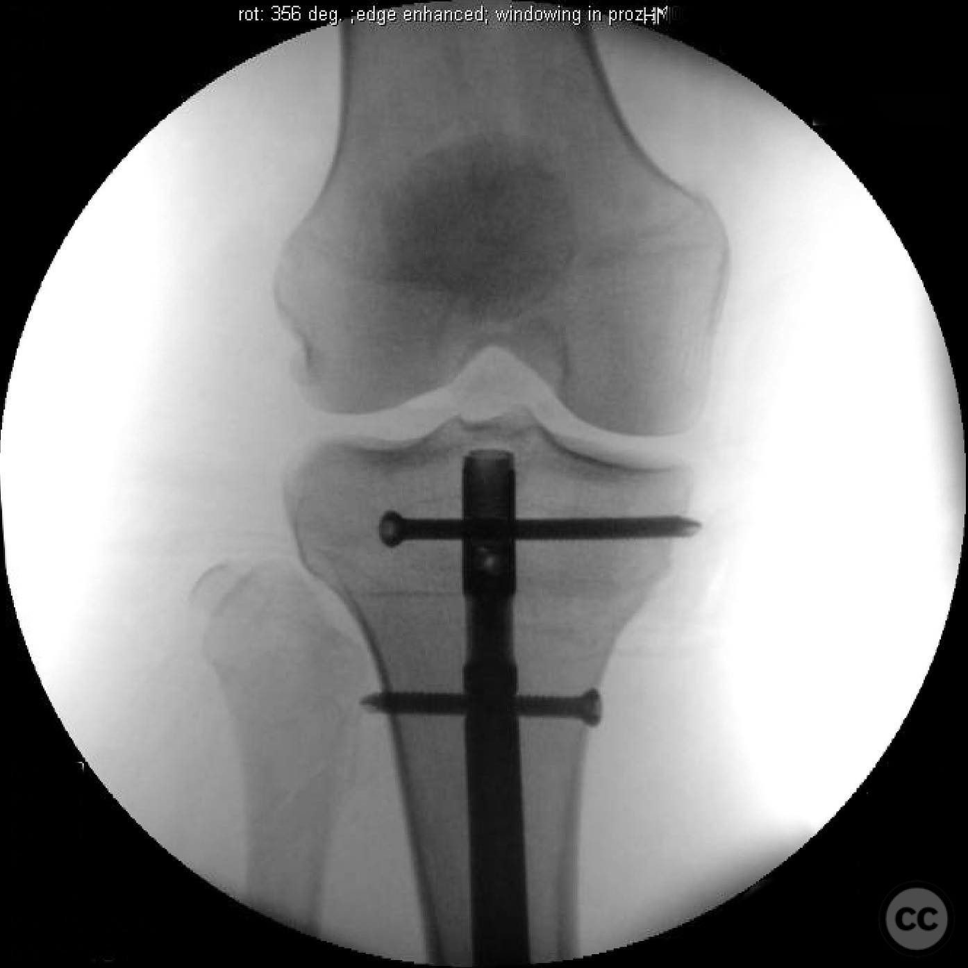
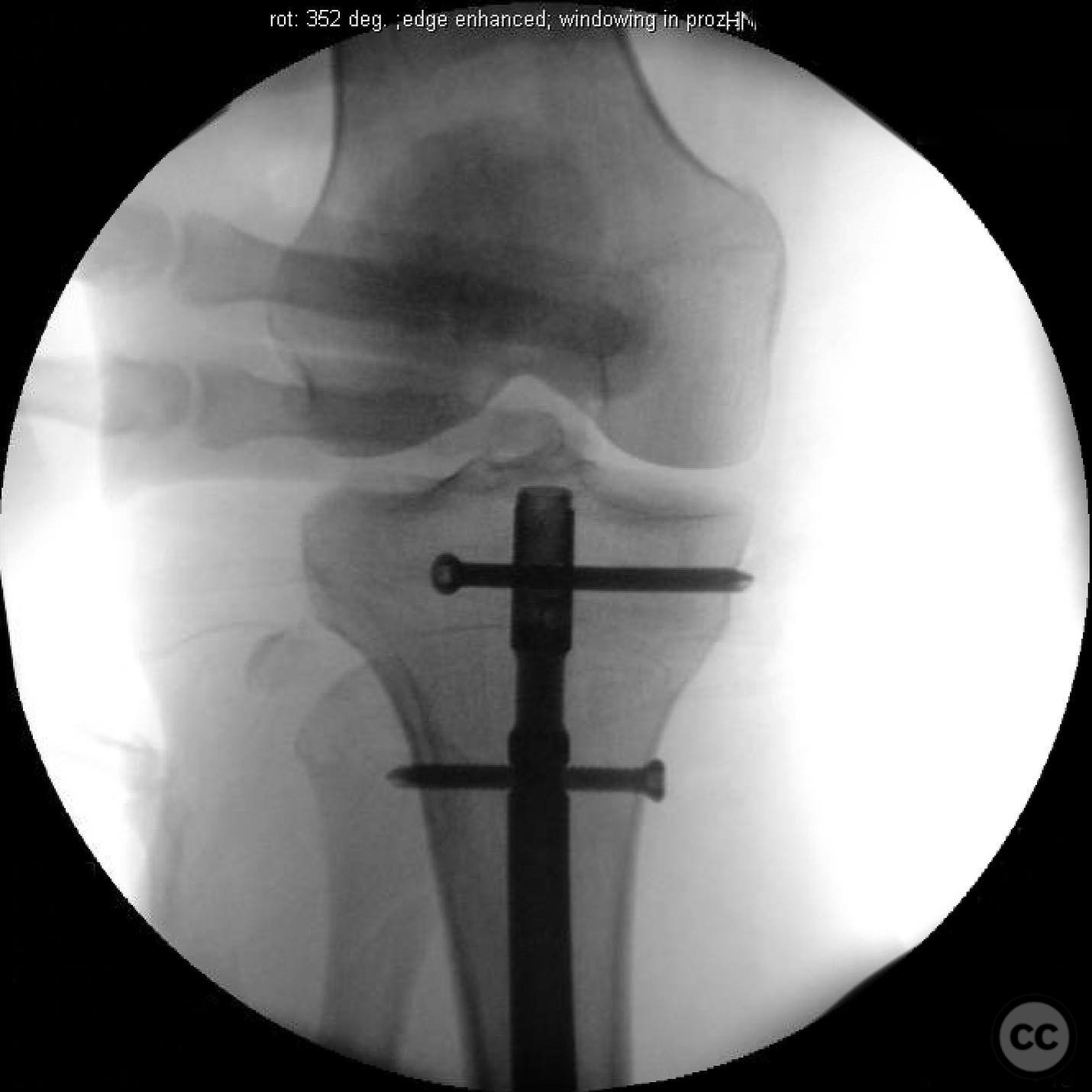
Planning remarks: The preoperative plan included urgent fasciotomy due to marked swelling and concern for possible vascular injury rather than classic compartment syndrome. Although the patient had a normal ABI (>0.9) and palpable distal pulses (DP and PT), the rapid onset of massive swelling within 30 minutes, coupled with moderate pain not exacerbated by passive toe stretch, suggested active arterial bleeding — likely from the anterior tibial or peroneal artery — rather than elevated compartment pressures. This decision was based on the recognition that compartment syndrome typically evolves over hours, whereas vascular blowout can cause acute swelling in minutes. Following fasciotomy and hematoma evacuation, intramedullary nailing of the tibial fracture was planned. During the procedure, proximal tibiofibular joint instability was noted and required intraoperative assessment to determine the need for stabilization.
Surgical Discussion
Patient positioning: The patient was positioned supine on the operating table to facilitate both fasciotomy and intramedullary nailing procedures.
Anatomical surgical approach: A dual incision fasciotomy was performed to decompress all four compartments of the leg. The tibial fracture was addressed via a standard medial parapatellar approach for intramedullary nailing. Upon completion of nailing, the proximal tibiofibular joint was assessed for instability.
Operative remarks:Intraoperative findings confirmed significant bleeding from the anterior tibial and peroneal arteries at the fracture site, necessitating vascular control measures. The proximal tibiofibular joint exhibited abnormal mobility upon stress testing, indicating dislocation. Given the rarity and complexity of this injury, stabilization of the joint was performed to restore native anatomy and potentially prevent future knee kinematic dysfunction.
Postoperative protocol: Postoperative rehabilitation included non-weight bearing on the affected limb for 6 weeks, followed by gradual weight-bearing as tolerated. Range of motion exercises for the knee and ankle were initiated early to prevent stiffness.
Follow up: Not specified.
Orthopaedic implants used: Intramedullary nail, locking screws for proximal tibiofibular joint stabilization.
Search for Related Literature

orthopaedic_trauma
- United States , Seattle
- Area of Specialty - General Trauma
- Position - Specialist Consultant

Industry Sponsership
contact us for advertising opportunities
Article viewed 310 times
16 Jul 2025
Add to Bookmarks
Full Citation
Cite this article:
Surname, Initial. (2025). Looks Like Compartment… But It’s Not: The Hidden Vascular Blowout + A Wobbly PTFJ. Journal of Orthopaedic Surgery and Traumatology. Case Report 44022495 Published Online Jul 16 2025.