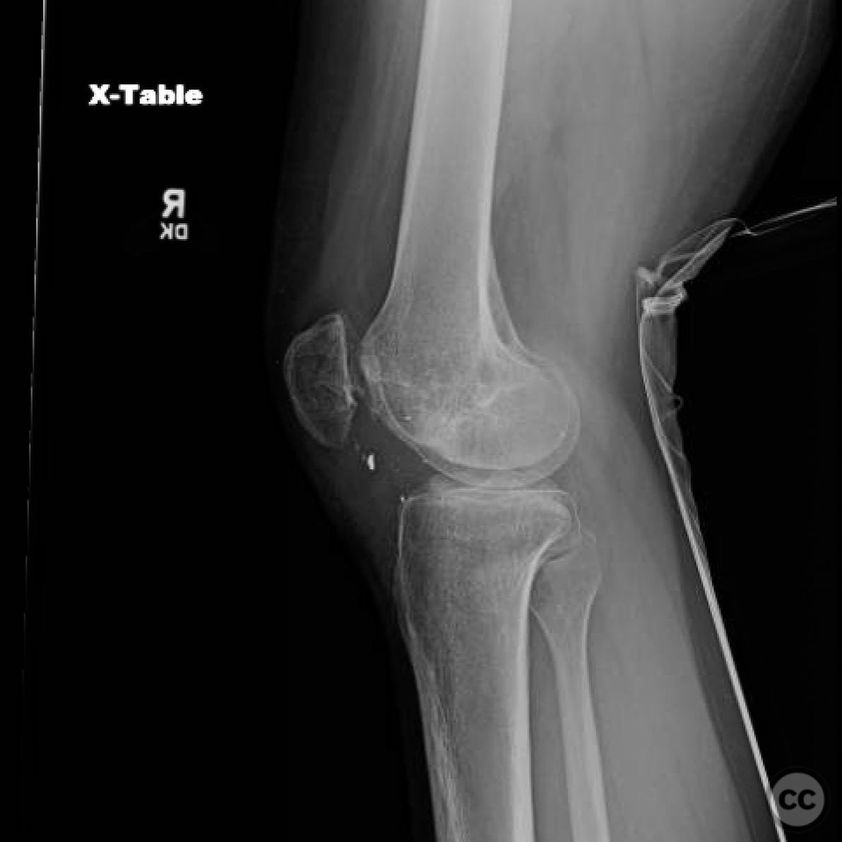
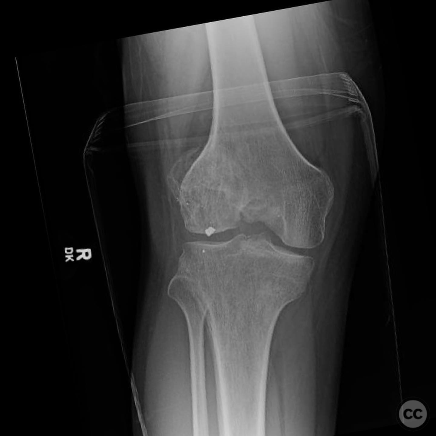
Complex intra-articular malunion of the lateral femoral condyle with patellar subluxation.
Score and Comment on this Case
Clinical Details
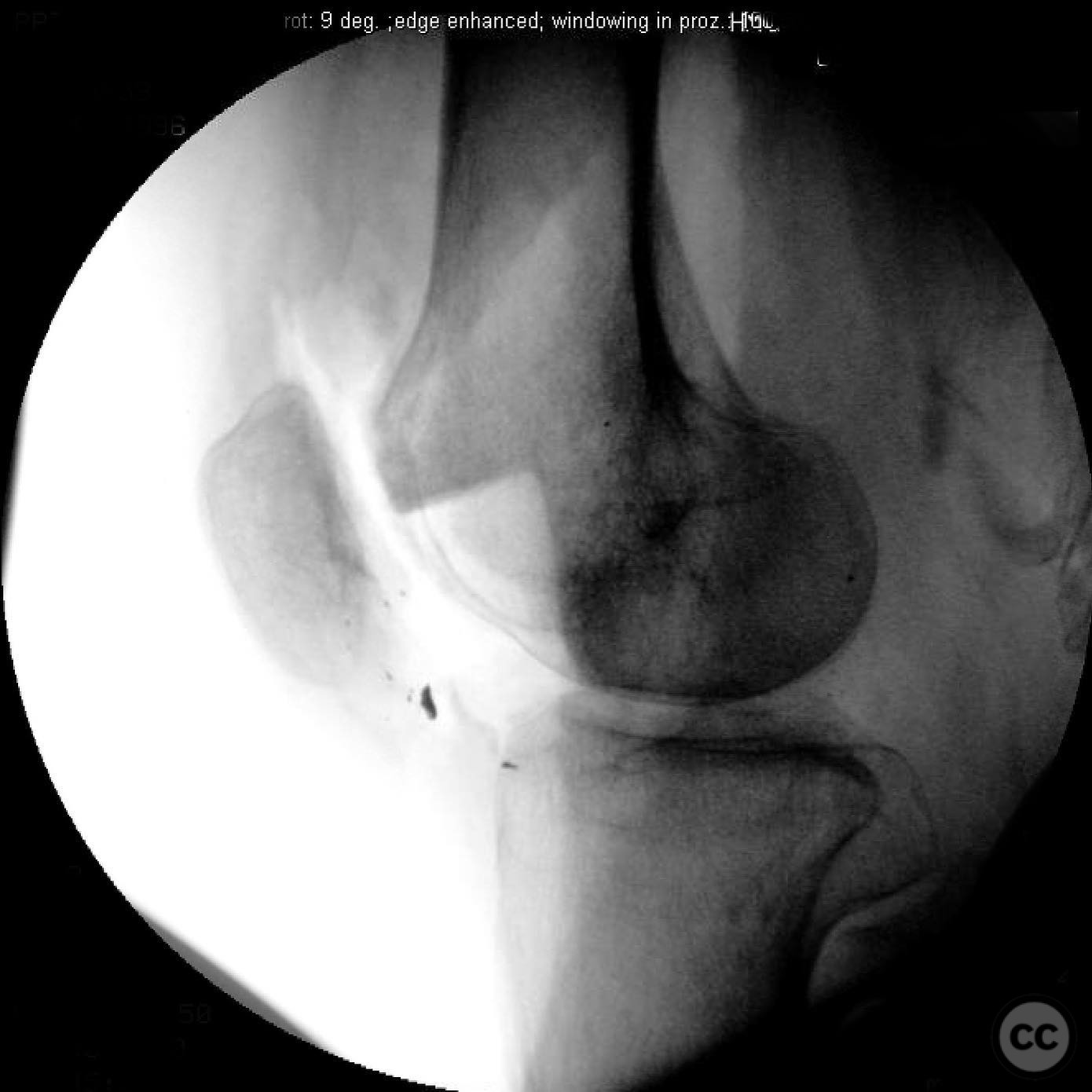
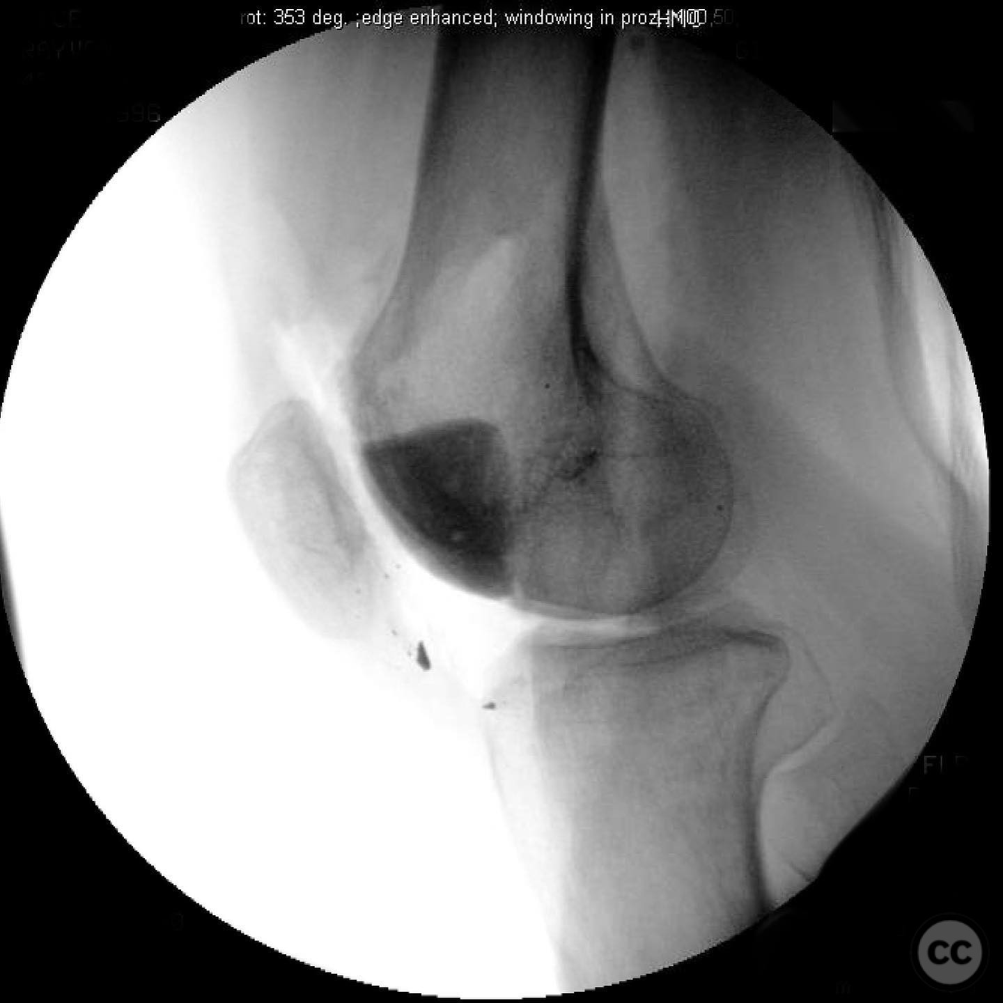
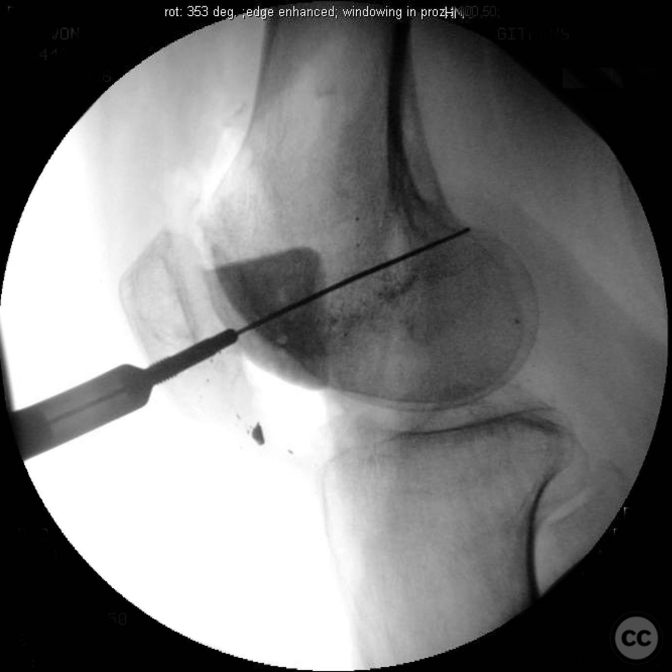
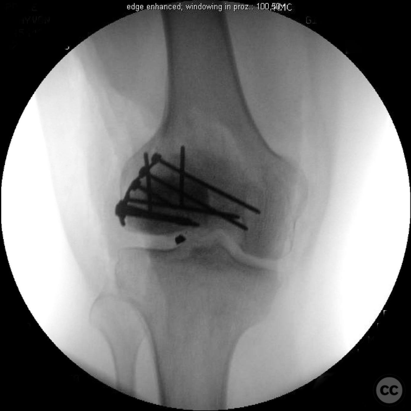
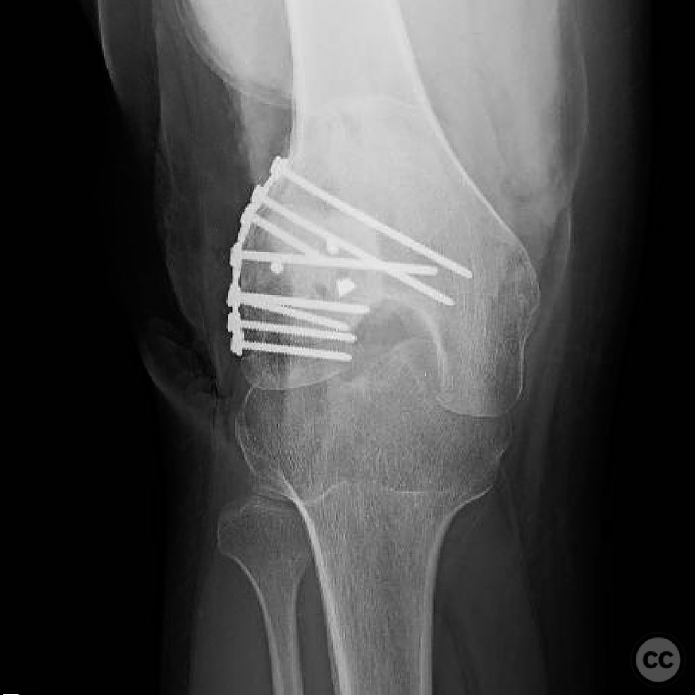
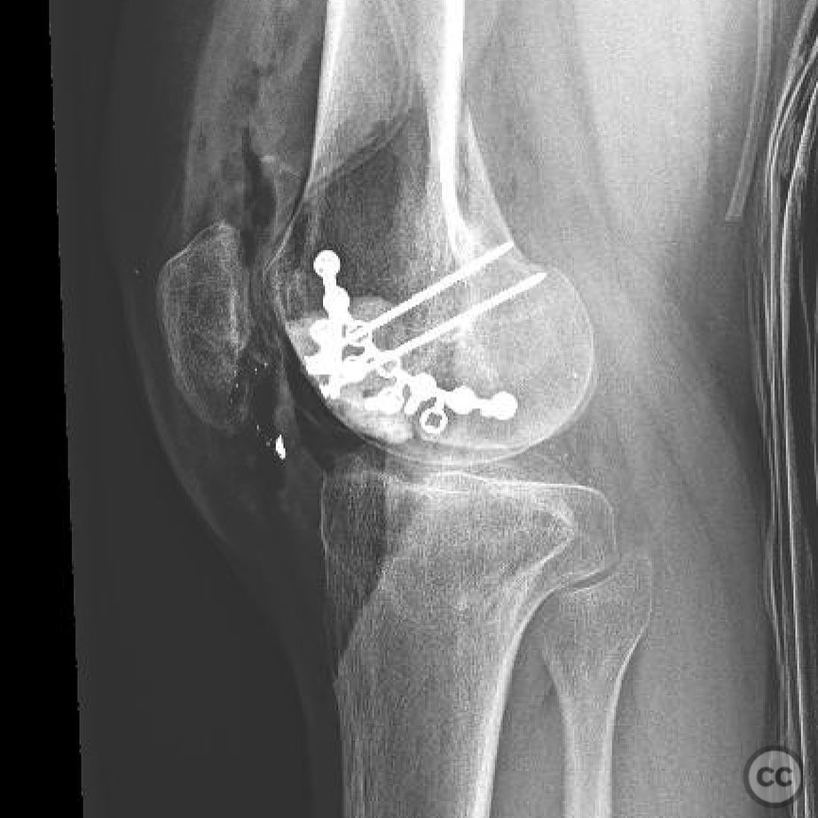
Clinical and radiological findings: A 20-year-old male presented 7 months post low-velocity gunshot wound to the knee, initially treated with neglect at an outside hospital. The patient reported severe knee pain exacerbated by motion, with a range of motion limited to 10-30 degrees. Clinical examination revealed moderate effusion and no signs of infection. Radiological assessment indicated a nonunion of the lateral femoral condyle, significant articular bone loss primarily affecting the patellofemoral joint, and patellar subluxation with the patella lodged in a lateral defect.
Preoperative Plan
Planning remarks: The preoperative plan involved a two-stage surgical approach. The first stage aimed to restore knee motion and correct patellar tracking through an open lysis of adhesions (LOA), potential quadricepsplasty, and reconstruction of the trochlea using polymethylmethacrylate (PMMA) with medial imbrication. The second stage was planned for anatomical osteochondral block allograft to address the osteochondral defect, contingent on the patient's postoperative progress and symptomatology.
Surgical Discussion
Patient positioning: Supine position with the affected knee supported to allow access for anterior and lateral approaches.
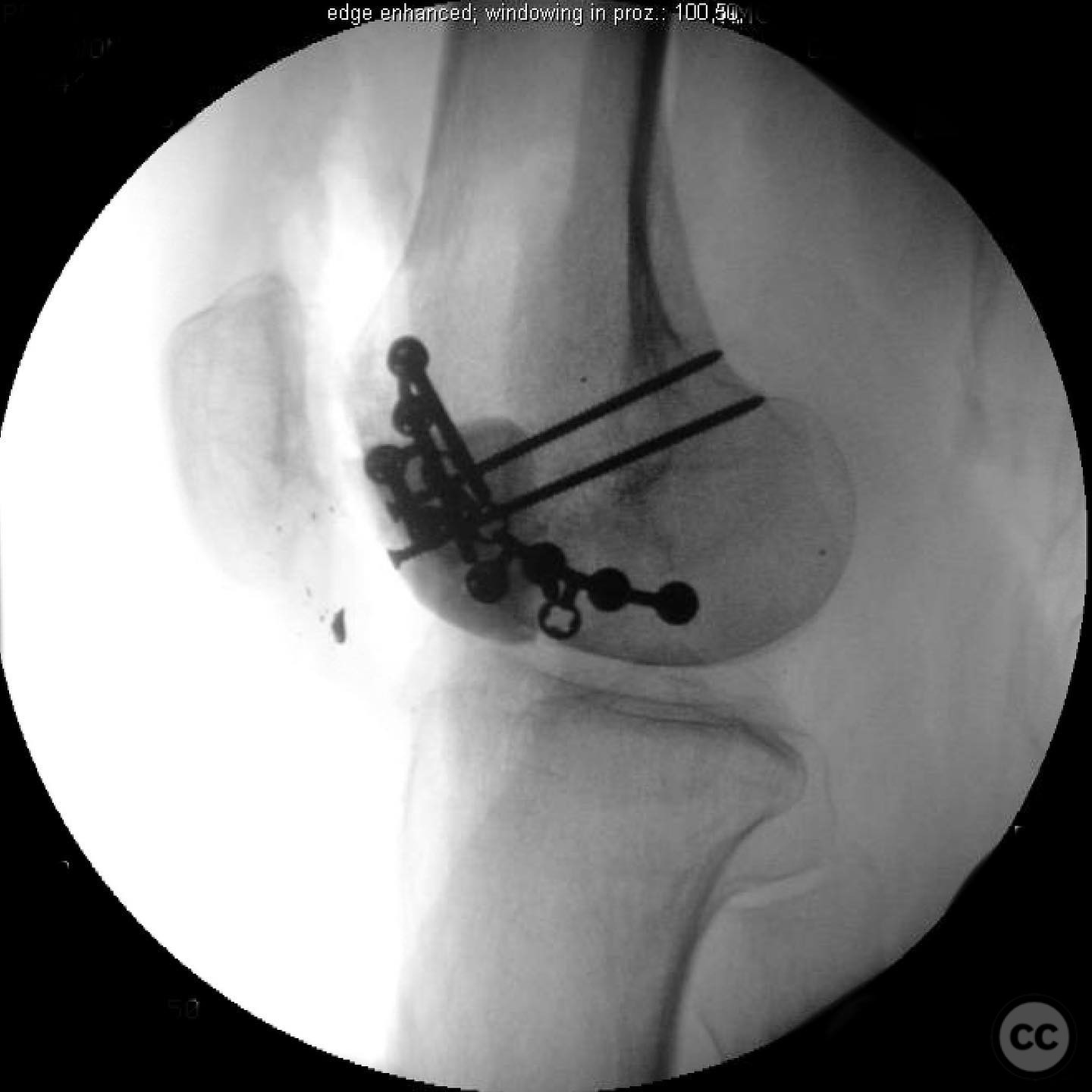
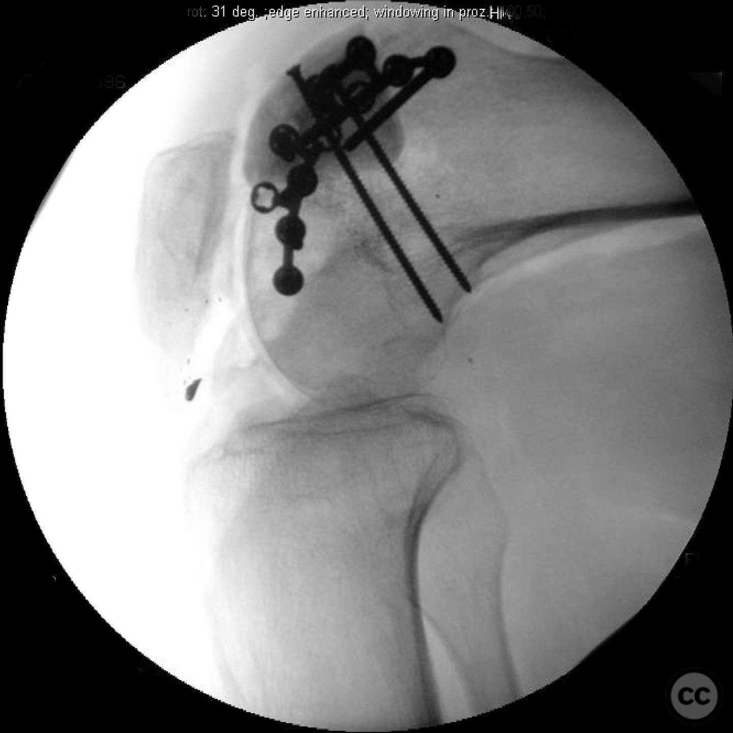
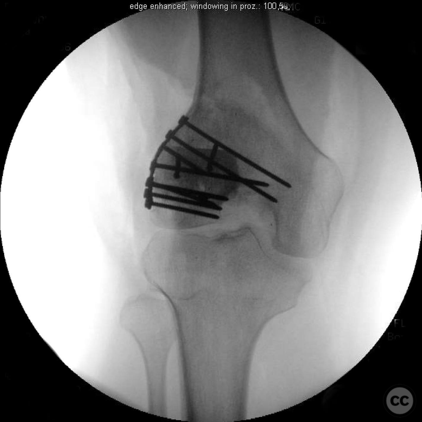
Anatomical surgical approach: An anterior midline incision was made, extending from the superior pole of the patella to the tibial tuberosity. Subcutaneous tissues were dissected to expose the extensor mechanism. Extensive lateral release was performed to free the patella from its lateral defect. The trochlea was reconstructed using PMMA, and medial imbrication was executed to stabilize patellar tracking.
Operative remarks:The surgeon noted that achieving adequate patellar tracking required extensive soft tissue releases and trochlear reconstruction with PMMA. Despite the initial plan for staged management, the patient expressed satisfaction with the functional outcome post-trochlear reconstruction and opted against proceeding with the second stage osteochondral allograft at that time.
Postoperative protocol: Postoperative rehabilitation focused on aggressive physiotherapy to improve range of motion and strengthen the quadriceps. Weight-bearing as tolerated was encouraged, with emphasis on patellar mobilization exercises and gradual progression of flexion-extension activities.
Follow up: Not specified.
Orthopaedic implants used: Polymethylmethacrylate (PMMA) for trochlear reconstruction.
Search for Related Literature

orthopaedic_trauma
- United States , Seattle
- Area of Specialty - General Trauma
- Position - Specialist Consultant

Industry Sponsership
contact us for advertising opportunities
Article viewed 159 times
14 Jul 2025
Add to Bookmarks
Full Citation
Cite this article:
Surname, Initial. (2025). Complex intra-articular malunion of the lateral femoral condyle with patellar subluxation.. Journal of Orthopaedic Surgery and Traumatology. Case Report 43750222 Published Online Jul 14 2025.