
Maisonneuve Fracture with Medial Plafond Impaction and Deltoid Ligament Rupture.
Score and Comment on this Case
Clinical Details
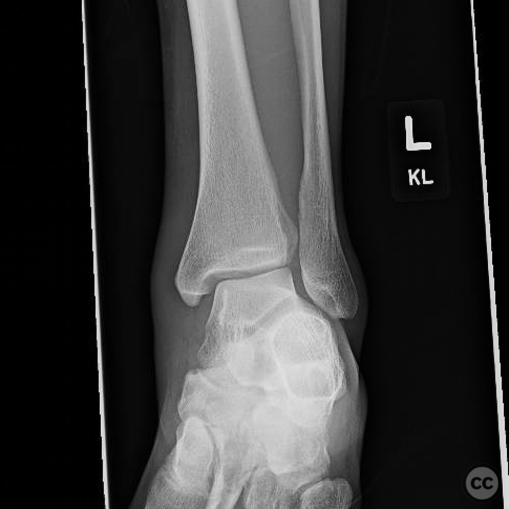
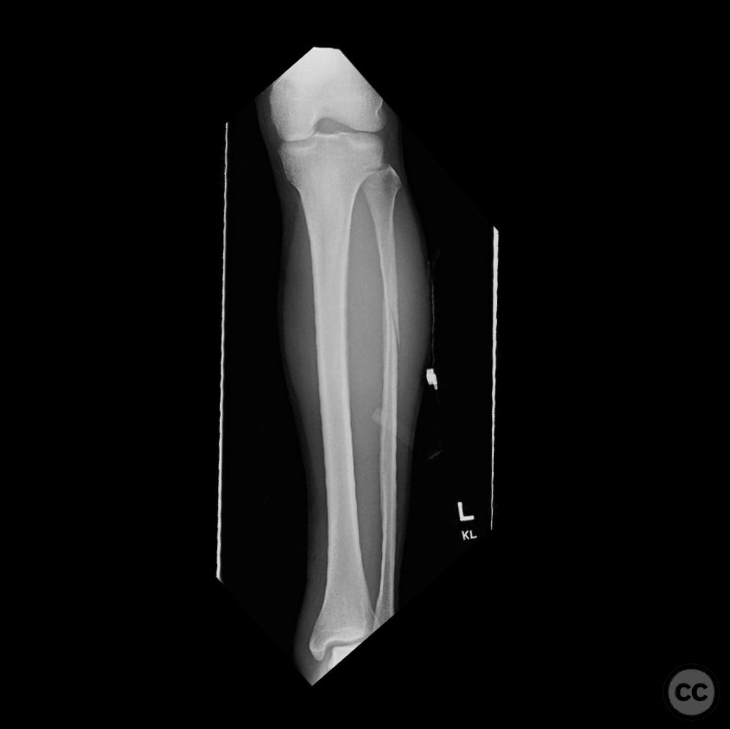
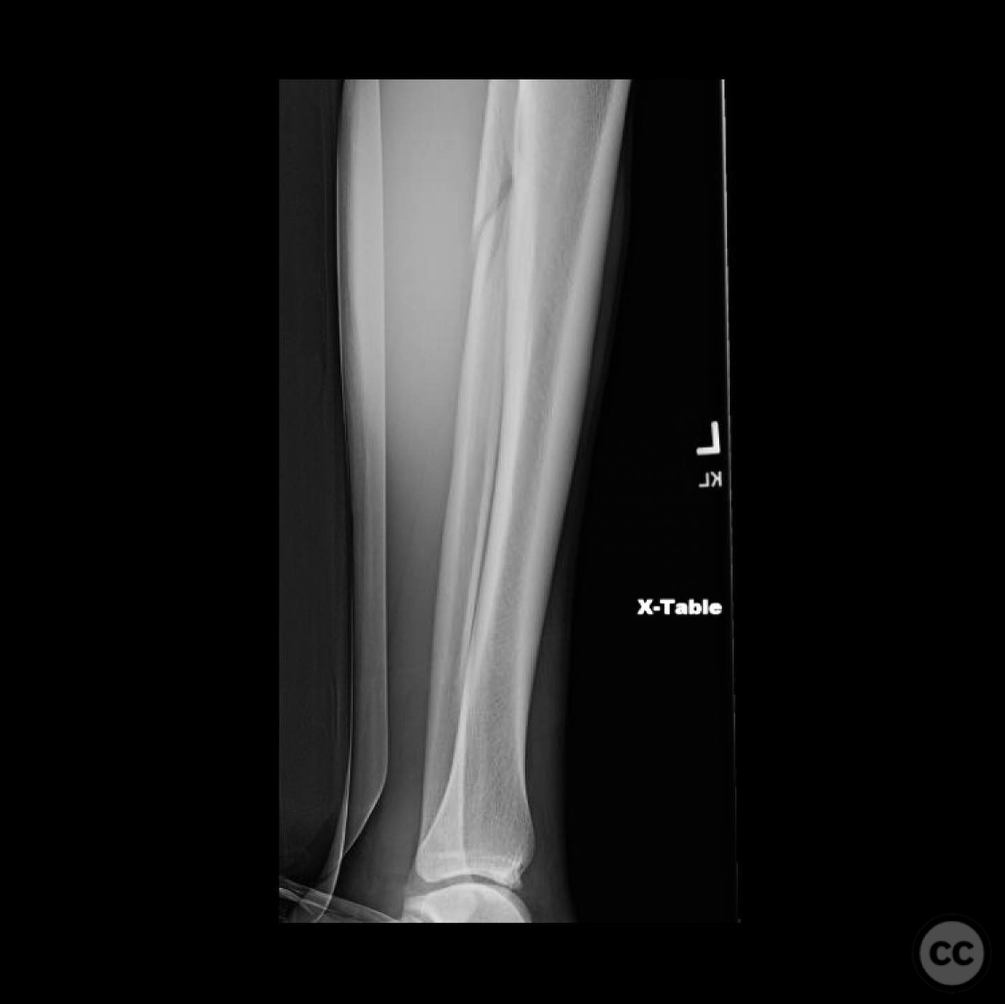
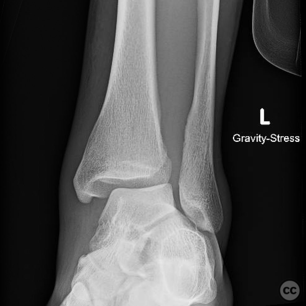
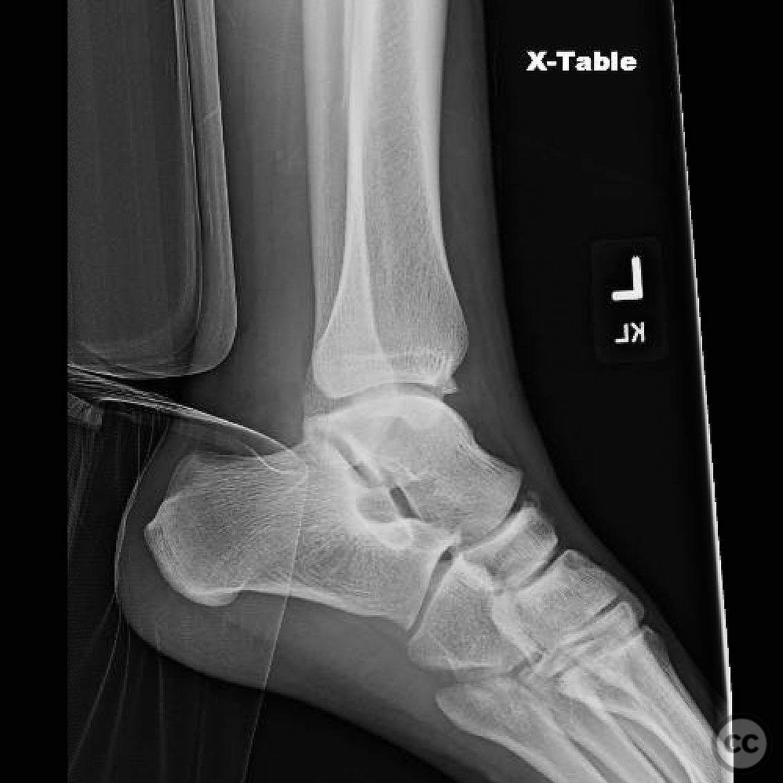
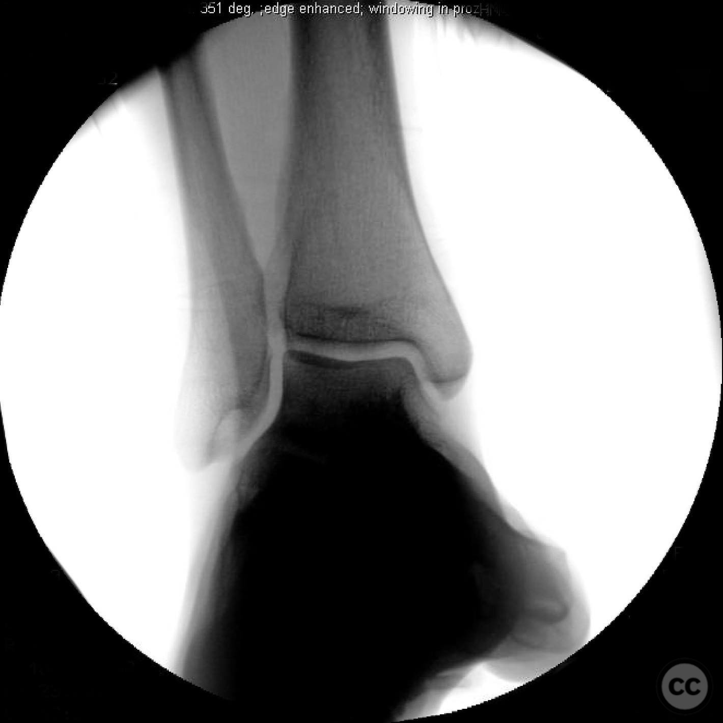
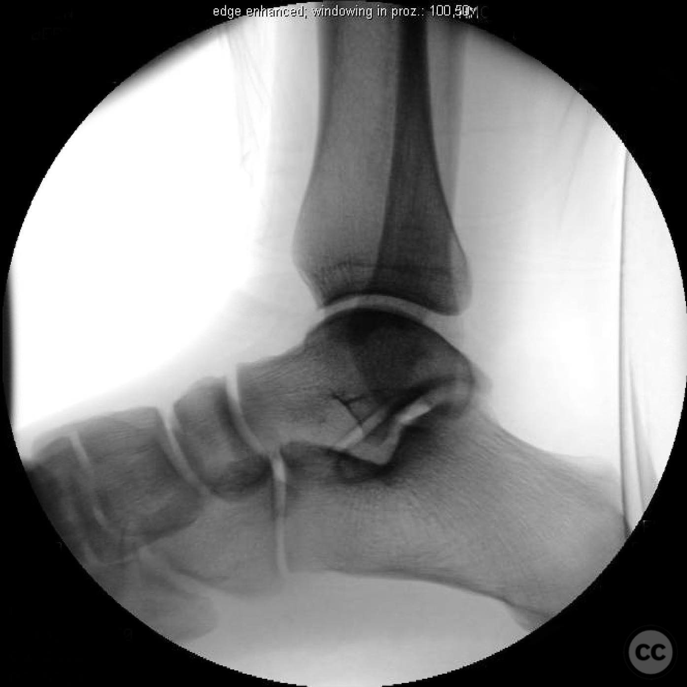
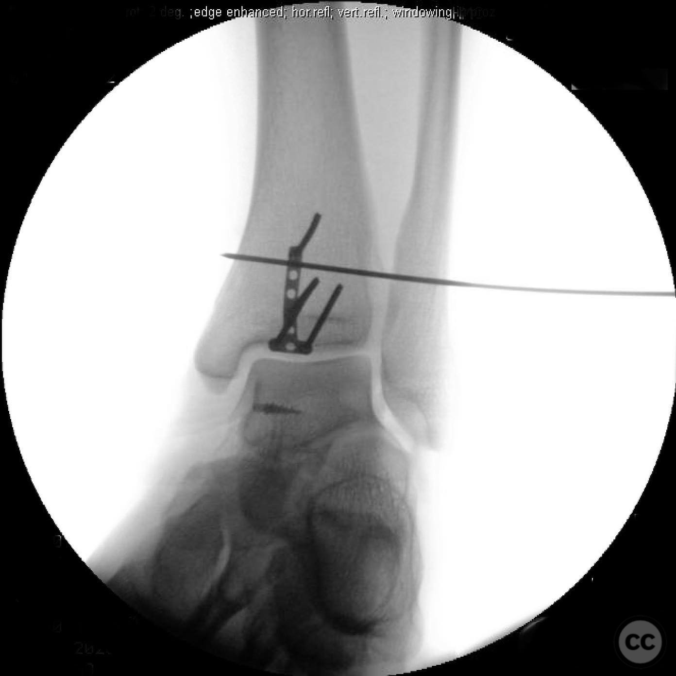
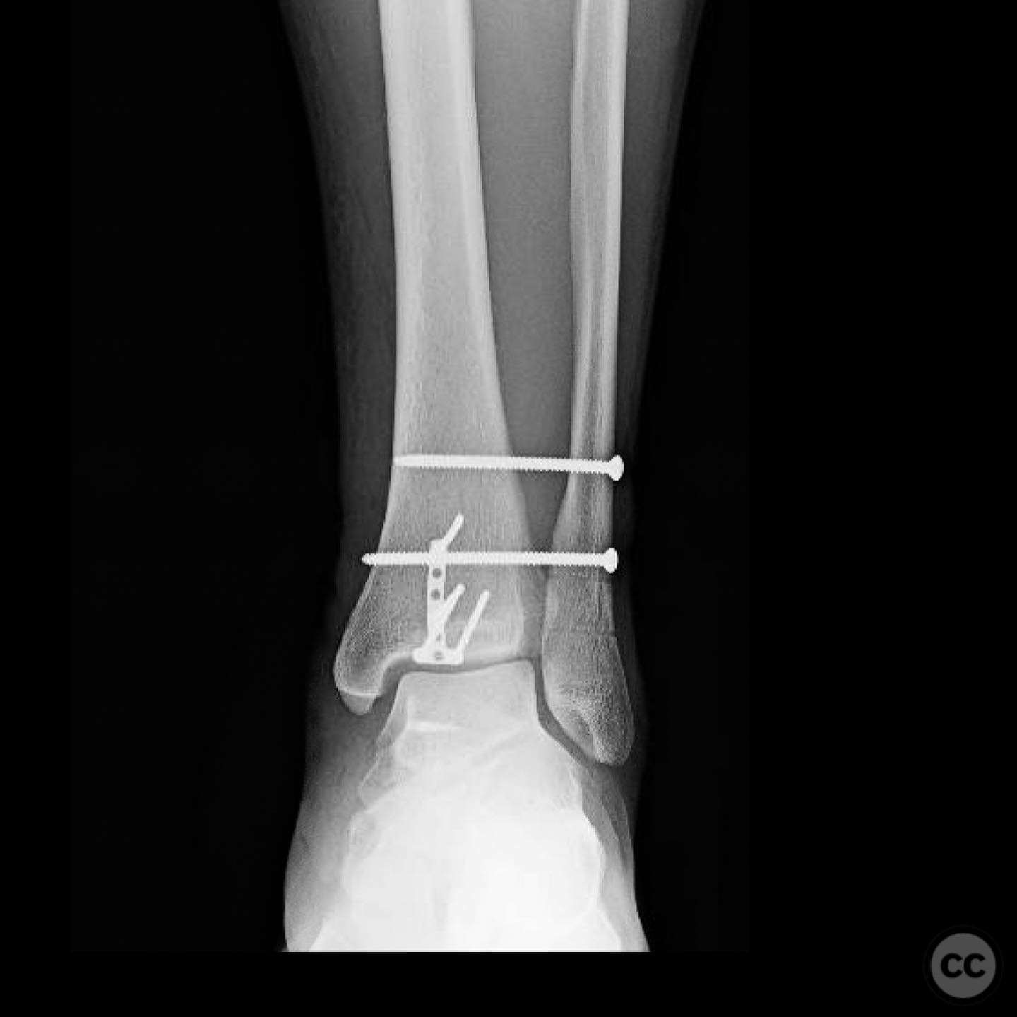
Clinical and radiological findings: A 31-year-old male skier sustained a Maisonneuve fracture after his left ski caught in a tree well, resulting in an external rotation force. Initial radiographs revealed a proximal fibular fracture consistent with a Maisonneuve injury, accompanied by a dislocated ankle joint. A small anterior plafond rim fragment was visible on X-ray. Upon surgical exploration, an impacted dime-sized medial plafond fragment and a 5x10mm anterior plafond rim fragment were identified. The deltoid ligament was found to be ruptured at the talar attachment.
Preoperative Plan
Planning remarks: The preoperative plan included a medial approach to repair the deltoid ligament, disimpact and graft the medial plafond, and fix the articular fragments. Syndesmotic stabilization was anticipated, with contralateral imaging for comparison and fluoroscopic guidance for reduction assessment.
Surgical Discussion
Patient positioning: Supine position with the affected limb externally rotated and supported to allow access to the medial aspect of the ankle.
Anatomical surgical approach: A medial approach to the ankle was utilized, involving an incision over the medial malleolus. The deltoid ligament was exposed, revealing its avulsion from the talus. The plafond impaction was addressed through this approach, followed by bone grafting and fixation of the articular fragments.
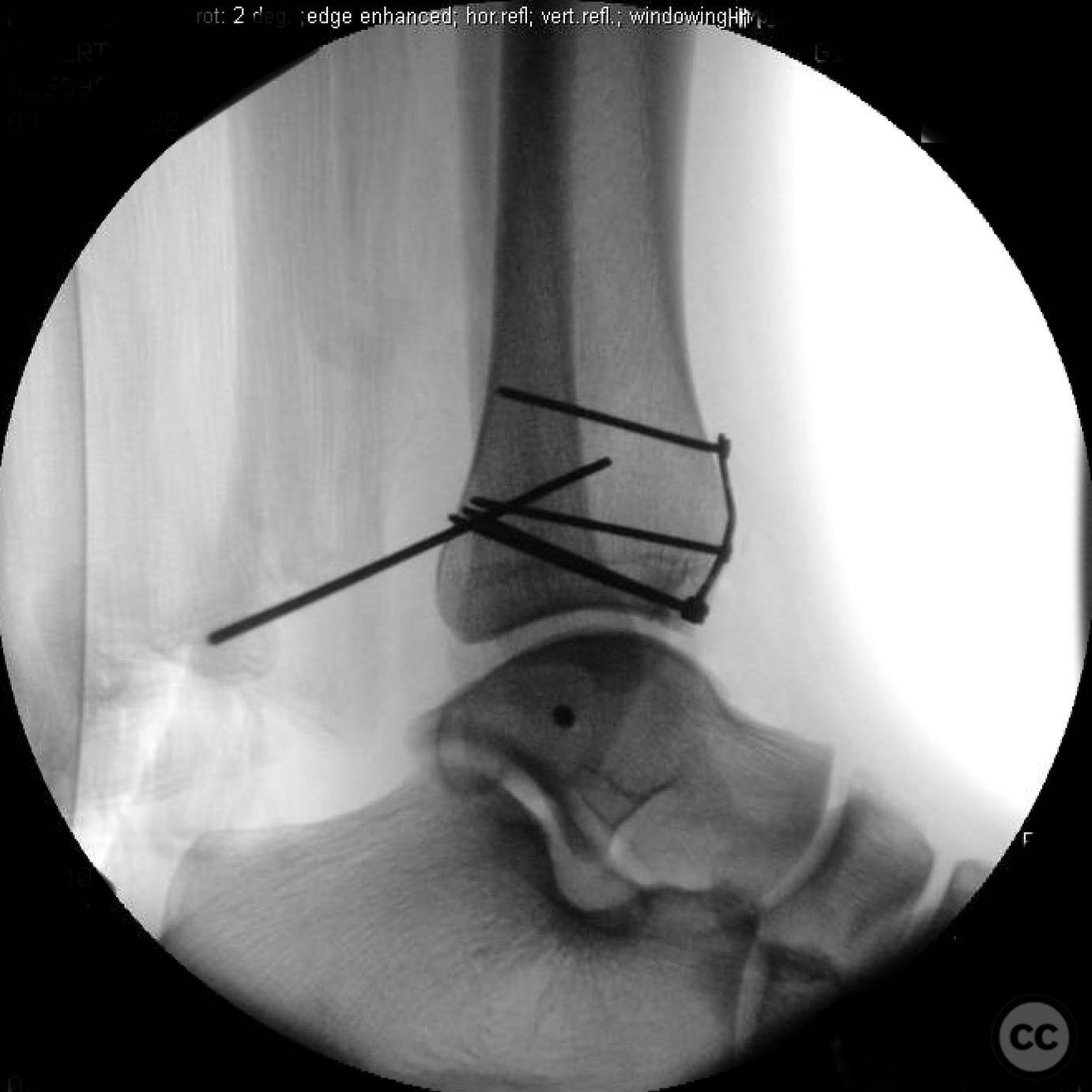
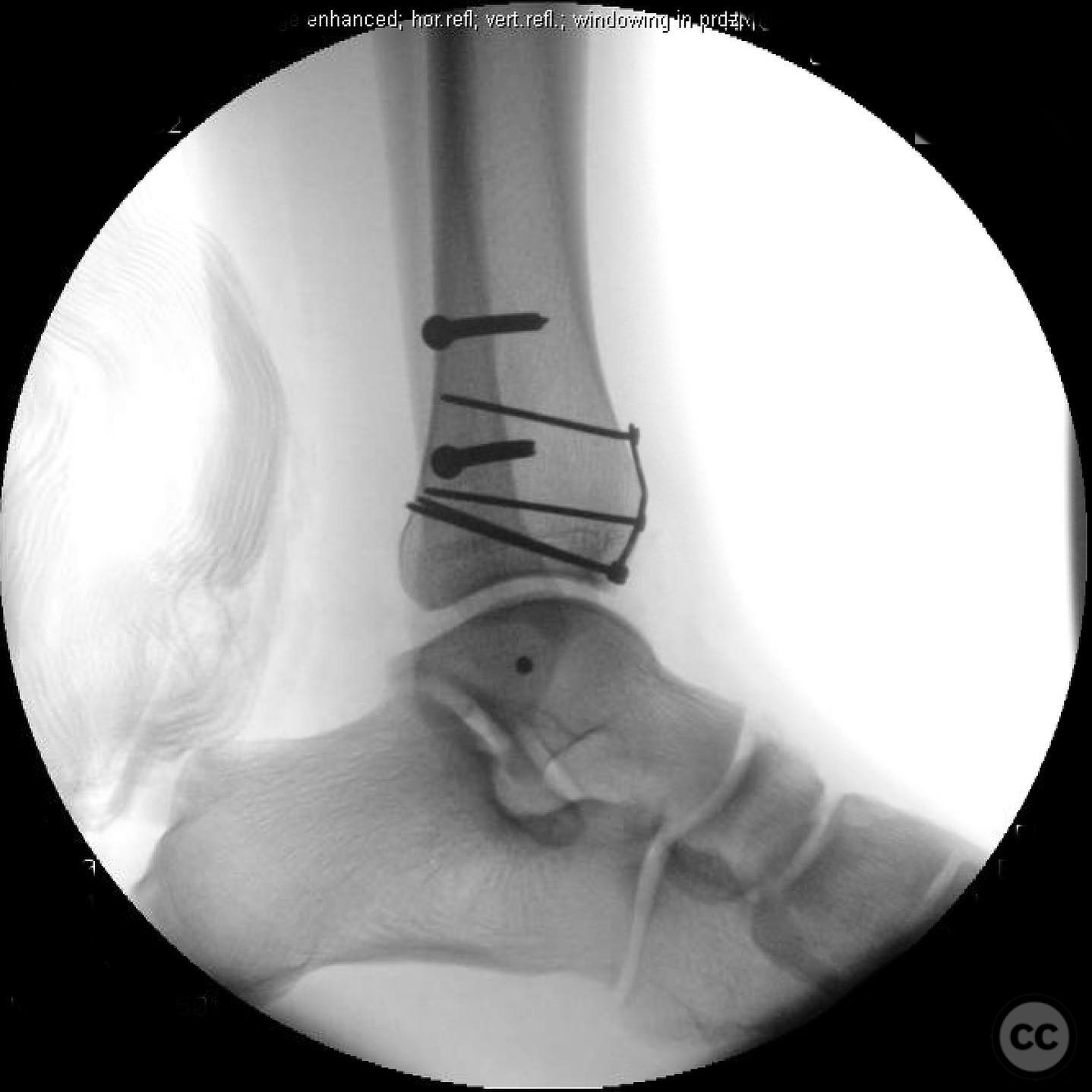
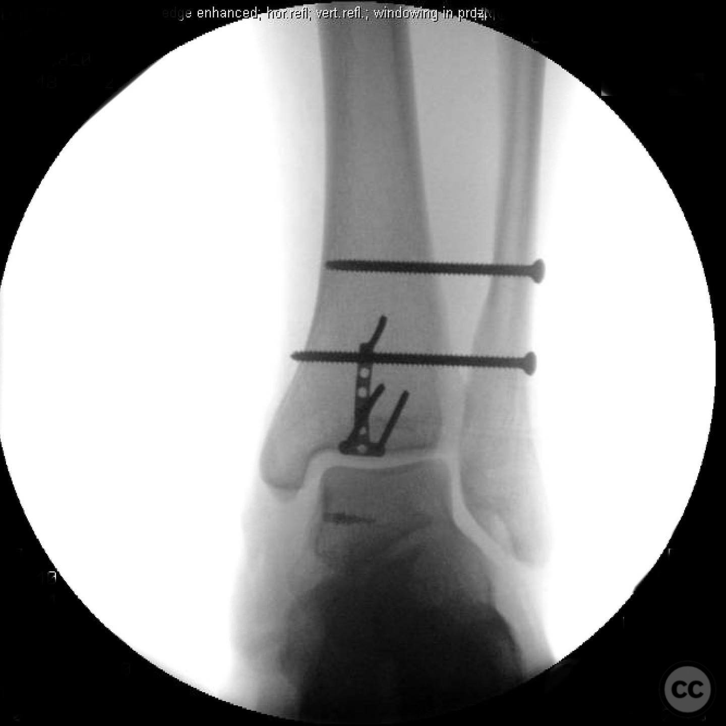
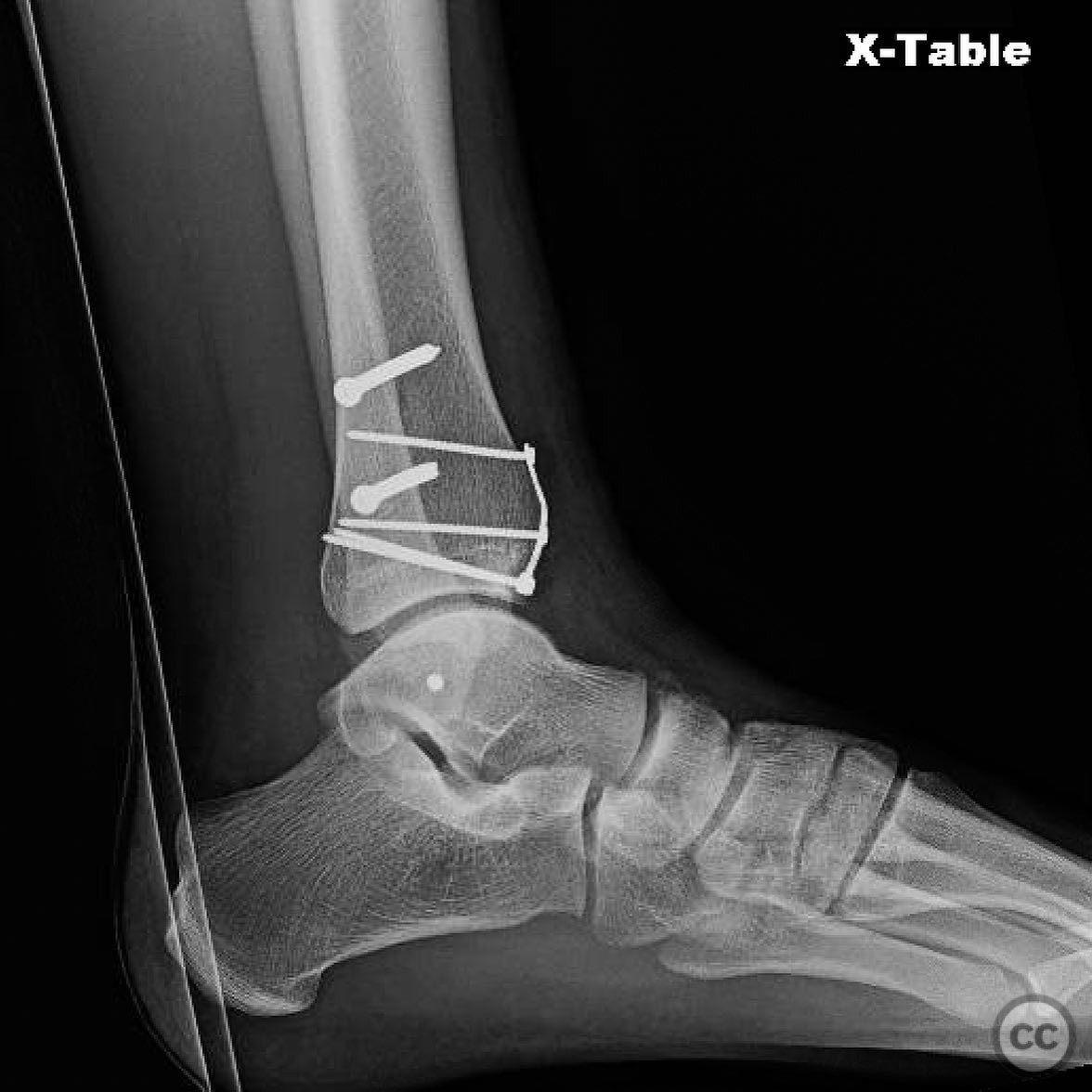
Operative remarks:The surgeon noted the deltoid ligament avulsion from the talus, which was repaired using an anchor placed at the talar bare spot. The medial plafond impaction was successfully disimpacted and grafted. Syndesmotic reduction was performed with attention to fibular length and rotation, using contralateral imaging for reference. The importance of fluoroscopic assessment of fibular alignment with the tibia and talus was emphasized.
Postoperative protocol: Non-weight bearing with crutches for 6 weeks, followed by progressive weight bearing as tolerated. Range of motion exercises initiated postoperatively to maintain joint mobility.
Follow up: Not specified.
Orthopaedic implants used: Syndesmotic screw fixation, bone anchor for deltoid ligament repair, bone graft material.
Search for Related Literature

orthopaedic_trauma
- United States , Seattle
- Area of Specialty - General Trauma
- Position - Specialist Consultant

Industry Sponsership
contact us for advertising opportunities
Article viewed 300 times
16 Jul 2025
Add to Bookmarks
Full Citation
Cite this article:
Surname, Initial. (2025). Maisonneuve Fracture with Medial Plafond Impaction and Deltoid Ligament Rupture.. Journal of Orthopaedic Surgery and Traumatology. Case Report 42487232 Published Online Jul 16 2025.