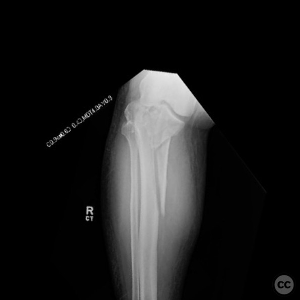
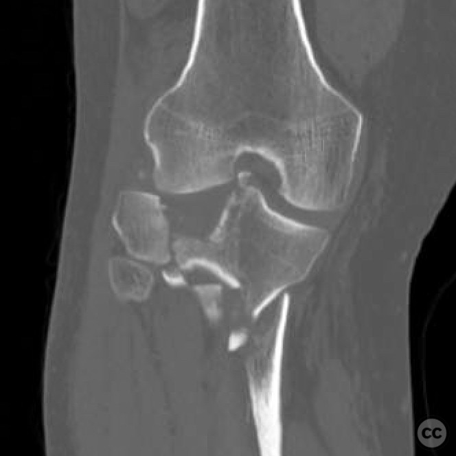
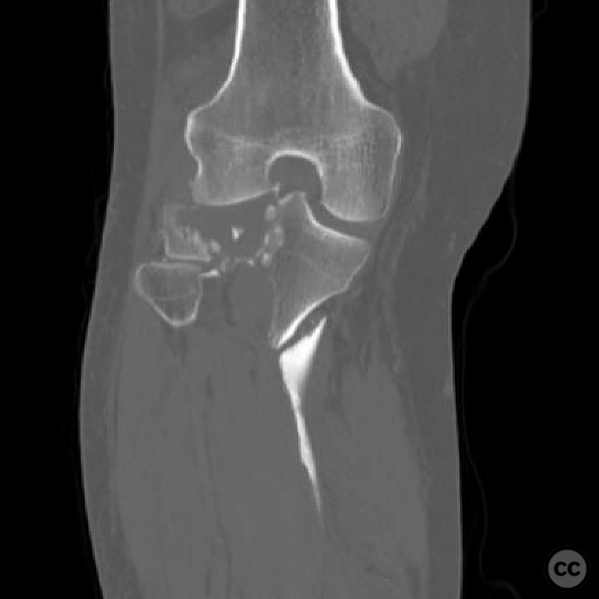
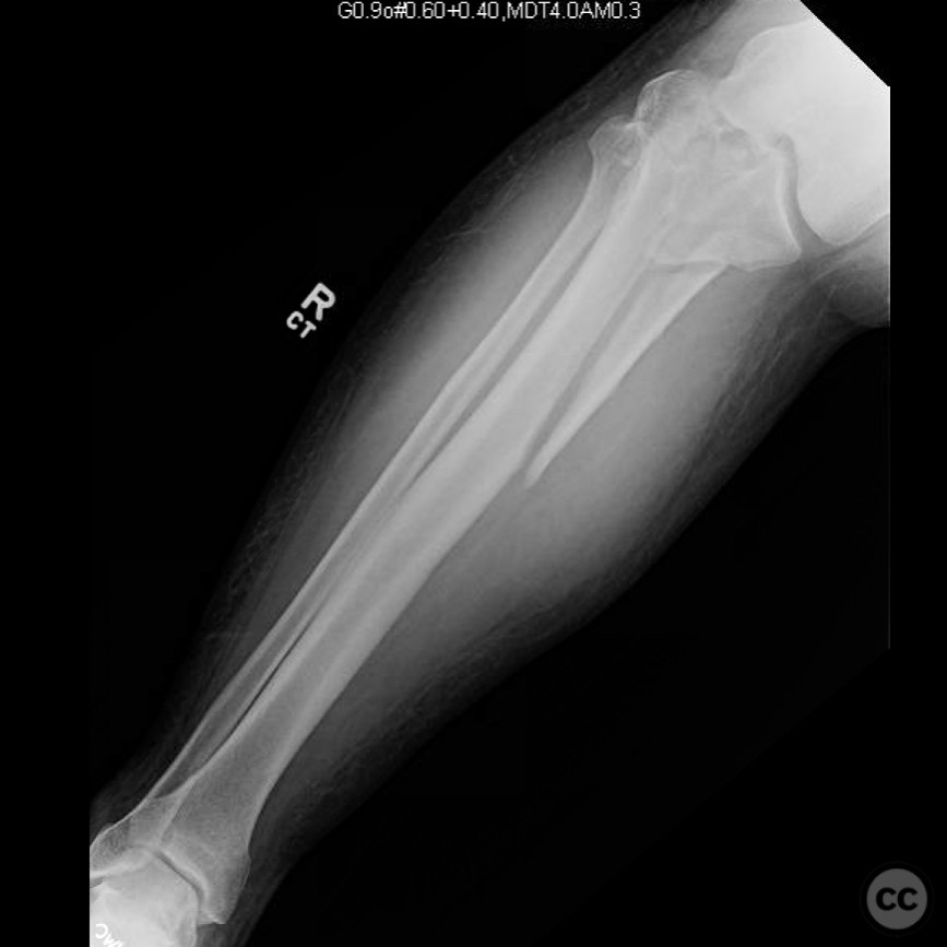
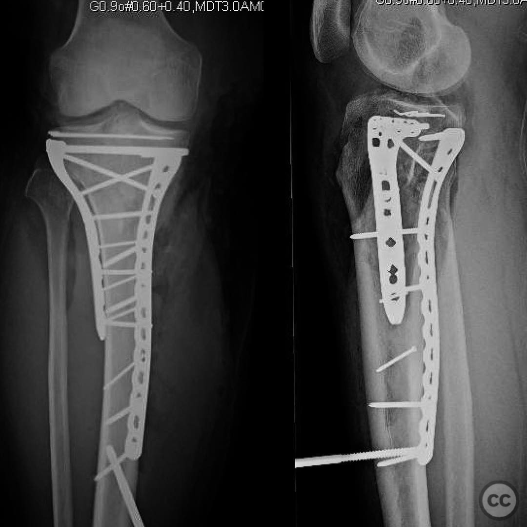
Bicondylar tibial plateau fracture with diaphyseal extension.
Score and Comment on this Case
Clinical Details
Clinical and radiological findings: A male patient involved in a high-speed motorcycle collision with a deer, traveling at 60 mph. The patient sustained a bicondylar tibial plateau fracture with diaphyseal extension. Initial clinical examination should be vigilant for compartment syndrome due to the mechanism of injury. Radiological assessment, including X-rays and CT scans, confirmed the fracture pattern. Neurovascular examination was crucial to rule out any compromise.
Preoperative Plan
Planning remarks: The preoperative plan involved dual plating for stabilization of the bicondylar fracture. A posteromedial approach was planned for the placement of a posteromedial plate, and an anterolateral approach for the anterolateral plate. The primary surgical goal was to restore the mechanical axis, with articular reduction being of secondary importance.
Surgical Discussion
Patient positioning: The patient was positioned supine on the operating table, with a radiolucent table used to facilitate intraoperative imaging. A bump was placed under the ipsilateral hip to allow for slight external rotation of the limb.
Anatomical surgical approach: The surgical approach included a posteromedial incision along the medial border of the tibia, with careful dissection to protect the saphenous nerve and vein. Subperiosteal elevation was performed to expose the posteromedial aspect of the tibia for plate placement. An anterolateral incision was made, extending from Gerdy's tubercle distally, splitting the iliotibial band and performing a sub-meniscal arthrotomy for visualization of the lateral plateau.
Operative remarks:The surgeon emphasized the importance of avoiding varus malalignment during fixation. Careful attention was given to restoring the mechanical axis of the limb. Articular reduction was achieved as much as possible, but it was considered secondary to achieving correct alignment. Compartment pressures were monitored intraoperatively due to the high-energy nature of the injury.
Postoperative protocol: Postoperative rehabilitation included non-weight bearing for 6 weeks, followed by progressive weight bearing as tolerated. Range of motion exercises were initiated early to prevent stiffness, with a focus on achieving full extension and flexion.
Follow up: Not specified.
Orthopaedic implants used: Posteromedial plate, anterolateral plate.
Search for Related Literature

orthopaedic_trauma
- United States , Seattle
- Area of Specialty - General Trauma
- Position - Specialist Consultant

Industry Sponsership
contact us for advertising opportunities
Article viewed 247 times
26 Jul 2025
Add to Bookmarks
Full Citation
Cite this article:
Surname, Initial. (2025). Bicondylar tibial plateau fracture with diaphyseal extension.. Journal of Orthopaedic Surgery and Traumatology. Case Report 42065116 Published Online Jul 26 2025.