 a(.jpg)
Supracondylar Intercondylar Distal Humerus Fracture with Fishtail Deformity in an Adolescent.
Score and Comment on this Case
Clinical Details
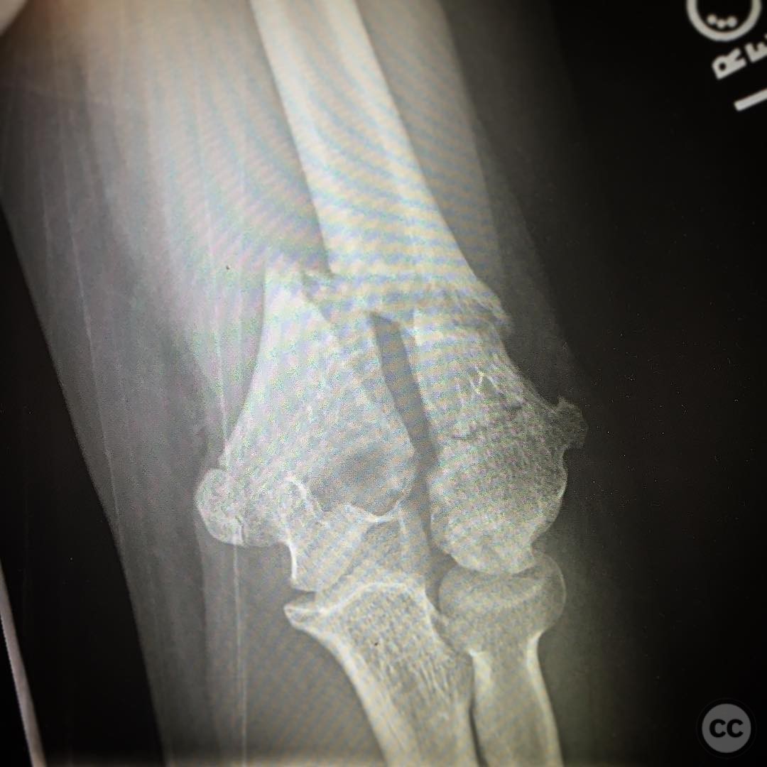
Clinical and radiological findings: A 14-year-old male presented with a supracondylar intercondylar distal humerus fracture. The patient had a history of a lateral condyle fracture treated operatively 9 years prior, resulting in a "fishtail" deformity due to avascular necrosis of the lateral trochlea and cubitus varus from lateral condyle overgrowth. Radiographs revealed distorted distal humeral morphology, complicating fracture reduction and stable fixation. The absence of the lateral trochlea precluded the use of lag screws across the columns at the articular surface.
Preoperative Plan
Planning remarks: The preoperative plan involved a posterior approach to the distal humerus, avoiding an olecranon osteotomy. Reduction was planned using metaphyseal reads due to the lack of articular reads.
Surgical Discussion
Patient positioning: The patient was positioned prone on the operating table with the arm supported on a padded arm board to allow optimal access to the posterior aspect of the elbow.
Anatomical surgical approach: A posterior midline incision was made over the distal humerus. The triceps was split in line with its fibers, providing access to the distal humerus without performing an olecranon osteotomy. Subperiosteal dissection was carried out to expose the fracture site.
Operative remarks:The distorted distal humeral morphology presented challenges in achieving stable reduction and fixation. Metaphyseal reads were utilized for fracture reduction due to the absence of articular reads. The absence of the lateral trochlea necessitated alternative fixation strategies, as lag screws across the columns at the articular surface were not feasible.
Postoperative protocol: Postoperative rehabilitation included immobilization in a posterior splint for 2 weeks, followed by gradual range of motion exercises. Weight-bearing activities were restricted until radiographic evidence of healing was observed.
Follow up: Not specified.
Orthopaedic implants used: Orthopaedic implants used: Not specified.
Search for Related Literature

orthopaedic_trauma
- United States , Seattle
- Area of Specialty - General Trauma
- Position - Specialist Consultant

Industry Sponsership
contact us for advertising opportunities
Article viewed 413 times
05 Aug 2025
Add to Bookmarks
Full Citation
Cite this article:
Surname, Initial. (2025). Supracondylar Intercondylar Distal Humerus Fracture with Fishtail Deformity in an Adolescent.. Journal of Orthopaedic Surgery and Traumatology. Case Report 39854042 Published Online Aug 05 2025.