Open Supracondylar Distal Femur Fracture
Score and Comment on this Case
Clinical Details
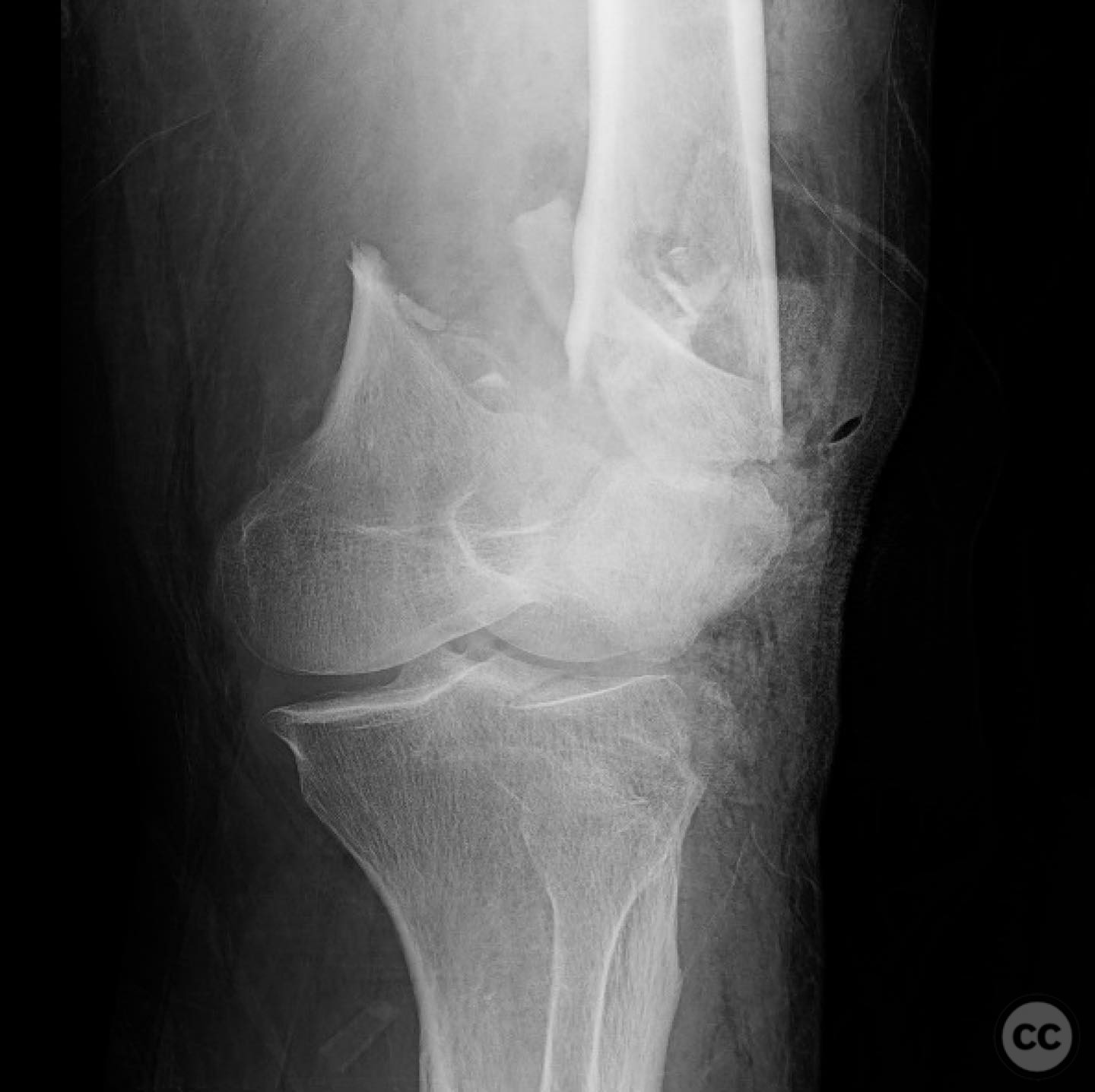
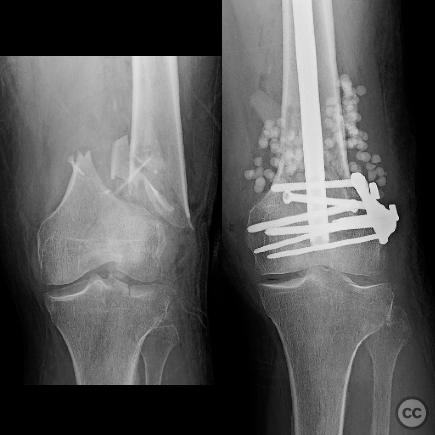
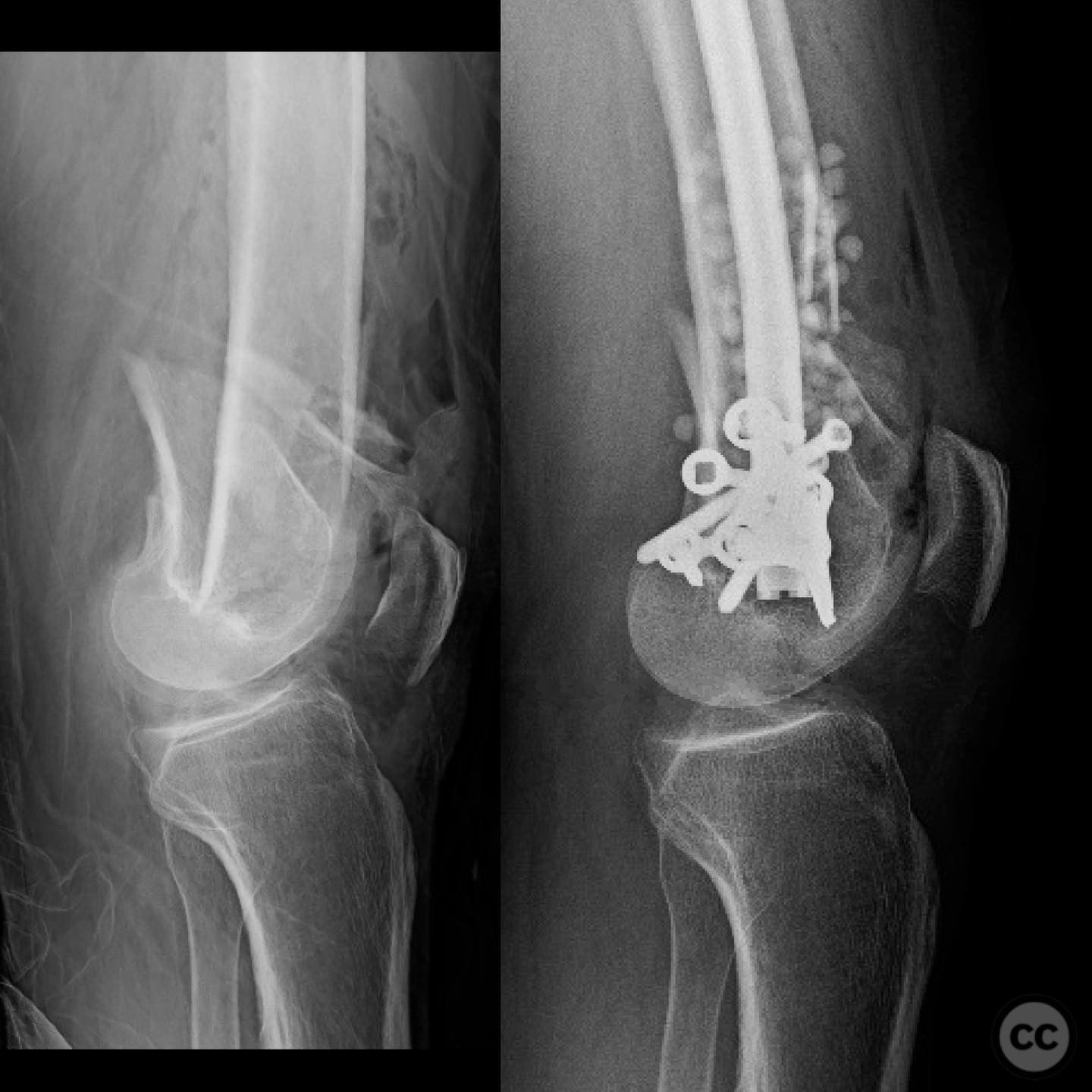
Clinical and radiological findings: A middle-aged male, estimated to be between 40-60 years old, presented intubated and sedated following a vehicular accident. Initial imaging revealed stable subarachnoid and subdural hemorrhages on serial CT scans, not necessitating intervention. The patient had no significant thoracic or abdominal injuries. Laboratory findings showed an initial lactate level of 4.8 mmol/L, which decreased to 2.0 mmol/L upon resuscitation. Orthopaedic evaluation identified an open supracondylar distal femur fracture with a 1 cm lateral skin wound. No other major skeletal injuries were noted. CT imaging confirmed the fracture as a rare supracondylar type without significant articular extension.
Preoperative Plan
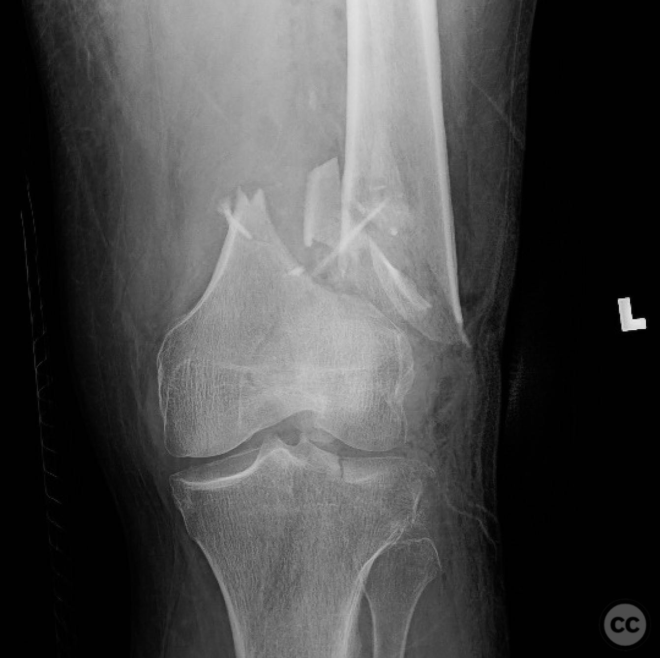
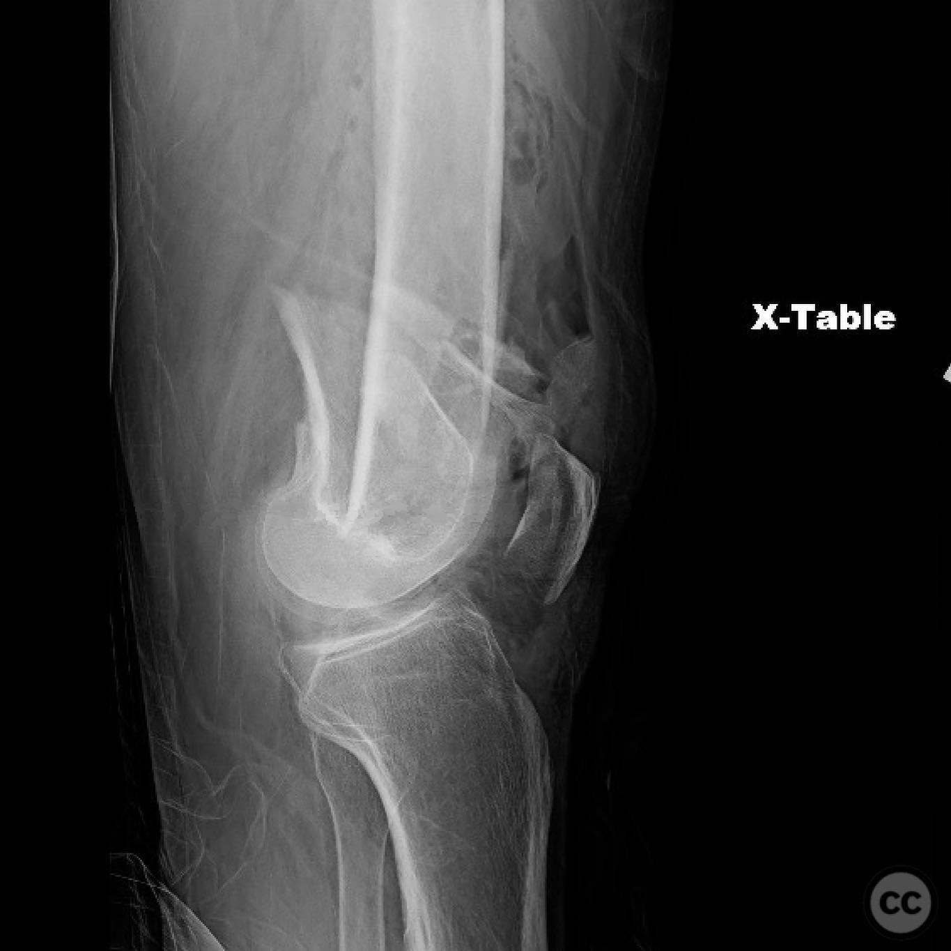
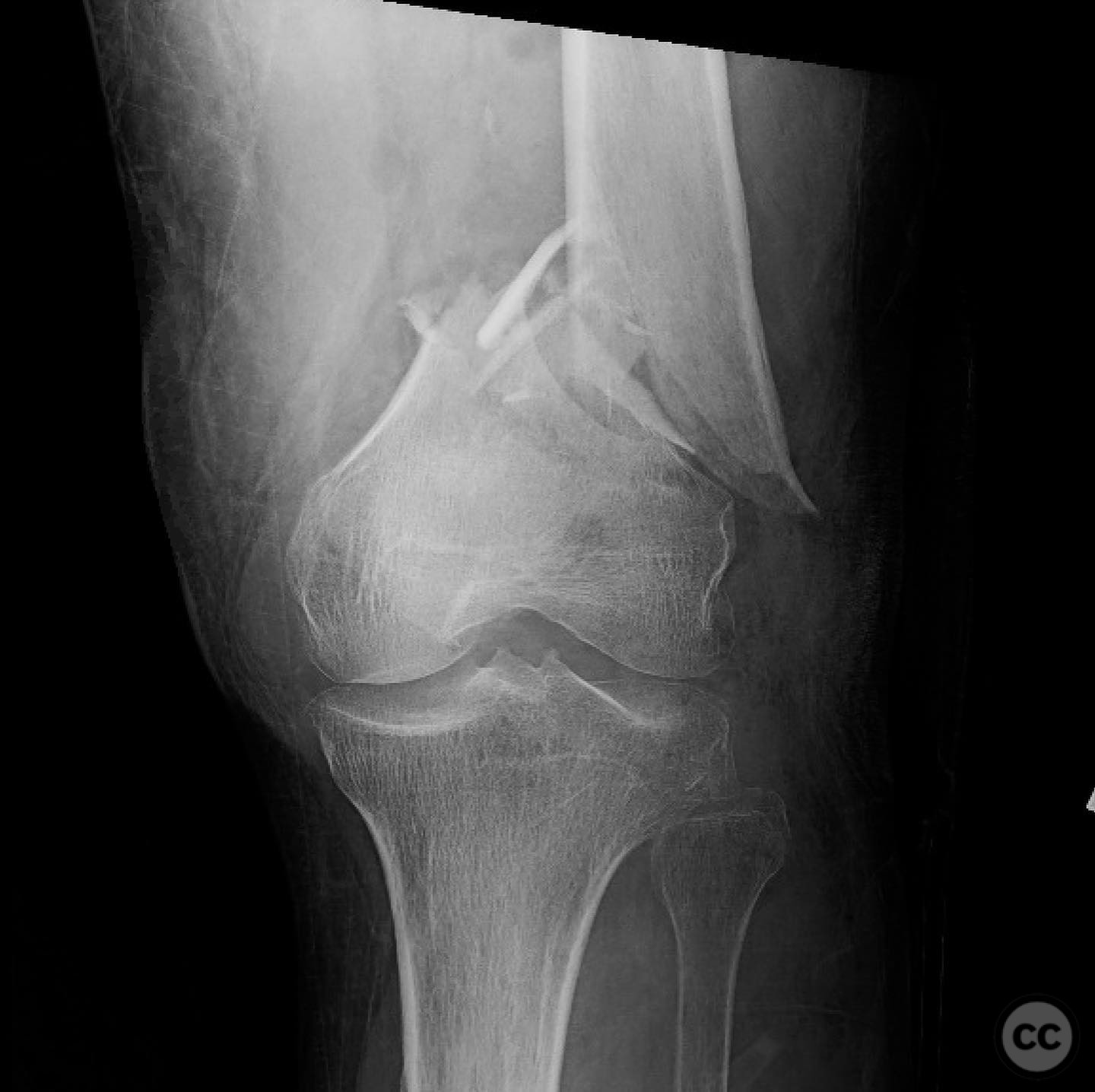
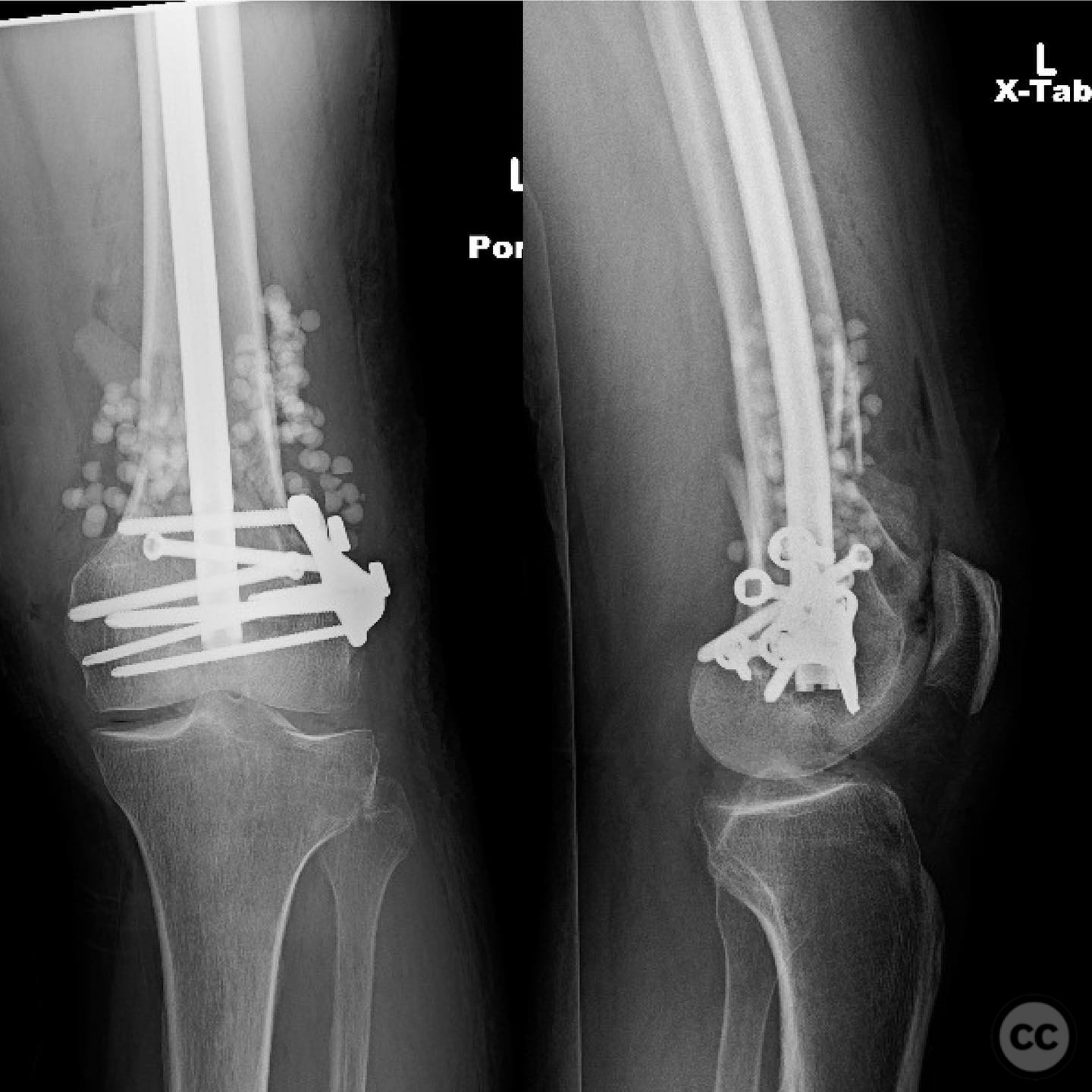
Planning remarks: The preoperative plan involved immediate intravenous antibiotic administration and tetanus prophylaxis due to the open nature of the fracture, classified as Gustilo-Anderson Type III. Surgical debridement was planned followed by definitive skeletal stabilization using a retrograde intramedullary nail with a fixed angle washer for additional fixation in the articular block.
Surgical Discussion
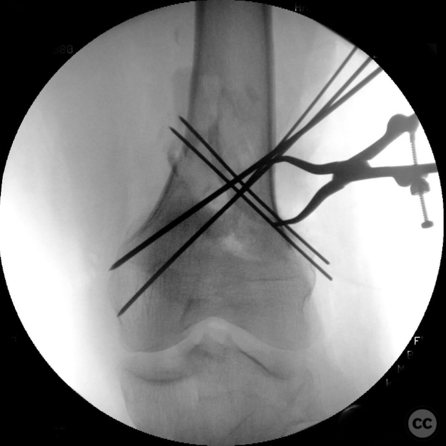
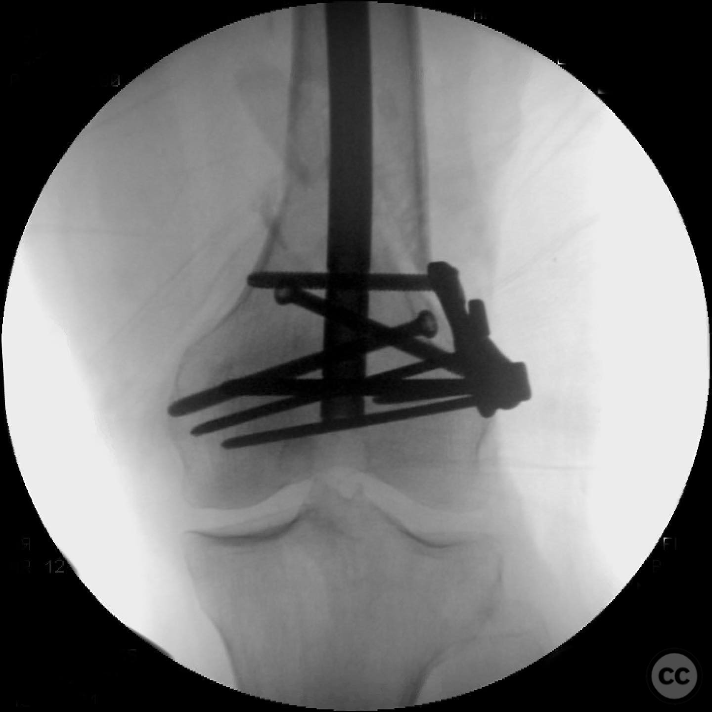
Patient positioning: The patient was positioned supine on the operating table to facilitate a retrograde intramedullary nailing approach.
Anatomical surgical approach: A direct lateral approach to the distal femur was utilized, involving careful dissection through the existing 1 cm skin wound. Subperiosteal elevation was performed to expose the fracture site, allowing for thorough irrigation and debridement of the open wound.
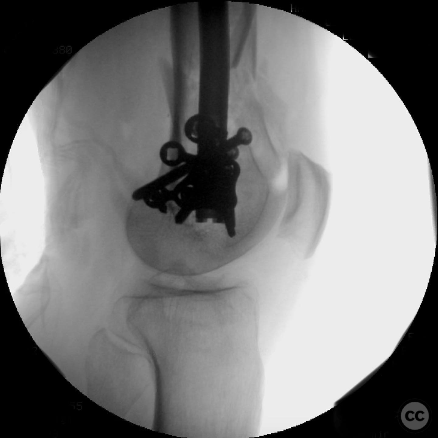
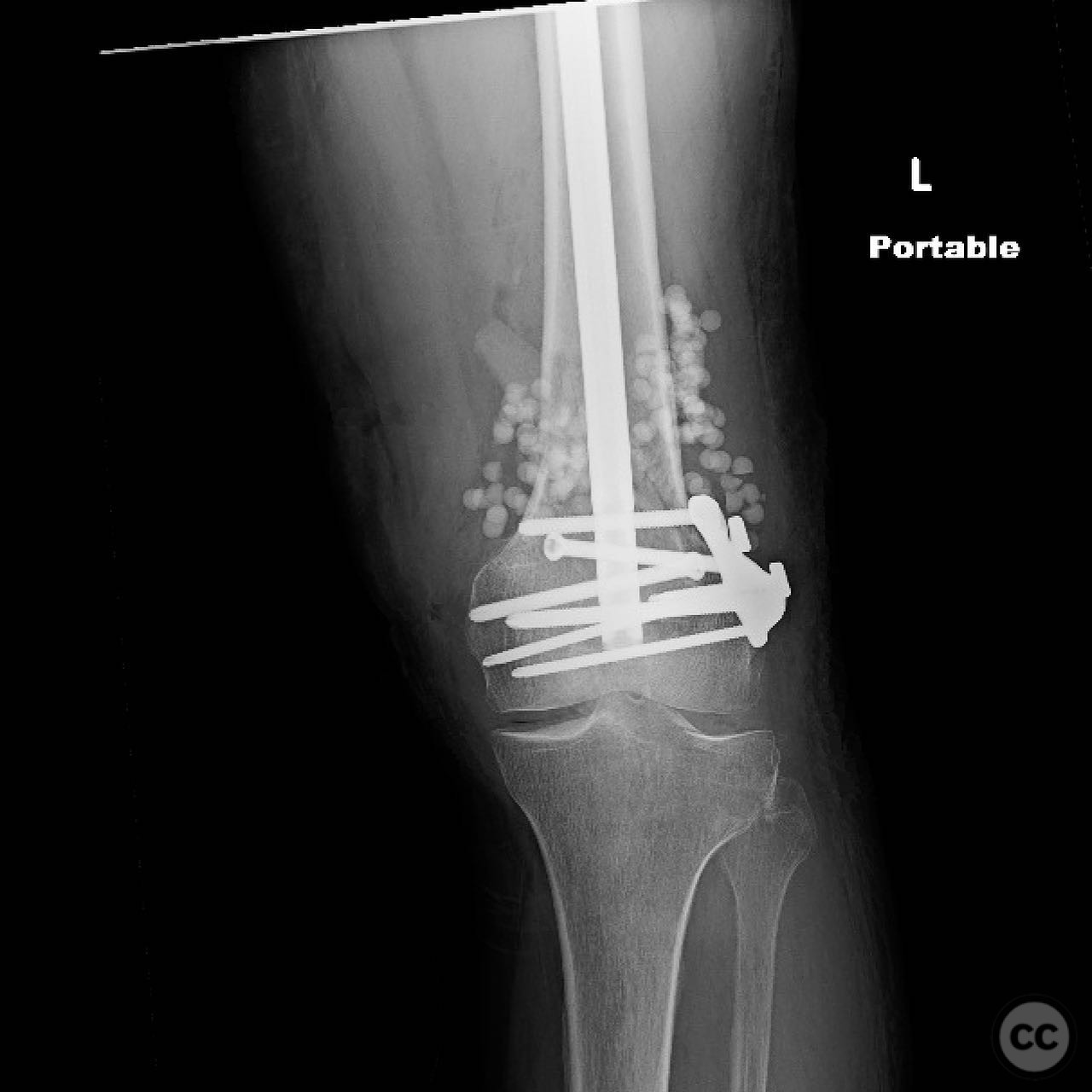
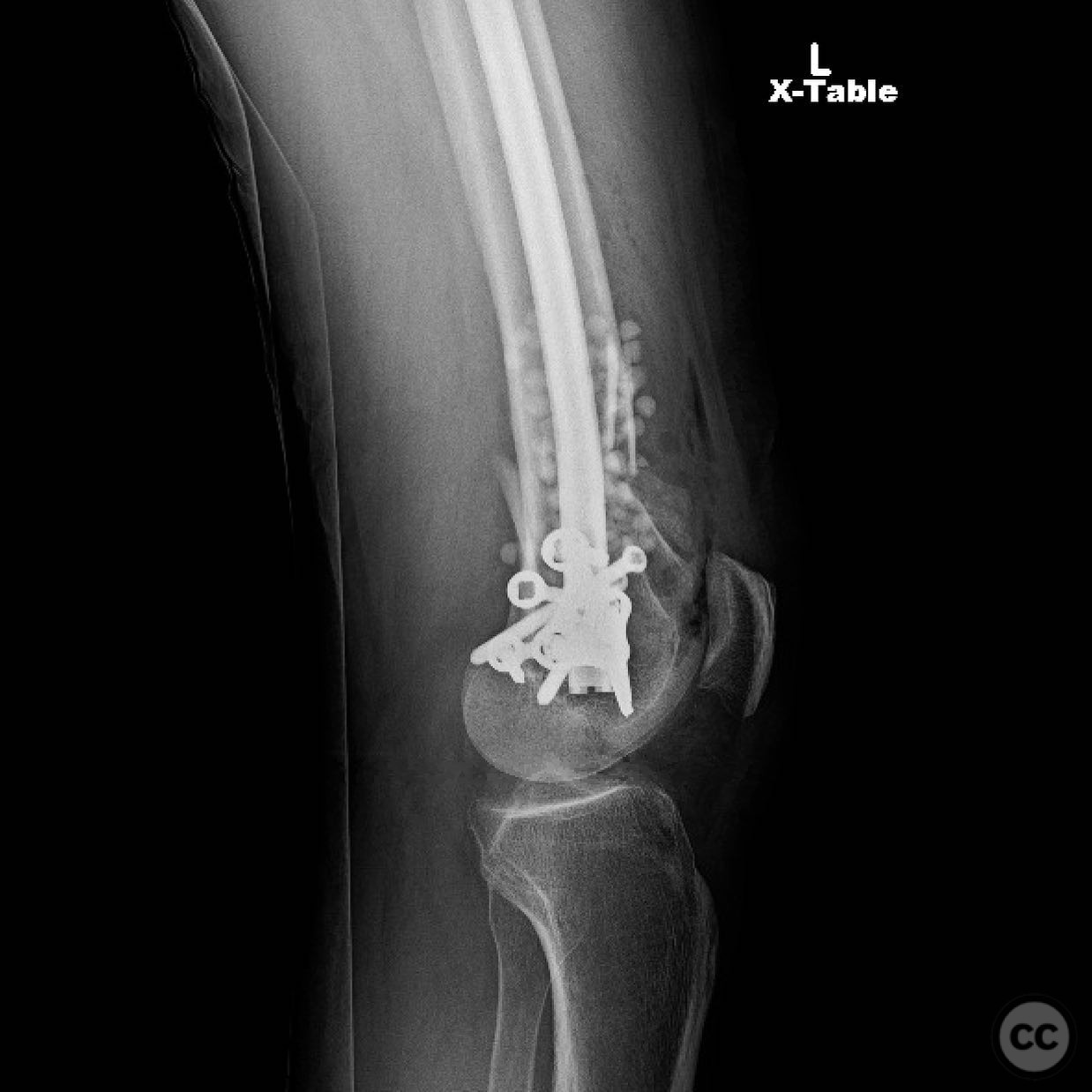
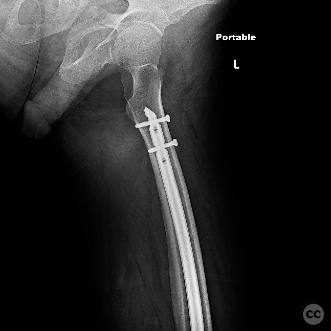
Operative remarks:The surgeon noted that achieving union and early mobilization was paramount, with relative stability and biologically friendly surgery being the goals. The use of a retrograde nail with a fixed angle washer was chosen to provide optimal fixation with a long working length, promoting endochondral ossification. Alternative fixation methods such as lateral locked plating, plate-nail combinations, and dual plating were considered but not utilized in this case.
Local antibiotic therapy included calcium sulfate (CaSO₄) beads impregnated with vancomycin and tobramycin, placed at the fracture site following debridemen
Postoperative protocol: Postoperative rehabilitation focused on early mobilization with weight-bearing as tolerated, progressing to full weight-bearing as clinical and radiological healing allowed.
Follow up: Not specified.
Orthopaedic implants used: Retrograde intramedullary nail, fixed angle washer, calcium sulfate beads with vancomycin and tobramycin.
Search for Related Literature

orthopaedic_trauma
- United States , Seattle
- Area of Specialty - General Trauma
- Position - Specialist Consultant

Industry Sponsership
contact us for advertising opportunities
Article viewed 267 times
13 Jul 2025
Add to Bookmarks
Full Citation
Cite this article:
Surname, Initial. (2025). Open Supracondylar Distal Femur Fracture. Journal of Orthopaedic Surgery and Traumatology. Case Report 39307974 Published Online Jul 13 2025.