Arthrodesis and Soft Tissue Release for Charcot Arthropathy with Tibiotalar Osteoarthritis
Score and Comment on this Case
Clinical Details
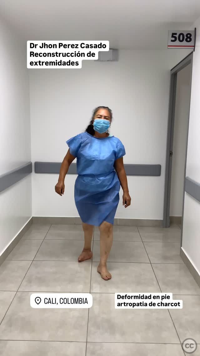
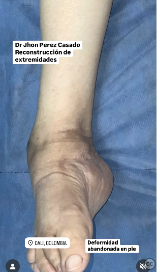
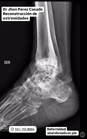
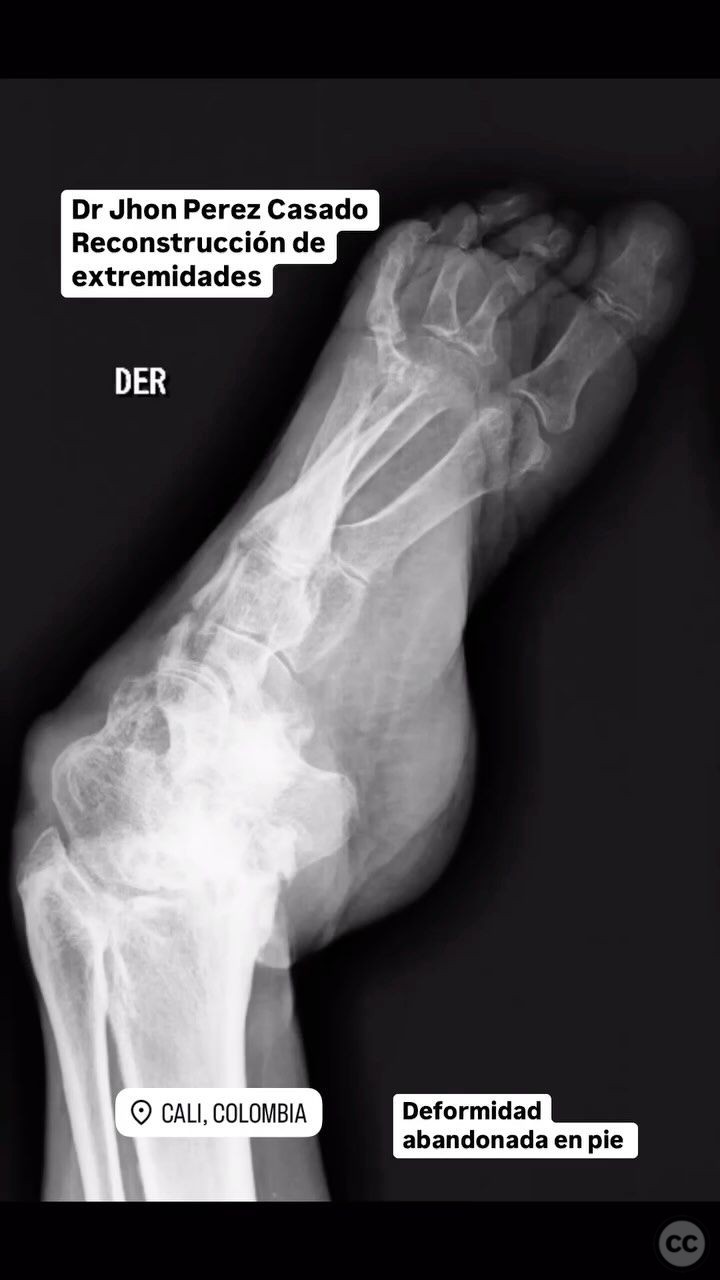
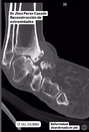
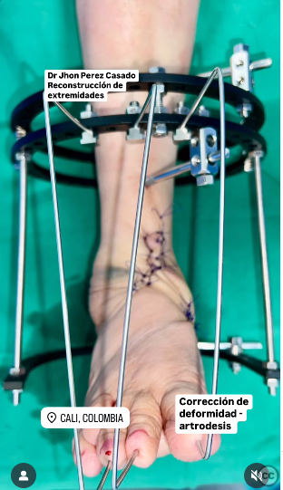
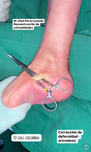
Clinical and radiological findings: The patient presented with an abandoned foot deformity and osteoarthritis of the tibiotalar joint, consistent with Charcot arthropathy. Radiological assessment confirmed severe degenerative changes in the tibiotalar joint with associated deformity.
Preoperative Plan
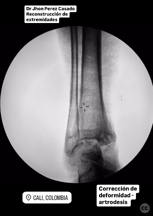
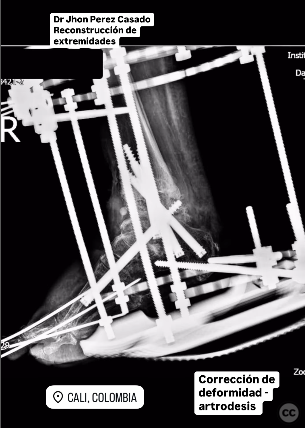
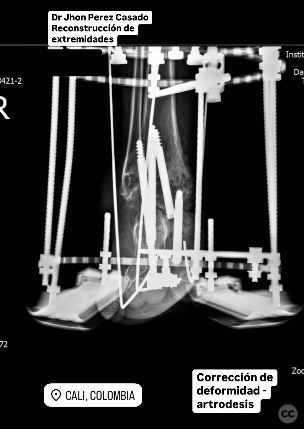
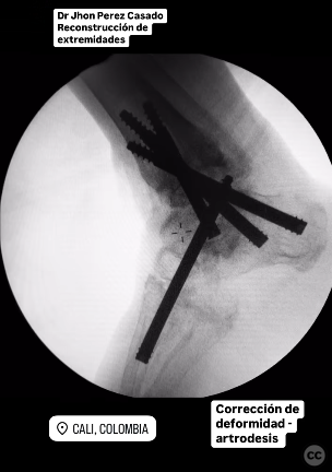
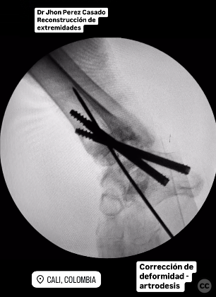
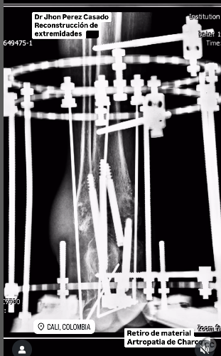
Planning remarks: The preoperative plan included multiple tenotomies: Achilles tendon, plantar fascia, posterior tibial tendon, and finger flexor tendons. The surgical goal was to correct the deformity through arthrodesis of the tibiotalar joint using cannulated screws, followed by stabilization with an Ilizarov external fixator.
Surgical Discussion
Patient positioning: The patient was positioned supine on the operating table, with the affected limb elevated and prepared for a sterile field.
Anatomical surgical approach: A longitudinal incision was made over the posterior aspect of the ankle to access the Achilles tendon for tenotomy. Subsequent incisions were made medially and plantarly to perform tenotomies of the posterior tibial tendon and plantar fascia, respectively. A dorsal approach was utilized for finger flexor tenotomy. For arthrodesis, a lateral approach to the ankle was employed to expose the tibiotalar joint, allowing for debridement and preparation of the joint surfaces before fixation with cannulated screws.
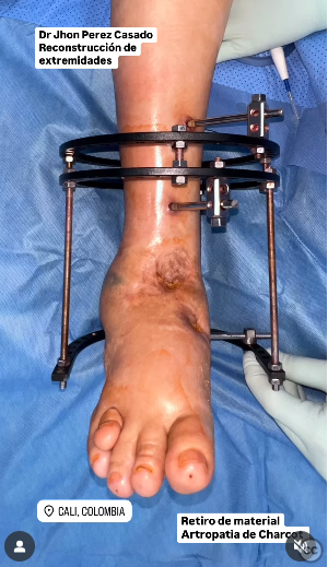
Operative remarks:The surgeon noted that the extensive soft tissue release facilitated correction of the deformity. The use of an Ilizarov external fixator provided necessary stability post-arthrodesis, accommodating any residual soft tissue contractures and ensuring proper alignment during the healing process.
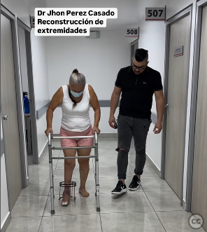
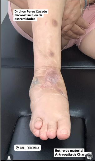
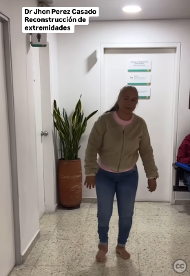
Postoperative protocol: Postoperatively, the patient underwent walking training with full support using crutches. Weight-bearing was gradually introduced as tolerated under supervision.
Follow up: Not specified.
Orthopaedic implants used: Cannulated screws, Ilizarov external fixator.
Search for Related Literature

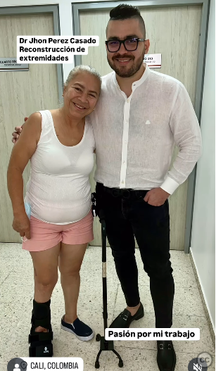
Jhon Perez Casado
- Colombia , Cali - Valle del Cauca
- Area of Specialty - Lower Limb
- Position - Specialist Consultant

Industry Sponsership
contact us for advertising opportunities
Article viewed 282 times
24 Jul 2025
Add to Bookmarks
Full Citation
Cite this article:
Perez Casado, J.J.. (2025). Arthrodesis and Soft Tissue Release for Charcot Arthropathy with Tibiotalar Osteoarthritis. Journal of Orthopaedic Surgery and Traumatology. Case Report 37977041 Published Online Jul 24 2025.