 of_2.jpg)
 of_3.jpg)
 of_1.jpg)
 of (.jpg)
 of_4.jpg)
 of_5.jpg)
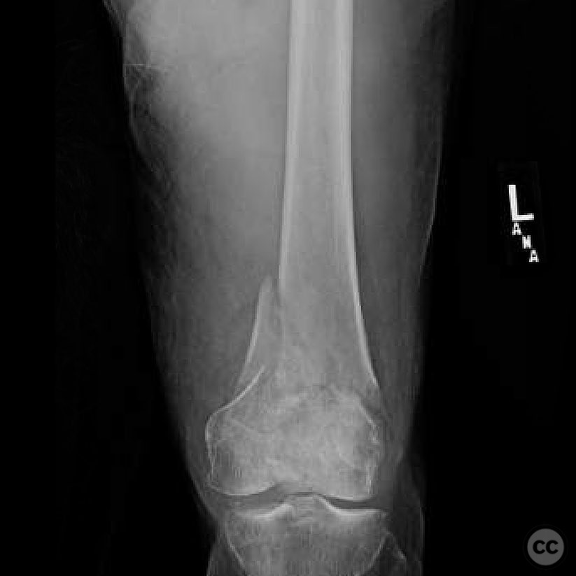
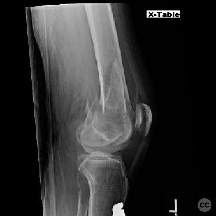
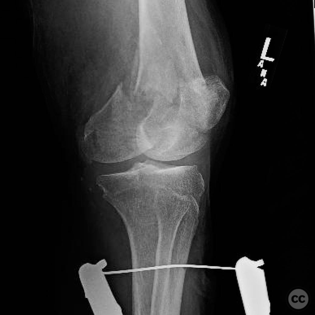
Complex 33C3 Distal Femur Fracture with Segmental Medial Hoffa.
Score and Comment on this Case
Clinical Details
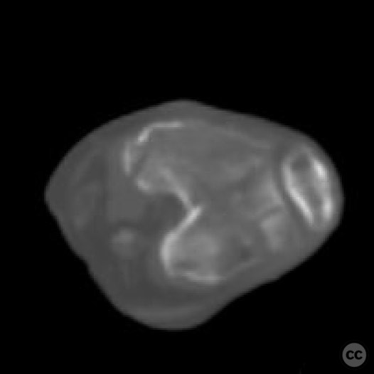
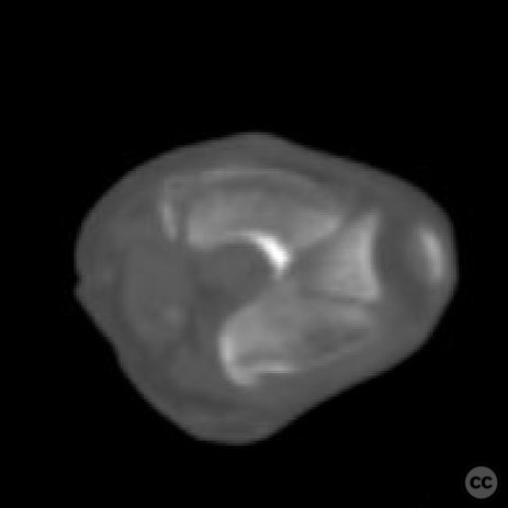
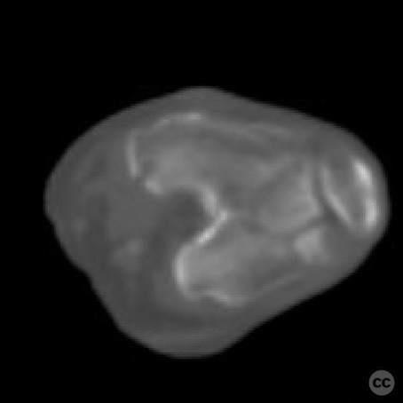
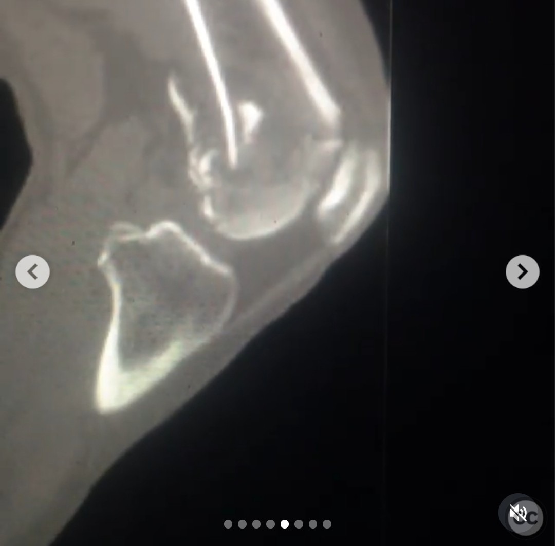
Clinical and radiological findings: A 73-year-old female involved in a motor vehicle accident presented with a complex distal femur fracture. The patient is otherwise healthy with no significant medical comorbidities and has an operative acetabulum fracture above. Initial radiographs appeared benign, but CT imaging revealed a complex articular pattern characterized by segmental articular comminution of the lateral condyle, a free notch fragment, and a segmental medial Hoffa fracture with both large and small components.
Preoperative Plan
Planning remarks: The preoperative plan involved a dual incision approach: an anterolateral incision for direct visualization of the lateral joint involvement and trochlear fragment, and a medial subvastus approach to address the medial Hoffa components. The surgical goal was to achieve anatomic reduction with interfragmentary compression for primary bone healing at the joint, and bridging of the comminuted metaphyseal segment for relative stability and secondary bone healing.
Surgical Discussion
Patient positioning: The patient was positioned supine with the knee in hyperflexion to facilitate access to the small type two Hoffa component.
Anatomical surgical approach: The surgical approach consisted of an anterolateral incision to expose the lateral condyle and trochlear fragment, followed by a medial subvastus approach to access the medial Hoffa fractures. The type one Hoffa component was buttressed, while screws were placed posterior to anterior for stabilization of the type two component.
Operative remarks:The surgeon noted the complexity of modulating construct stiffness in this geriatric distal femur fracture. The decision was made against using a distal femoral replacement due to the presence of an operative acetabulum above and anticipated three-month TTWB status. Instead, the focus was on achieving stable fixation through anatomic reduction and appropriate hardware selection.
Postoperative protocol: The postoperative rehabilitation protocol included toe-touch weight bearing (TTWB) for three months due to the operative acetabulum above.
Follow up: Not specified.
Orthopaedic implants used: Orthopaedic implants used included interfragmentary screws and buttress plates.
Search for Related Literature

orthopaedic_trauma
- United States , Seattle
- Area of Specialty - General Trauma
- Position - Specialist Consultant

Industry Sponsership
contact us for advertising opportunities
Article viewed 310 times
24 Jul 2025
Add to Bookmarks
Full Citation
Cite this article:
Surname, Initial. (2025). Complex 33C3 Distal Femur Fracture with Segmental Medial Hoffa.. Journal of Orthopaedic Surgery and Traumatology. Case Report 37755053 Published Online Jul 24 2025.