Complex Articular Humerus Fracture with Comminuted Diaphysis
Score and Comment on this Case
Clinical Details
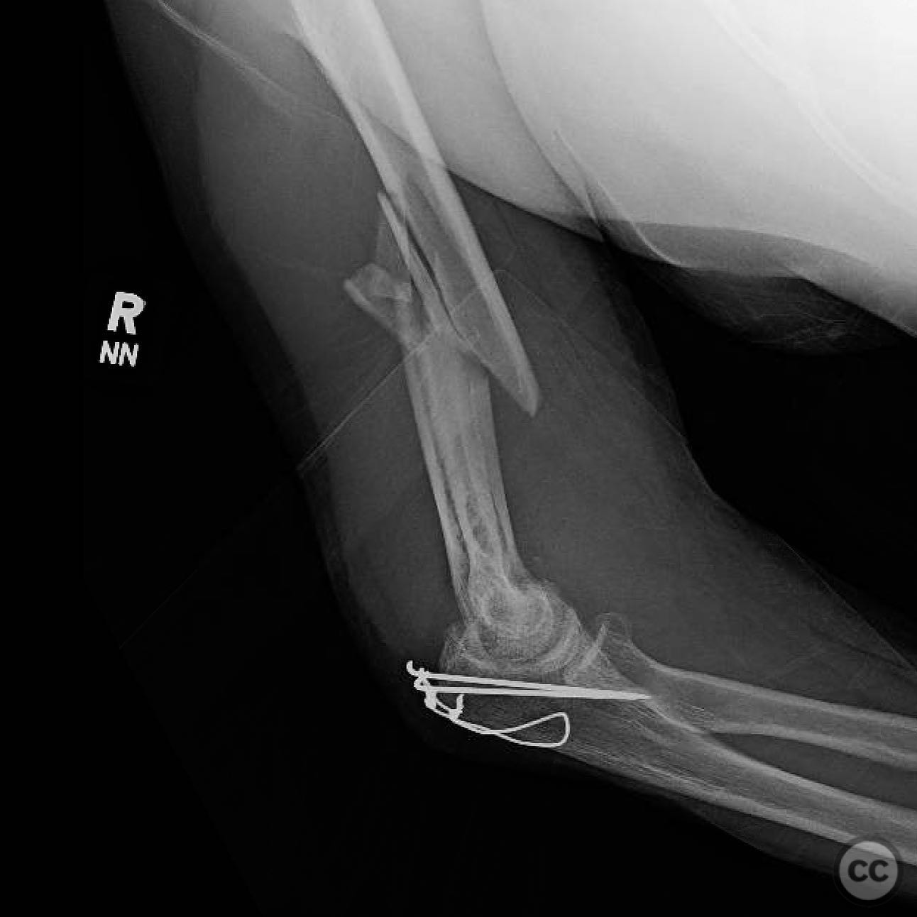
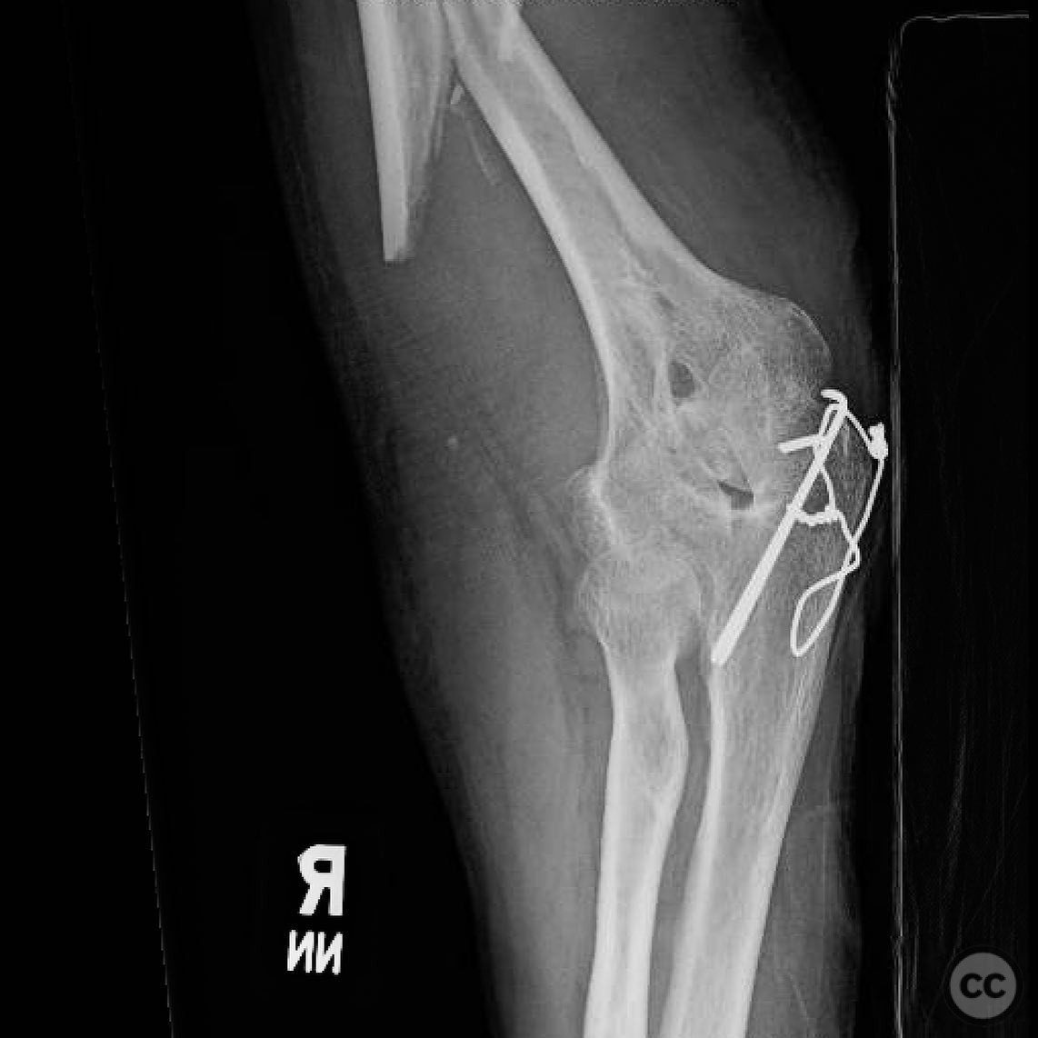
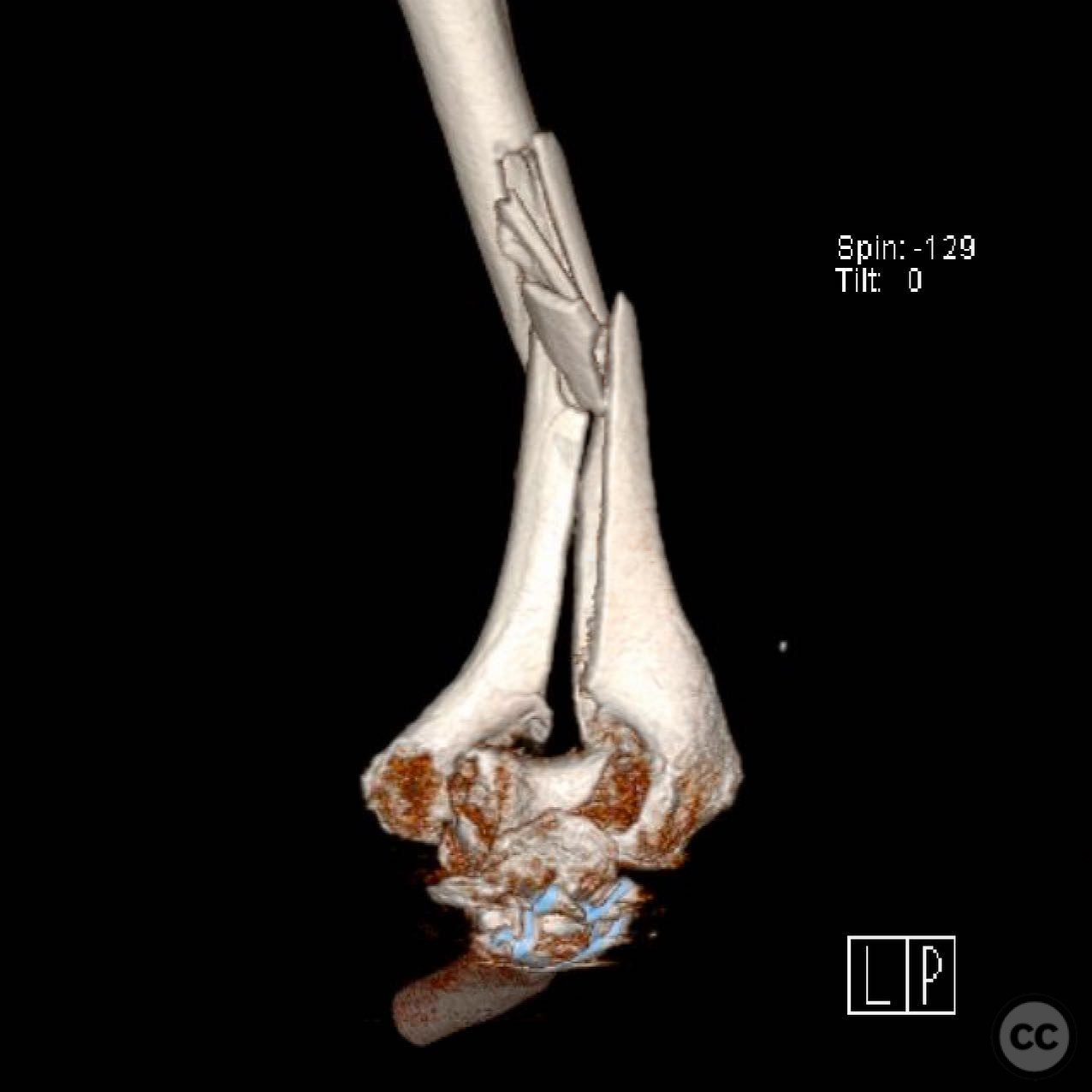
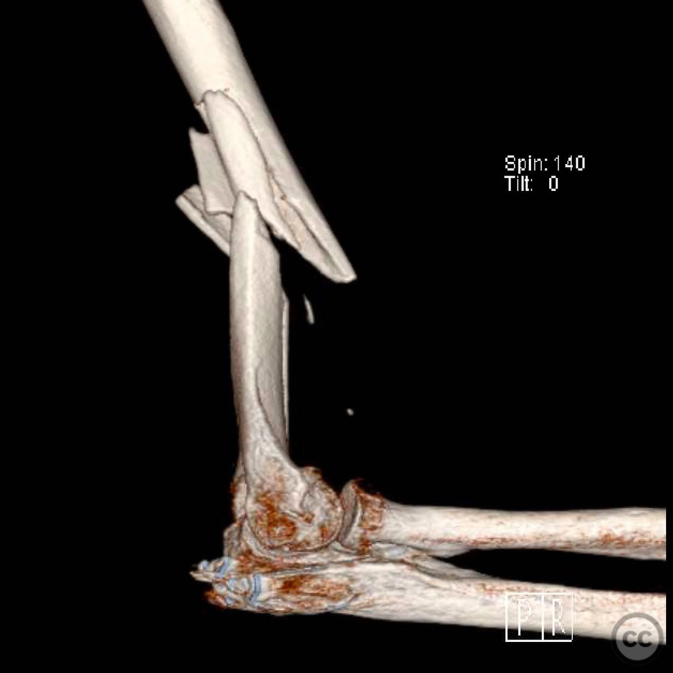
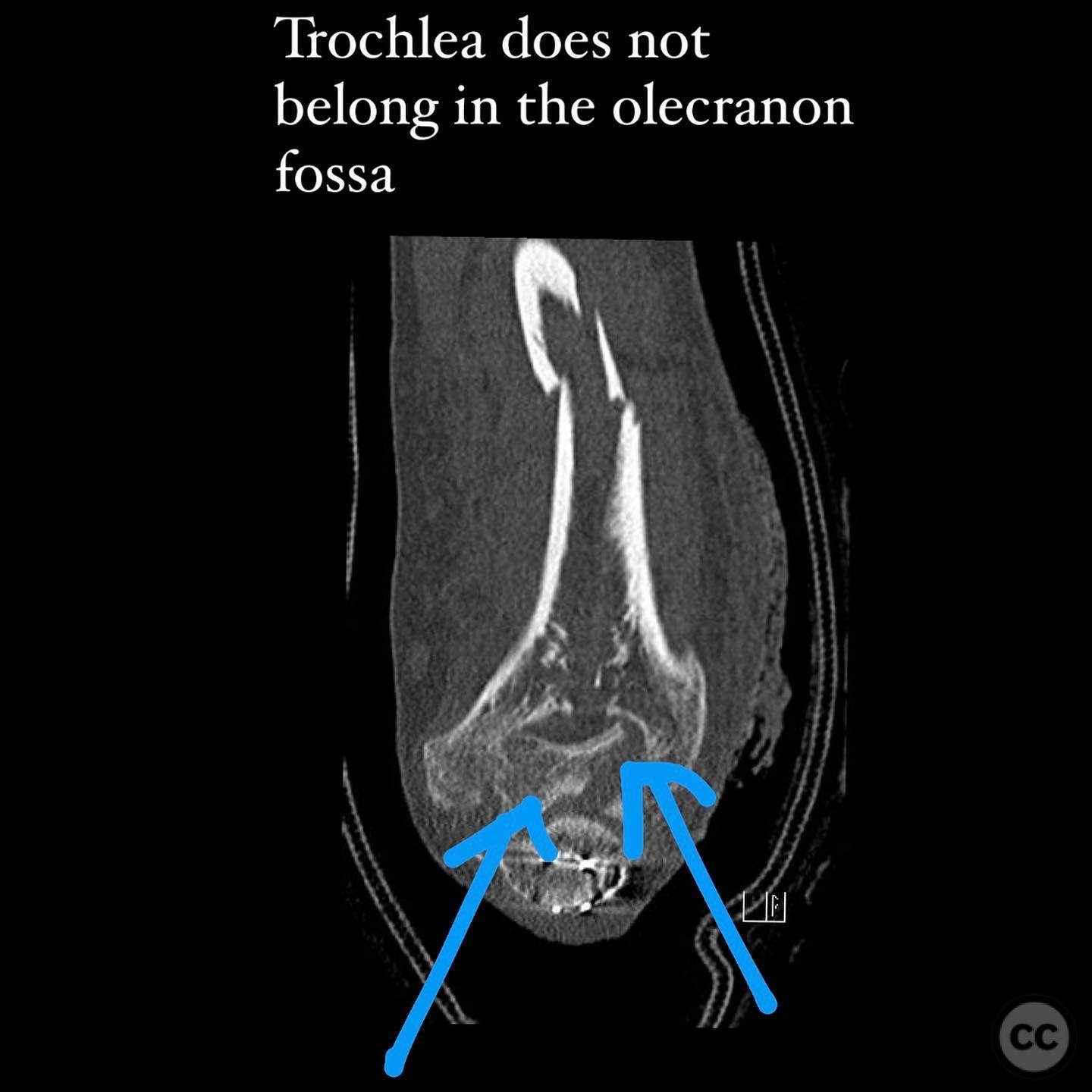
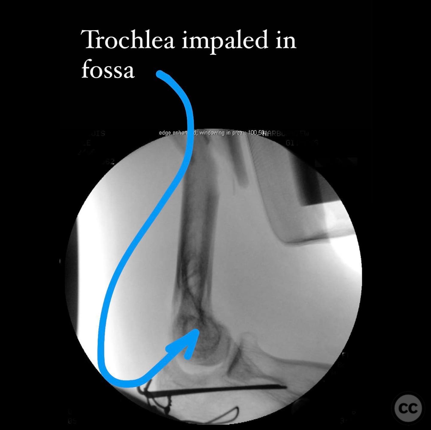
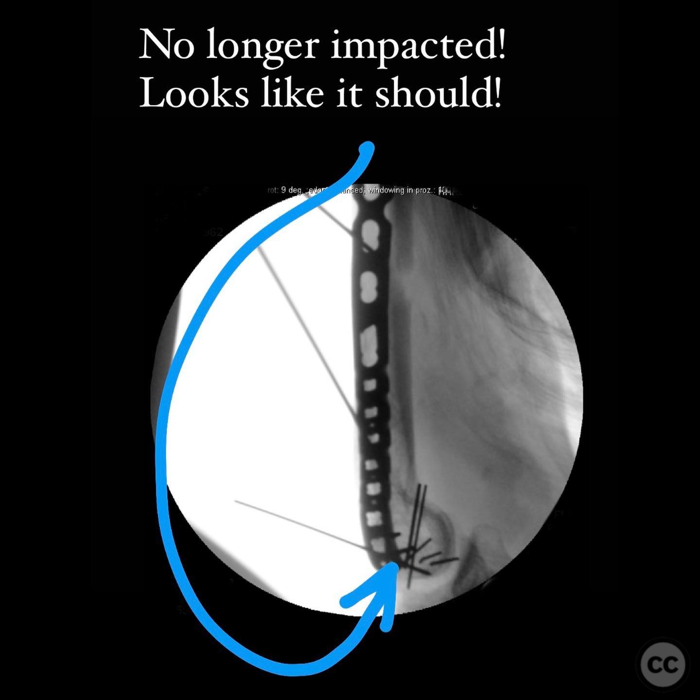
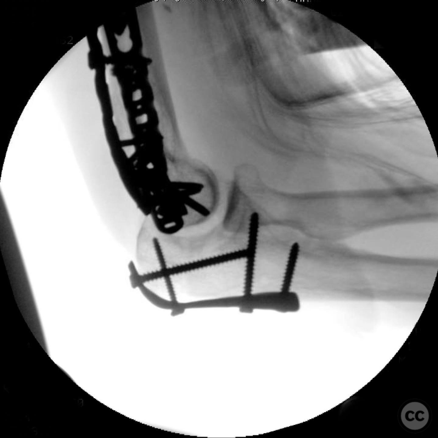
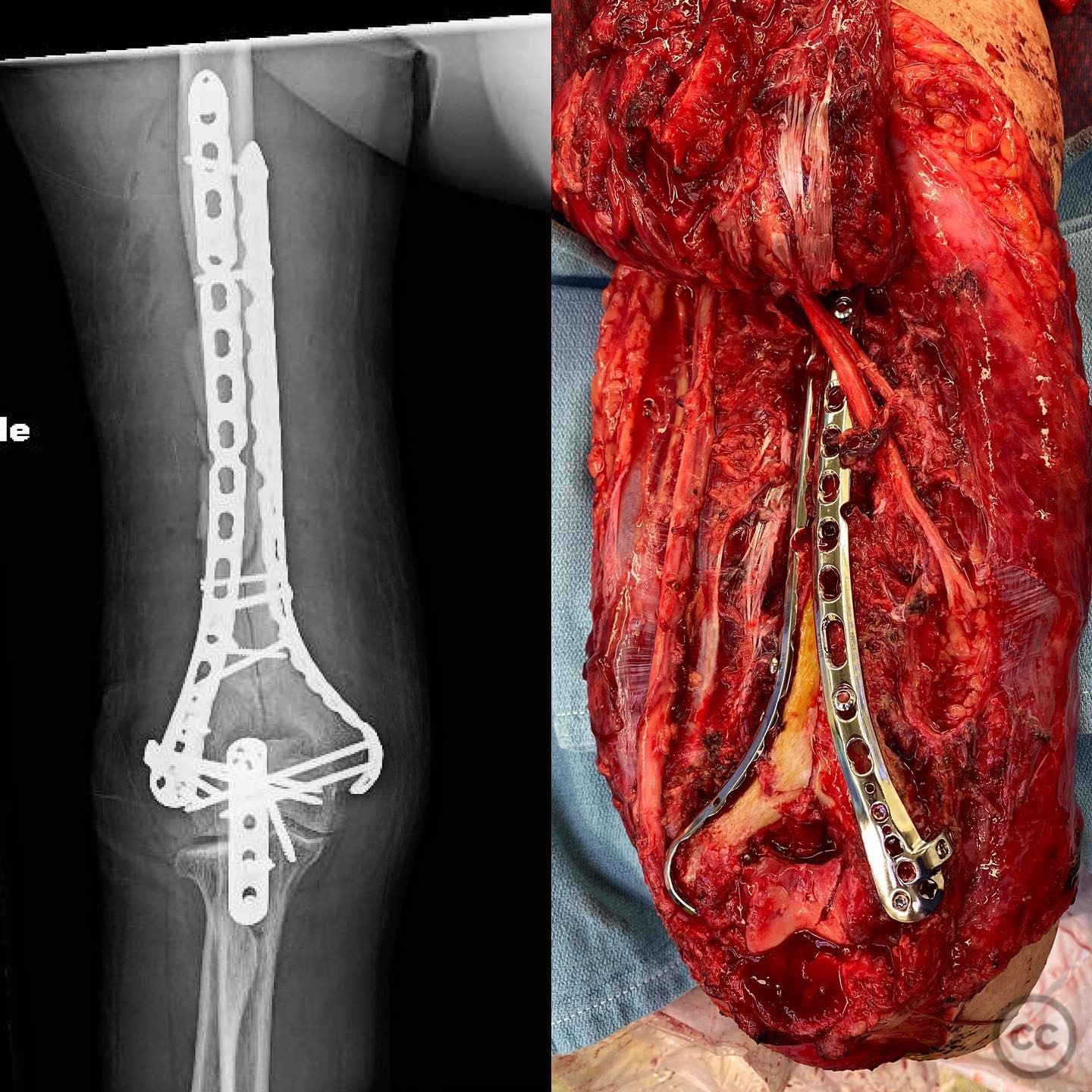
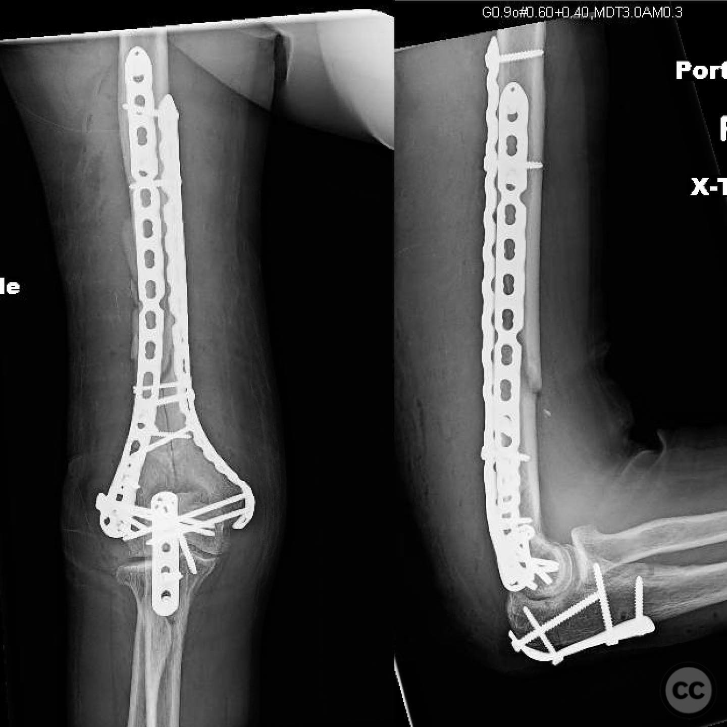
Clinical and radiological findings: A 49-year-old female sustained a fall from a 6-foot ledge while chasing her pet ferret, resulting in a type 1 open fracture of the humerus with a 1 cm wound on the anterolateral arm. Initial radiological assessment revealed a complex articular injury characterized by a free trochlea deeply impacted on the olecranon fossa and a multifragmentary capitellum. The diaphyseal component was comminuted, initially misinterpreted as a simple articular pattern with diaphyseal comminution. The injury was classified as AO/OTA 13-C3.
Preoperative Plan
Planning remarks: The preoperative plan involved a direct anatomic reduction of the joint through an osteotomy, accompanied by capsular release and scar takedown due to previous elbow injury and arthrofibrosis. The comminuted diaphyseal fracture was to be managed with indirect reduction and relative stability to facilitate endochondral ossification.
Surgical Discussion
Patient positioning: The patient was positioned supine on the operating table with the affected arm draped across the chest to allow for optimal access to the humerus.
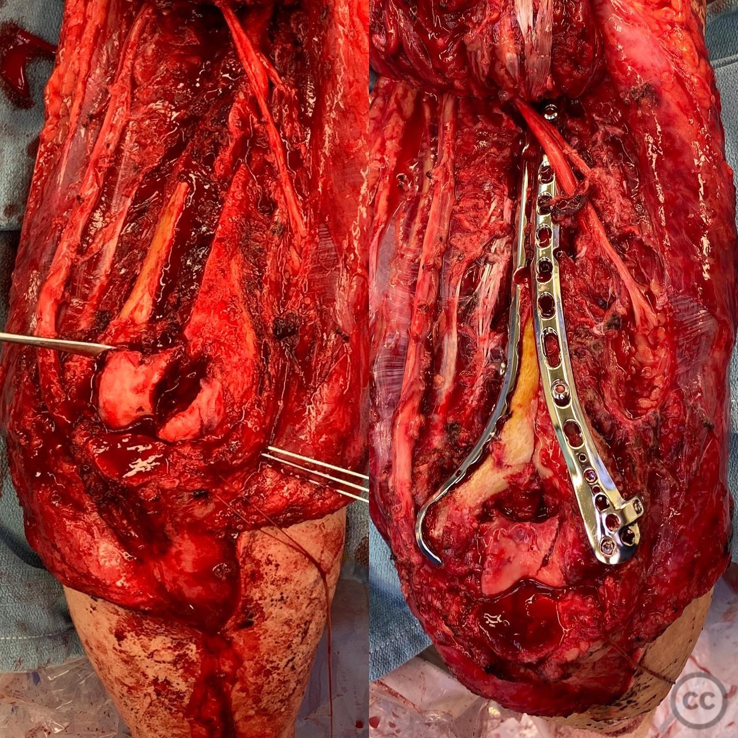
Anatomical surgical approach: A posterior approach to the elbow was utilized, involving an olecranon osteotomy to gain access to the distal humerus. Capsular release and scar takedown were performed to address arthrofibrosis. Careful mobilization of the ulnar nerve was conducted to prevent damage during exposure and fixation.
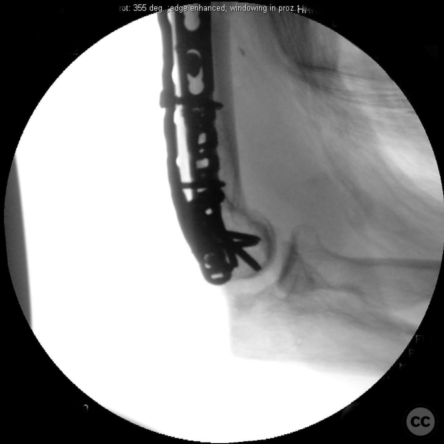
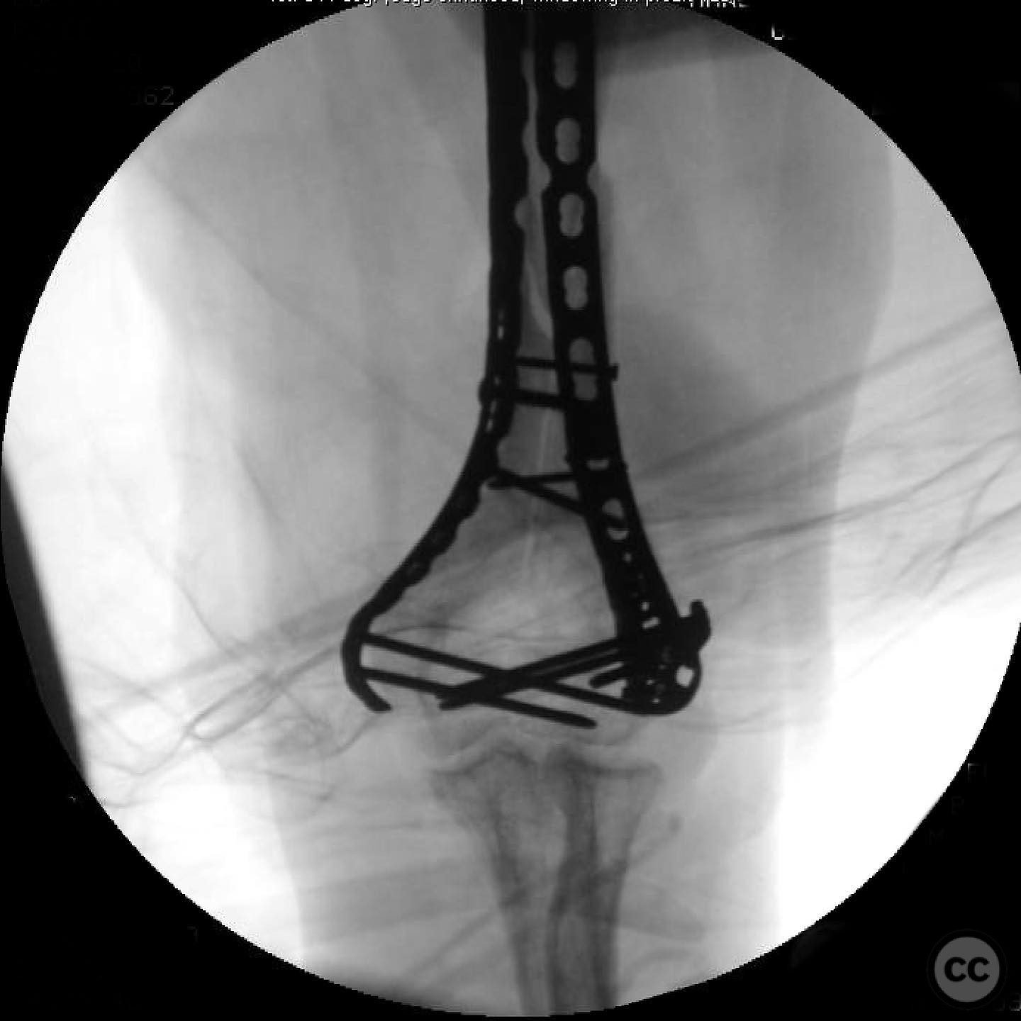
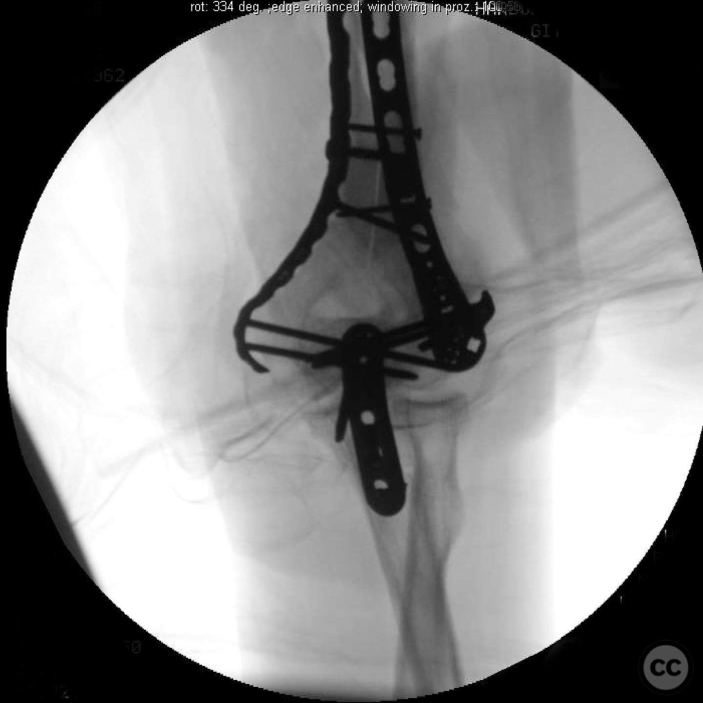
Operative remarks:The surgeon emphasized the importance of recognizing the complex articular nature of the injury, which necessitated a detailed approach to achieve anatomic reduction of the joint. The diaphyseal fracture was managed with bridge plating, ensuring relative stability through long plates with low screw density to promote callus formation. The indirect reduction technique preserved the biological environment of the diaphyseal fragments.
Postoperative protocol: Postoperatively, a soft dressing was applied, and early active and passive motion were encouraged to promote joint mobility and prevent stiffness.
Follow up: Not specified.
Orthopaedic implants used: Long bridge plates with low screw density for relative stability.
Search for Related Literature

orthopaedic_trauma
- United States , Seattle
- Area of Specialty - General Trauma
- Position - Specialist Consultant

Industry Sponsership
contact us for advertising opportunities
Article viewed 289 times
14 Jul 2025
Add to Bookmarks
Full Citation
Cite this article:
Surname, Initial. (2025). Complex Articular Humerus Fracture with Comminuted Diaphysis. Journal of Orthopaedic Surgery and Traumatology. Case Report 36954330 Published Online Jul 14 2025.