Comminuted Diaphyseal Femur Fracture in a 16-Year-Old
Score and Comment on this Case
Clinical Details
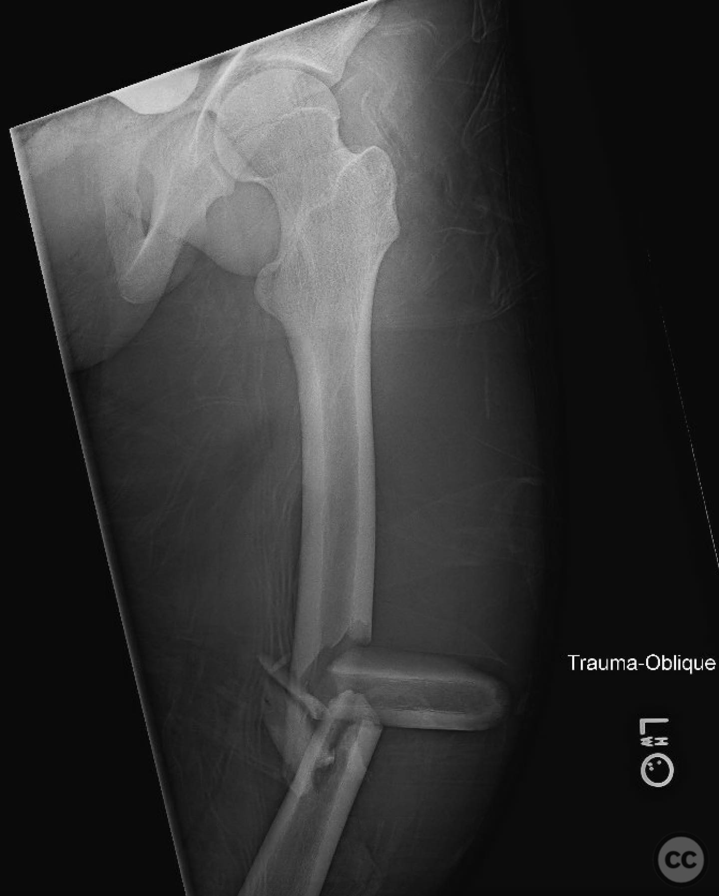
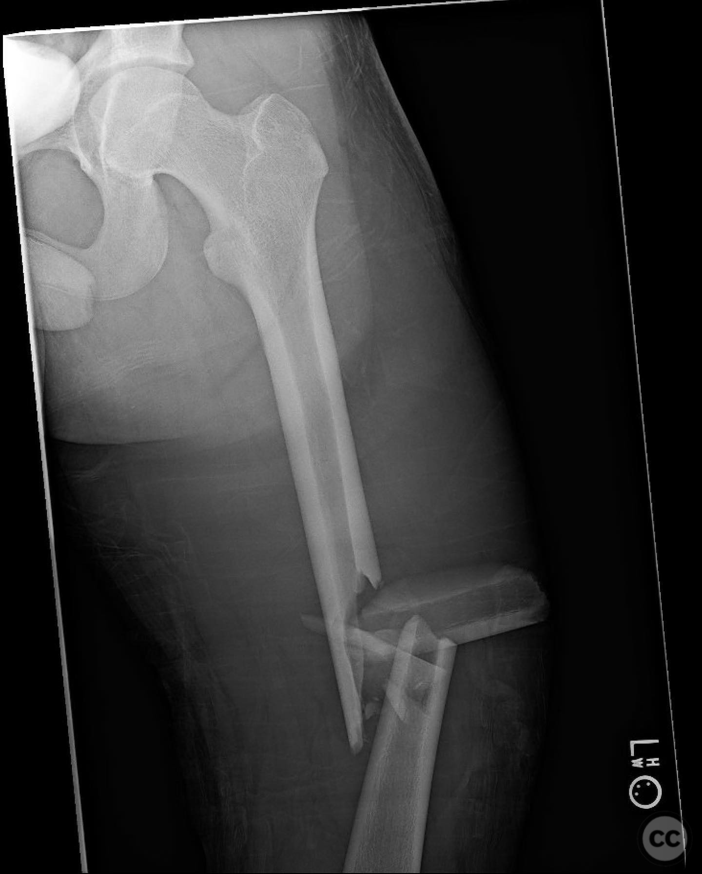
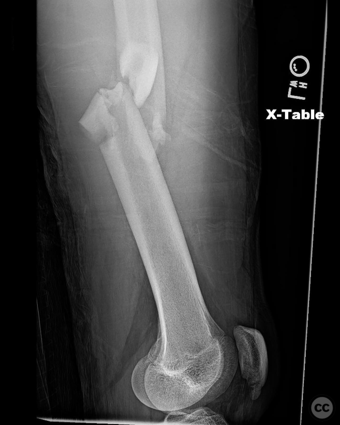
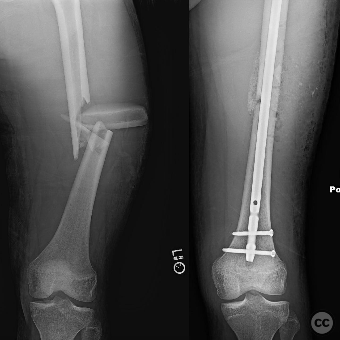
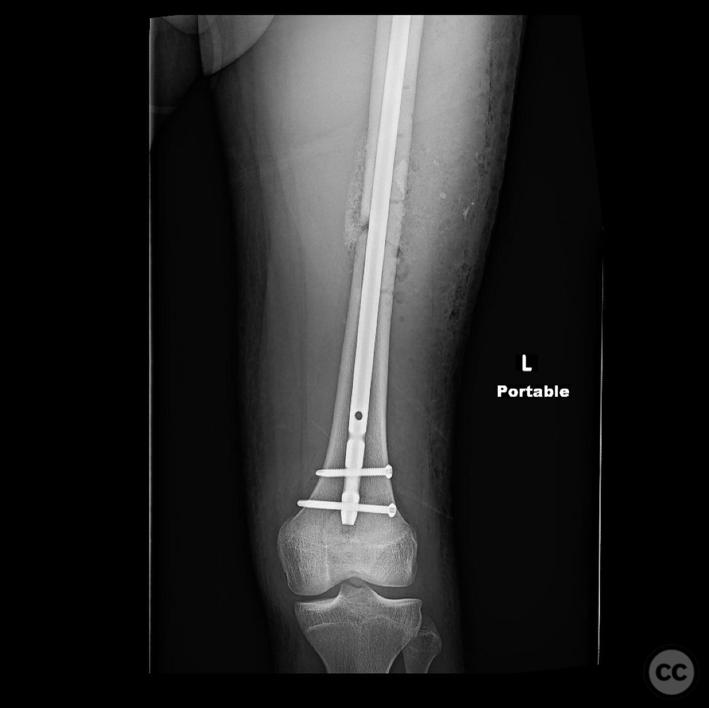
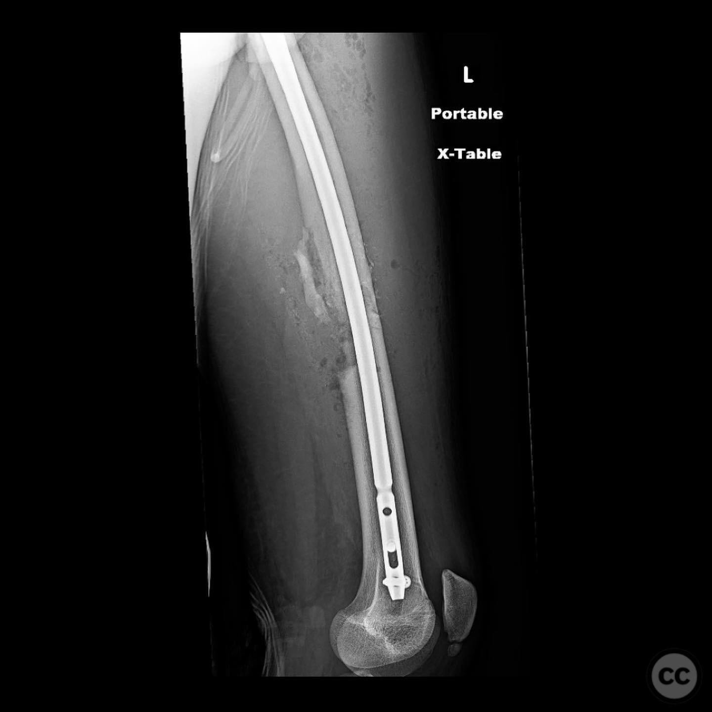
Clinical and radiological findings: A 16-year-old male sustained a comminuted diaphyseal femur fracture after a 50-foot fall while rock climbing. The patient presented with multiple injuries but was well resuscitated and cleared for surgical treatment of the femur fracture within 24 hours. The injury was closed, with no neurological or vascular compromise noted.
Preoperative Plan
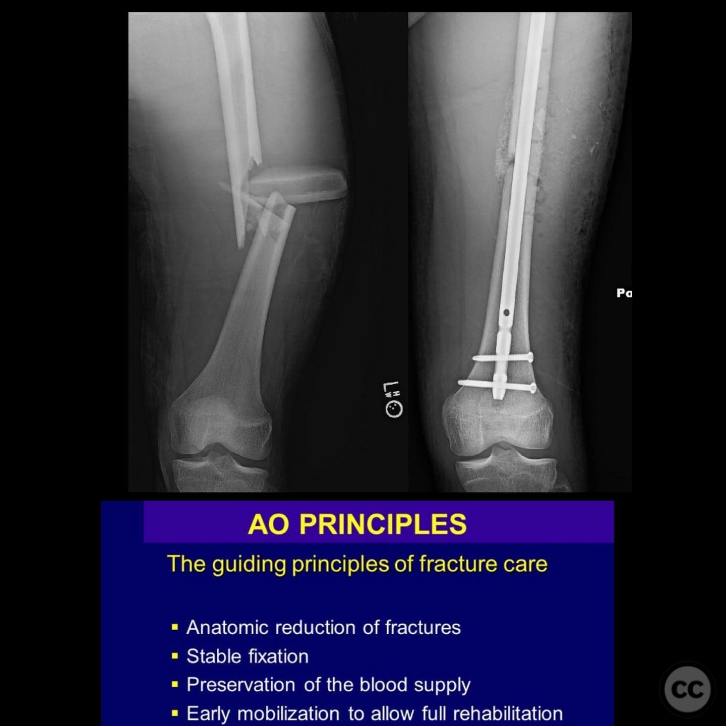
Planning remarks: The preoperative plan involved an open reduction and internal fixation (ORIF) due to the complexity of the fracture and the presence of a large incomplete napkin ring segment that could not be managed with closed nailing techniques. The anatomical approach was planned through a lateral incision to allow direct access to the fracture site for reduction and fixation.
Surgical Discussion
Patient positioning: The patient was positioned supine on a radiolucent table to facilitate intraoperative imaging and access to the femur.
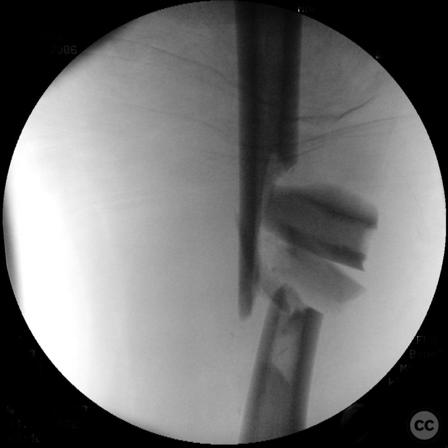
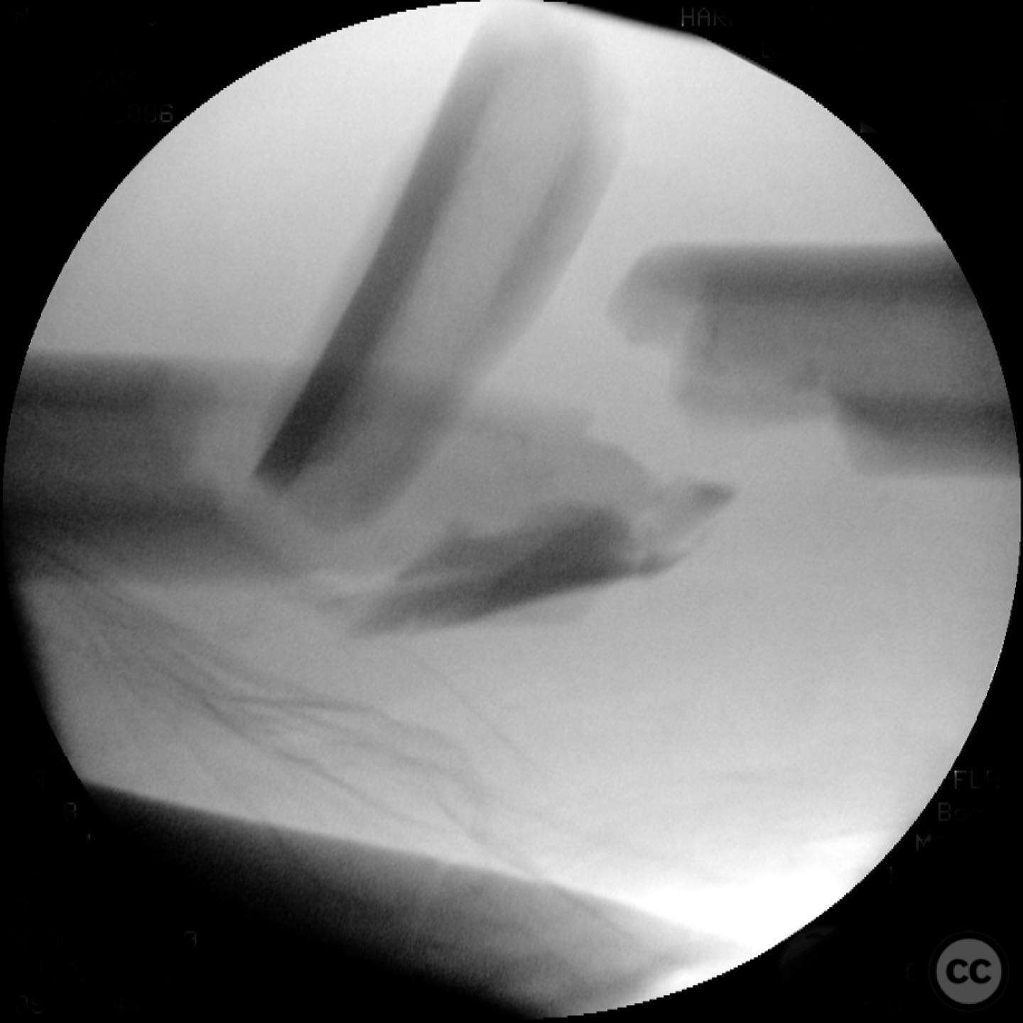
Anatomical surgical approach: A lateral approach to the femur was performed, involving an incision along the lateral aspect of the thigh. Subperiosteal dissection was carried out to expose the fracture site while preserving as much periosteum as possible. Care was taken not to disturb the medullary hematoma, and reamings were collected for later use in grafting.
Operative remarks:The surgeon noted that indirect reduction techniques were inadequate due to the fracture's complexity, necessitating an open reduction. Emphasis was placed on preserving fracture biology by minimizing periosteal stripping and avoiding suctioning of the medullary hematoma. Reamings were used as autograft material around the fracture site to enhance healing.
Postoperative protocol: Postoperative rehabilitation included early mobilization with toe-touch weight-bearing (TTWB) for 6 weeks, followed by progressive weight-bearing as tolerated. Range of motion exercises were initiated early to prevent joint stiffness.
Follow up: Not specified.
Orthopaedic implants used: Intramedullary nail, autologous bone graft from reamings.
Search for Related Literature

orthopaedic_trauma
- United States , Seattle
- Area of Specialty - General Trauma
- Position - Specialist Consultant

Industry Sponsership
contact us for advertising opportunities
Article viewed 332 times
11 Jul 2025
Add to Bookmarks
Full Citation
Cite this article:
Surname, Initial. (2025). Comminuted Diaphyseal Femur Fracture in a 16-Year-Old. Journal of Orthopaedic Surgery and Traumatology. Case Report 36207773 Published Online Jul 11 2025.