Coronal Shear Fracture of the Distal Humerus with Trochlear Extension
Score and Comment on this Case
Clinical Details
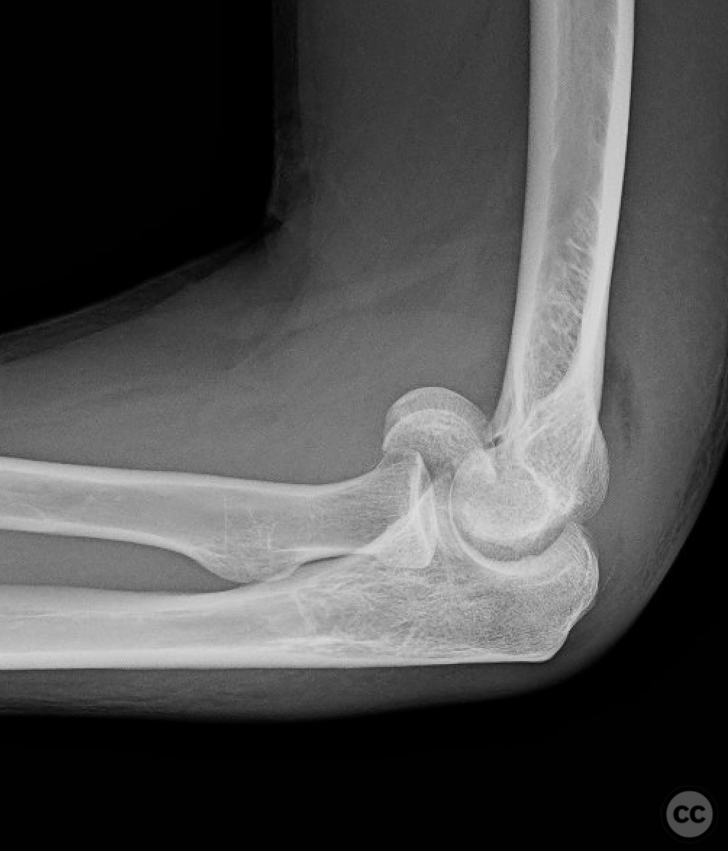
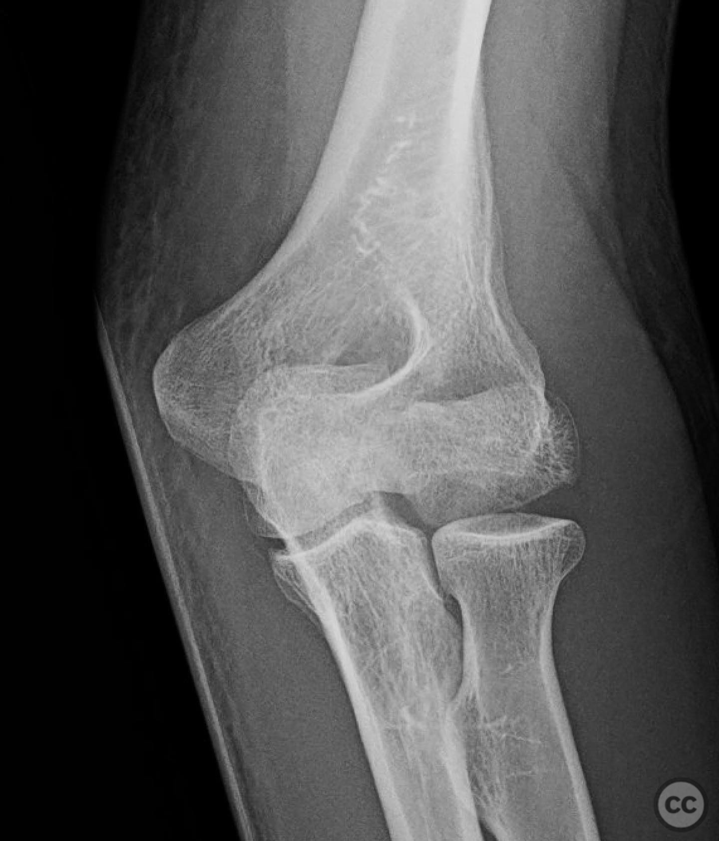
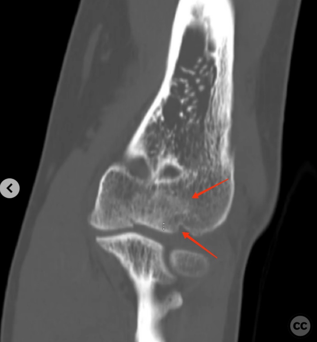
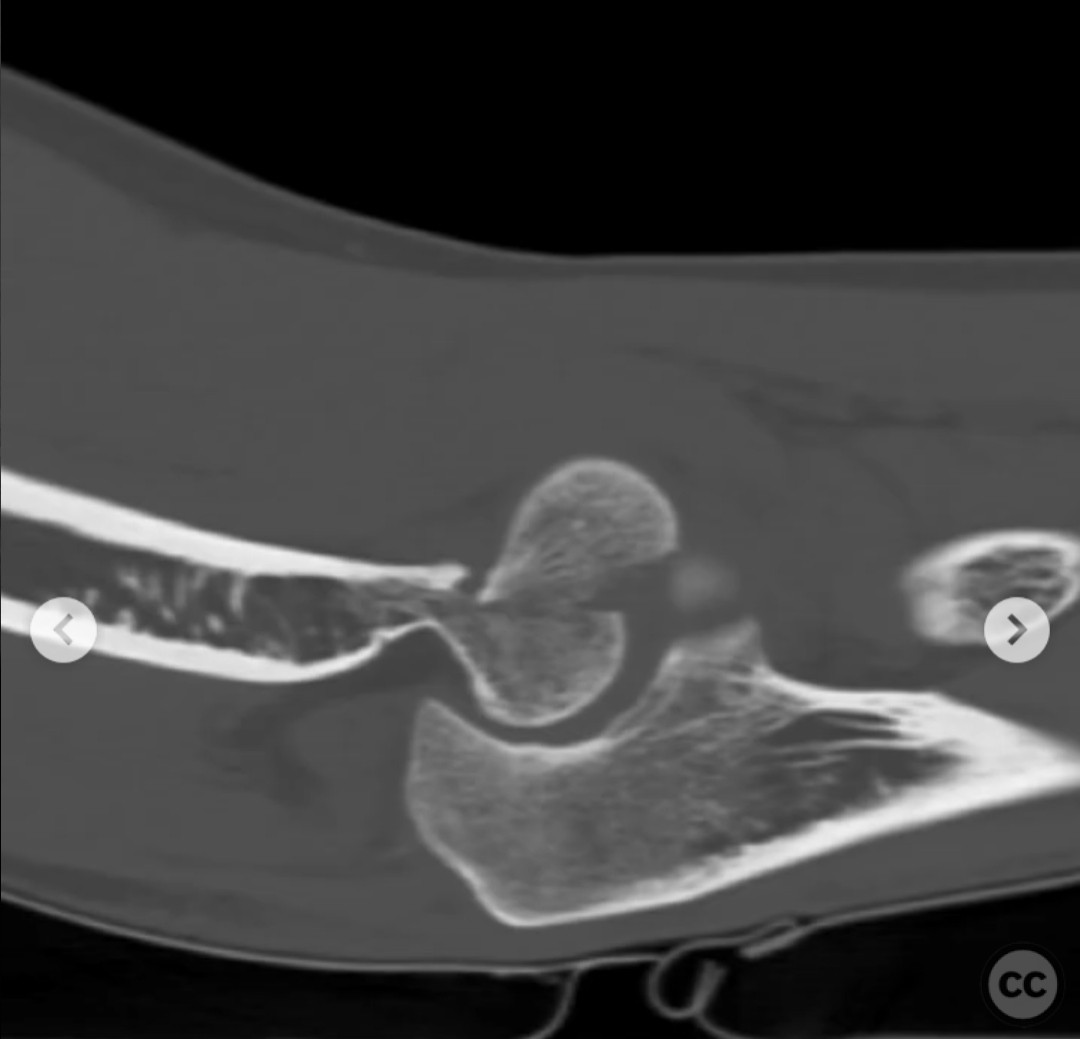
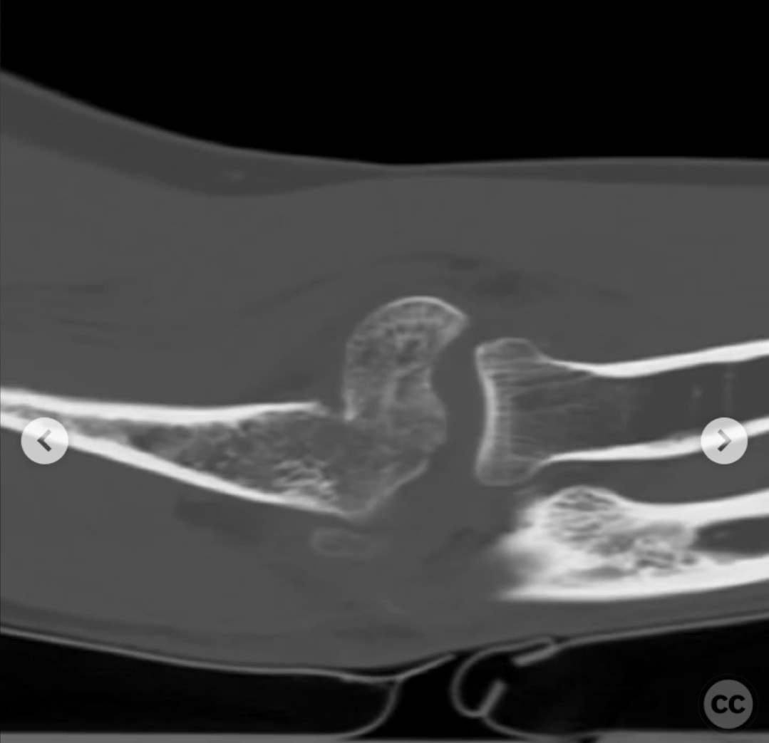
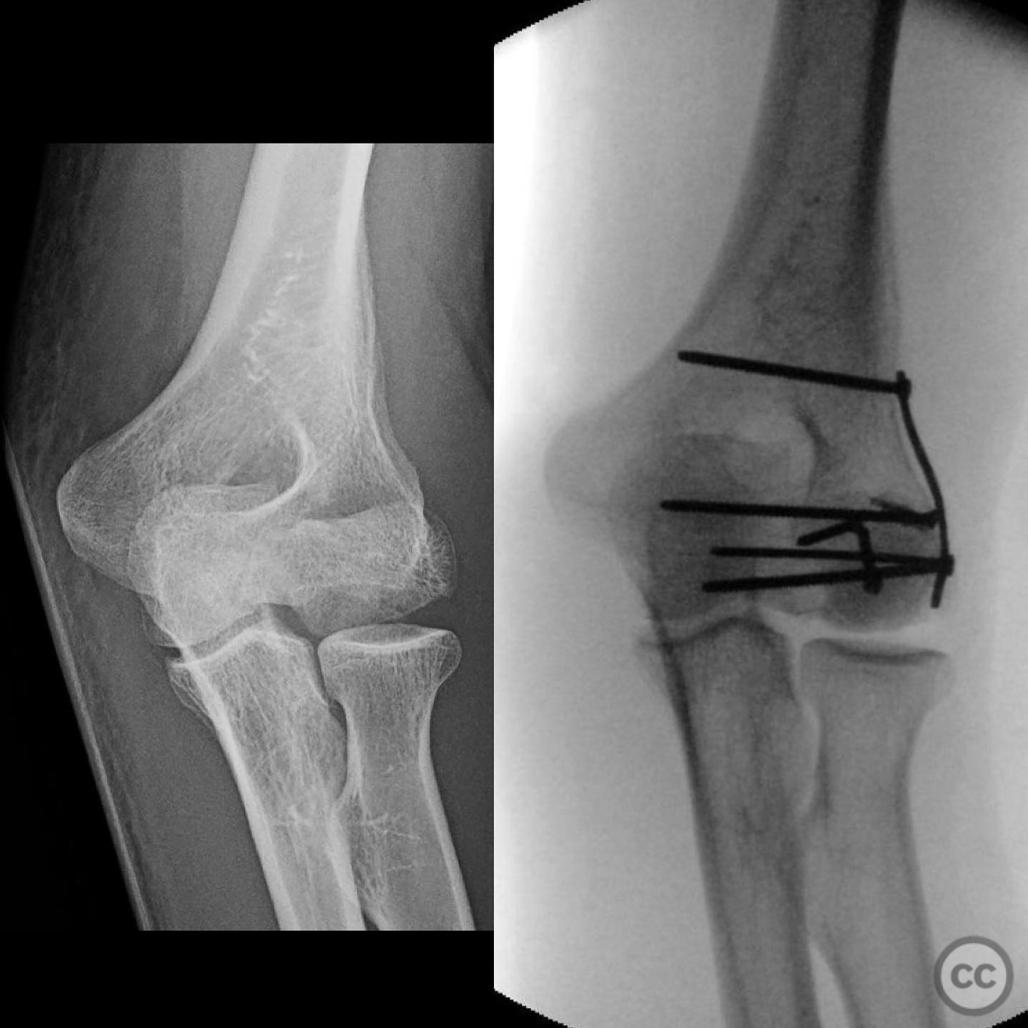
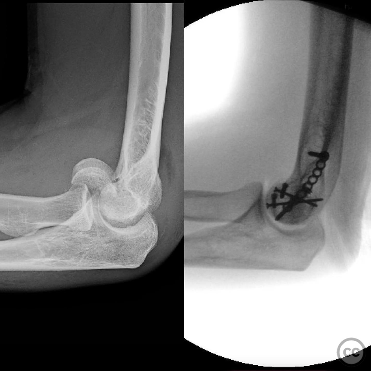
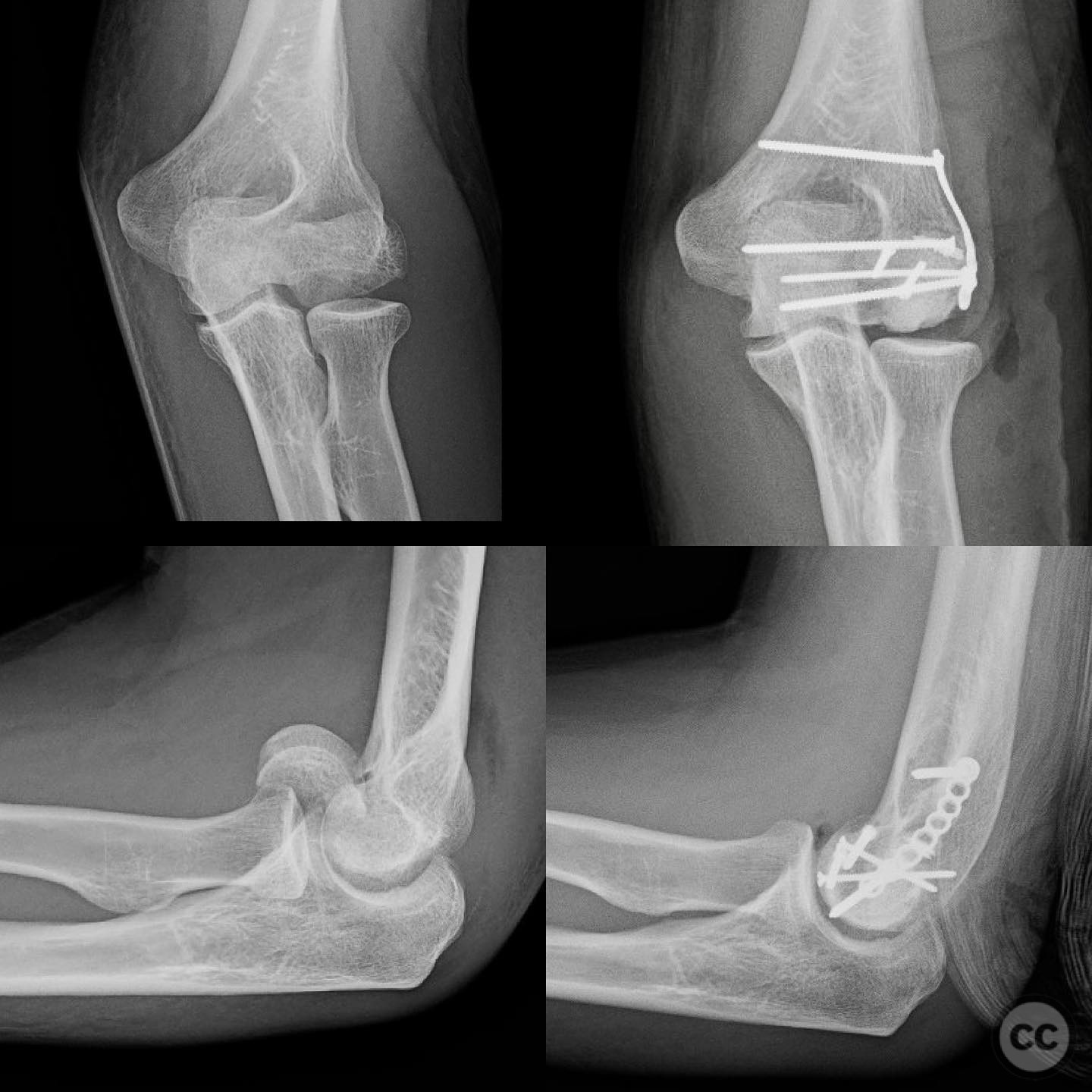
Clinical and radiological findings: A 36-year-old healthy, active male sustained a coronal plane shear fracture of the distal humerus while trail running, resulting in a fall on his outstretched arm. The fracture involved the capitellum and extended into the trochlea, with central impaction noted. No associated 13C injuries were reported.
Preoperative Plan
Planning remarks: The preoperative plan involved a lateral column approach using the Kocher interval, with modifications as necessary to address the fracture pattern. The plan included releasing the annular ligament and sleeving off the common extensor origin. If visualization and reduction were insufficient, a lateral epicondyle osteotomy was considered.
Surgical Discussion
Patient positioning: The patient was positioned supine for the procedure.
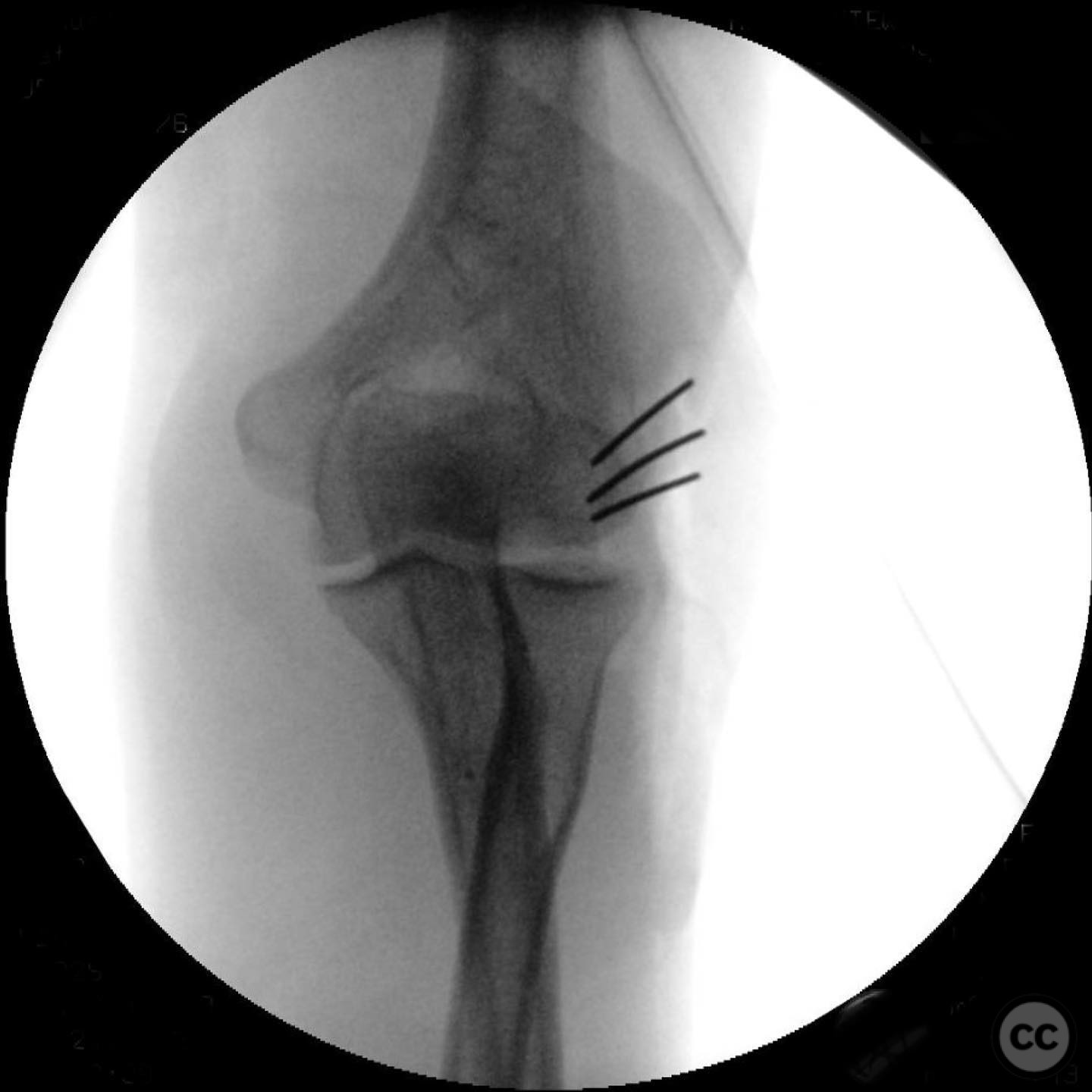
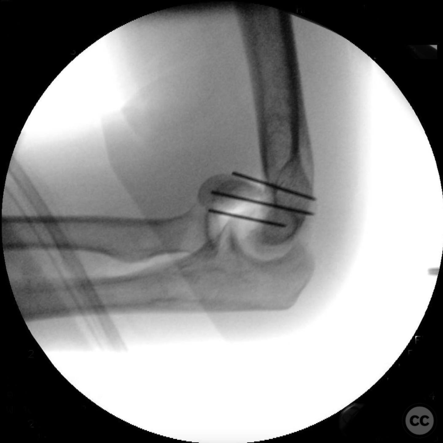
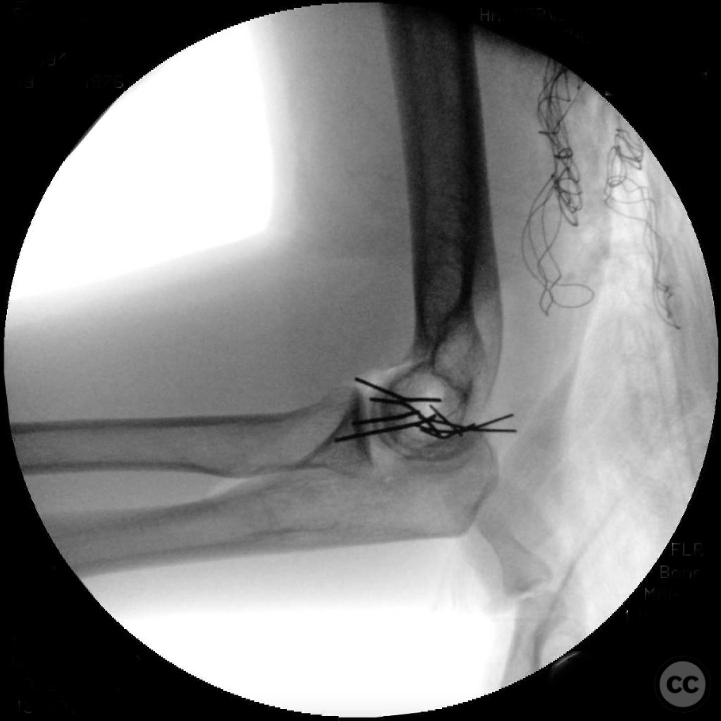
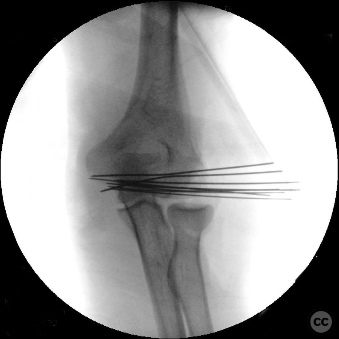
Anatomical surgical approach: A lateral column approach was utilized, entering through the Kocher interval. The annular ligament was released, and the common extensor origin was sleeved off to provide adequate exposure. A lateral epicondyle osteotomy was performed to improve visualization and access to the fracture site, with the lateral ulnar collateral ligament (LUCL) accompanying the osteotomy.
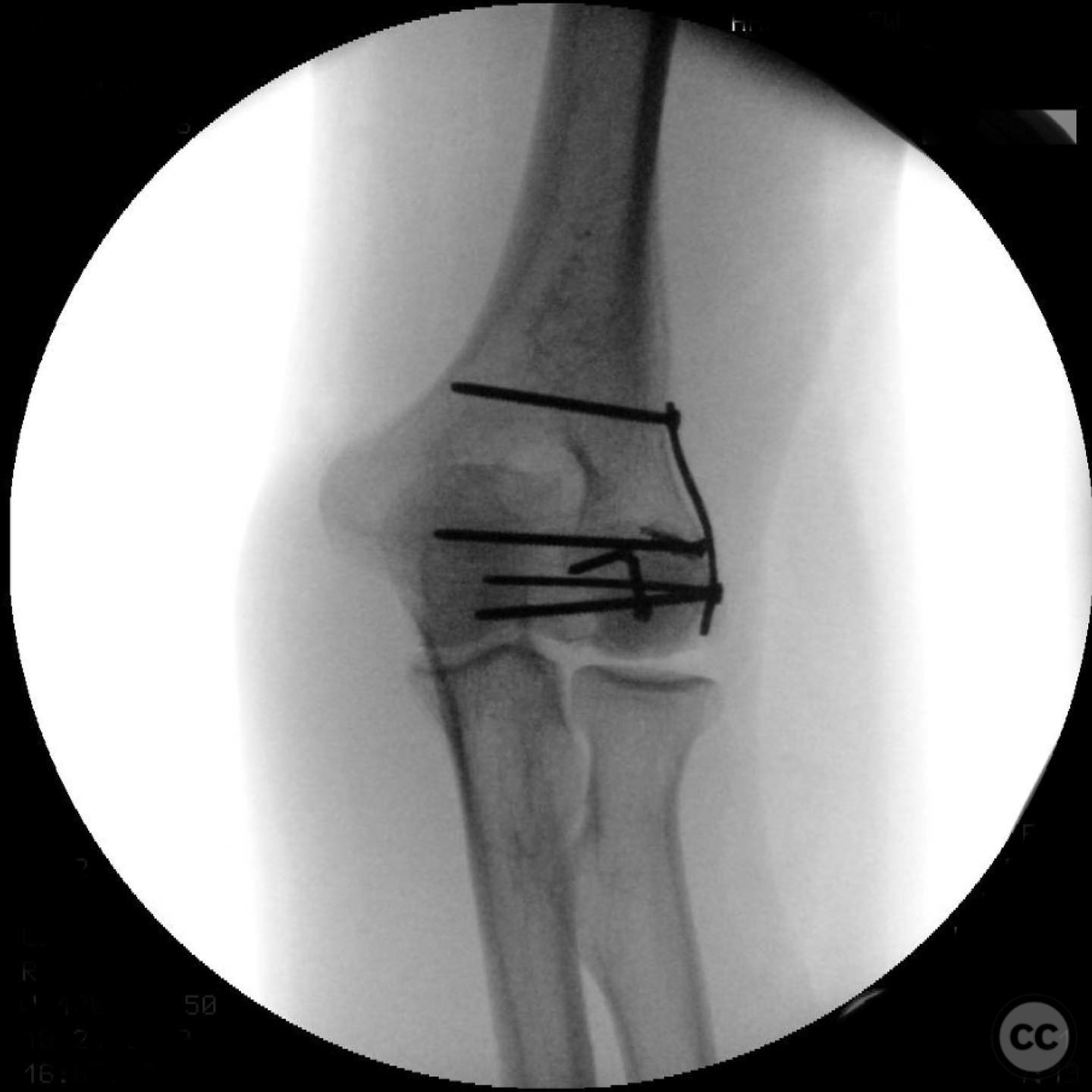
Operative remarks:The surgeon noted that the extension of the fracture into the trochlea complicated visualization. The central impaction required careful disimpaction without fragmenting the large articular segment. A synthetic bone void filler was used to address the capitellum impaction defect. Surgical destabilization of the elbow allowed for excellent access and facilitated stable fixation to permit early motion.
Postoperative protocol: Postoperative rehabilitation focused on achieving stable fixation to allow early motion. Specific details of the rehabilitation protocol were not provided.
Follow up: Not specified.
Orthopaedic implants used: Synthetic bone void filler.
Search for Related Literature

orthopaedic_trauma
- United States , Seattle
- Area of Specialty - General Trauma
- Position - Specialist Consultant

Industry Sponsership
contact us for advertising opportunities
Article viewed 282 times
11 Jul 2025
Add to Bookmarks
Full Citation
Cite this article:
Surname, Initial. (2025). Coronal Shear Fracture of the Distal Humerus with Trochlear Extension. Journal of Orthopaedic Surgery and Traumatology. Case Report 36007049 Published Online Jul 11 2025.