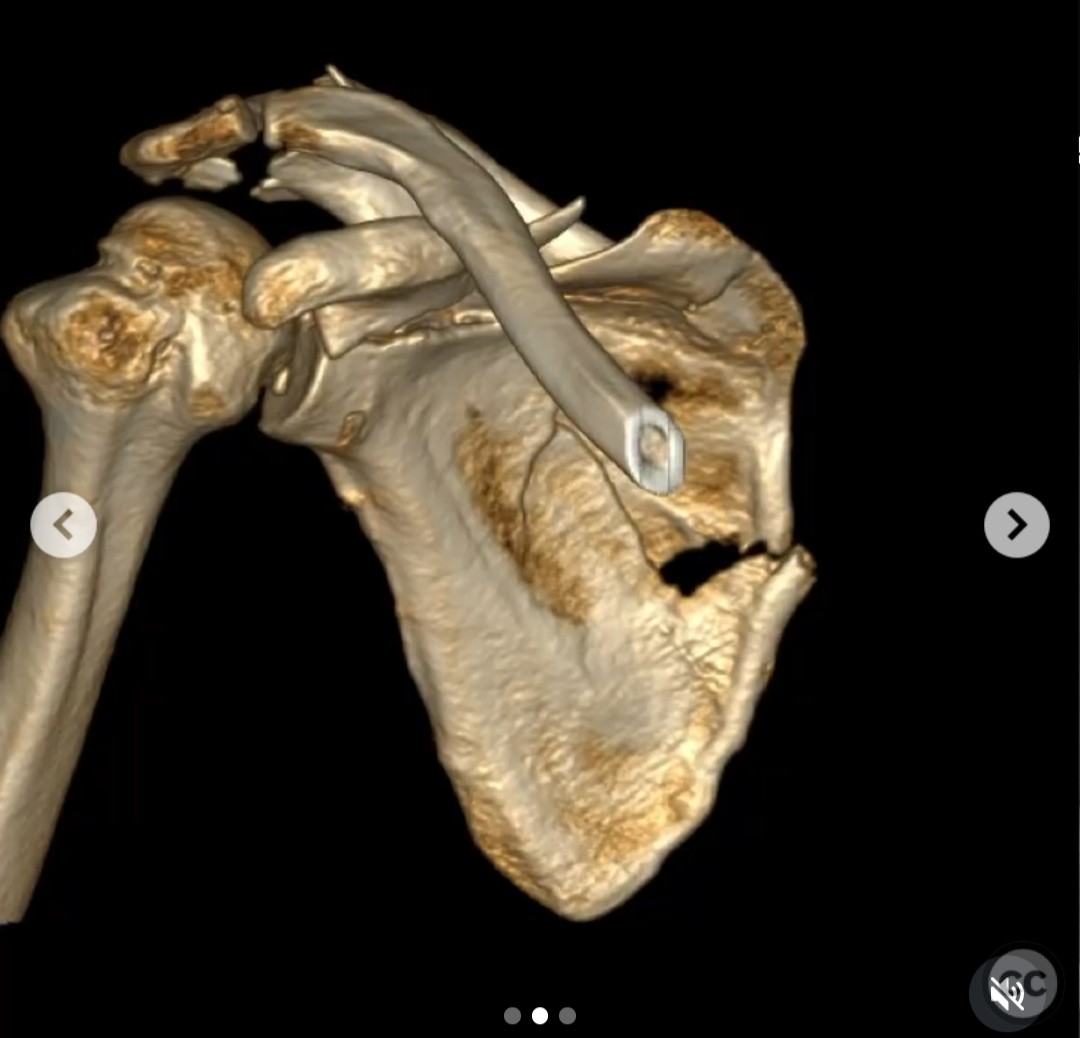
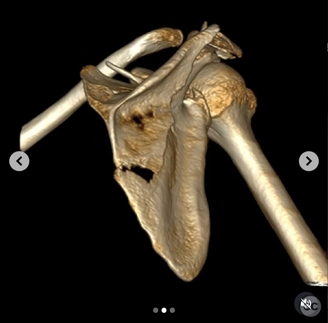
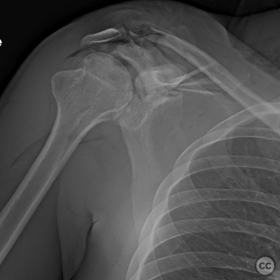
Open Ideberg Type III Scapula Fracture with Acromion and Spine Involvement.
Score and Comment on this Case
Clinical Details
Clinical and radiological findings: A 33-year-old male sustained a direct impact to the scapula from a truck's rearview mirror traveling at approximately 50 mph, resulting in a 3a open fracture. The injury presented as a longitudinal laceration along the scapular spine. Radiological assessment revealed an Ideberg Type III fracture involving the coracoid, cranial glenoid, acromion, and scapular spine with significant displacement. This injury represents a distal double or triple disruption of the shoulder's suspensatory complex.
Preoperative Plan
Planning remarks: The preoperative plan involved addressing the coracoid and glenoid through an anterior approach, and the acromion and scapular spine through a posterior approach. The plan included reducing and fixing the coracoid/glenoid, reducing and fixing the displaced acromion to prevent nonunion, and stabilizing the spine/suprascapular fossa fragment to facilitate proper acromion fixation.
Surgical Discussion
Patient positioning: Initial posterior approach was performed with the patient in a prone position utilizing the traumatic wound for access. For the anterior approach, the patient was repositioned supine.
Anatomical surgical approach: Posterior approach: Access through the traumatic wound along the scapular spine, exposing the supraspinatus fossa and spine fragment. Reduction of the lateralized supraspinatus fossa/spine fragment was achieved by medializing it to create a flexible hinge point at the medial border. The acromion was then reduced to the spine and fixed with a tension band plate. Anterior approach: Proximal deltopectoral incision with biceps tenotomy, subscapularis release, and arthrotomy to reduce the coracoid-glenoid fragment under direct visualization.
Operative remarks:The surgeon noted that addressing each lesion was crucial for optimizing shoulder function. The sequence began with restoring scapular morphology via the posterior approach, followed by addressing the coracoid-glenoid fragment anteriorly. The intact coracoclavicular ligaments provided stability, allowing for accurate reduction of the acromioclavicular joint once the coracoid-glenoid was fixed.
Postoperative protocol: Postoperative rehabilitation included immobilization in a shoulder sling for initial weeks, followed by gradual passive range of motion exercises. Active range of motion and strengthening exercises were introduced progressively as tolerated.
Follow up: Not specified.
Orthopaedic implants used: Tension band plate for acromion fixation Locking screws Cannulated screws Lag screws Mini-fragment plate Reconstruction plate
Search for Related Literature

orthopaedic_trauma
- United States , Seattle
- Area of Specialty - General Trauma
- Position - Specialist Consultant

Industry Sponsership
contact us for advertising opportunities


Article viewed 157 times
13 Jul 2025
Add to Bookmarks
Full Citation
Cite this article:
Surname, Initial. (2025). Open Ideberg Type III Scapula Fracture with Acromion and Spine Involvement.. Journal of Orthopaedic Surgery and Traumatology. Case Report 35600864 Published Online Jul 13 2025.