 ☠️ ._._._Subtroch femur fractures are notoriously easy to mess up. And in a very predictable way (th_7.jpg)
 ☠️ ._._._Subtroch femur fractures are notoriously easy to mess up. And in a very predictable way (th_6.jpg)
 ☠️ ._._._Subtroch femur fractures are notoriously easy to mess up. And in a very predictable way (th_5.jpg)
 ☠️ ._._._Subtroch femur fractures are notoriously easy to mess up. And in a very predictable way (th_2.jpg)
 ☠️ ._._._Subtroch femur fractures are notoriously easy to mess up. And in a very predictable way (th_3.jpg)
 ☠️ ._._._Subtroch femur fractures are notoriously easy to mess up. And in a very predictable way (th_1.jpg)
 ☠️ ._._._Subtroch femur fractures are notoriously easy to mess up. And in a very predictable way (thr(.jpg)
 ☠️ ._._._Subtroch femur fractures are notoriously easy to mess up. And in a very predictable way (th_4.jpg)
Subtrochanteric Femur Fracture: Challenges and Solutions
Score and Comment on this Case
Clinical Details
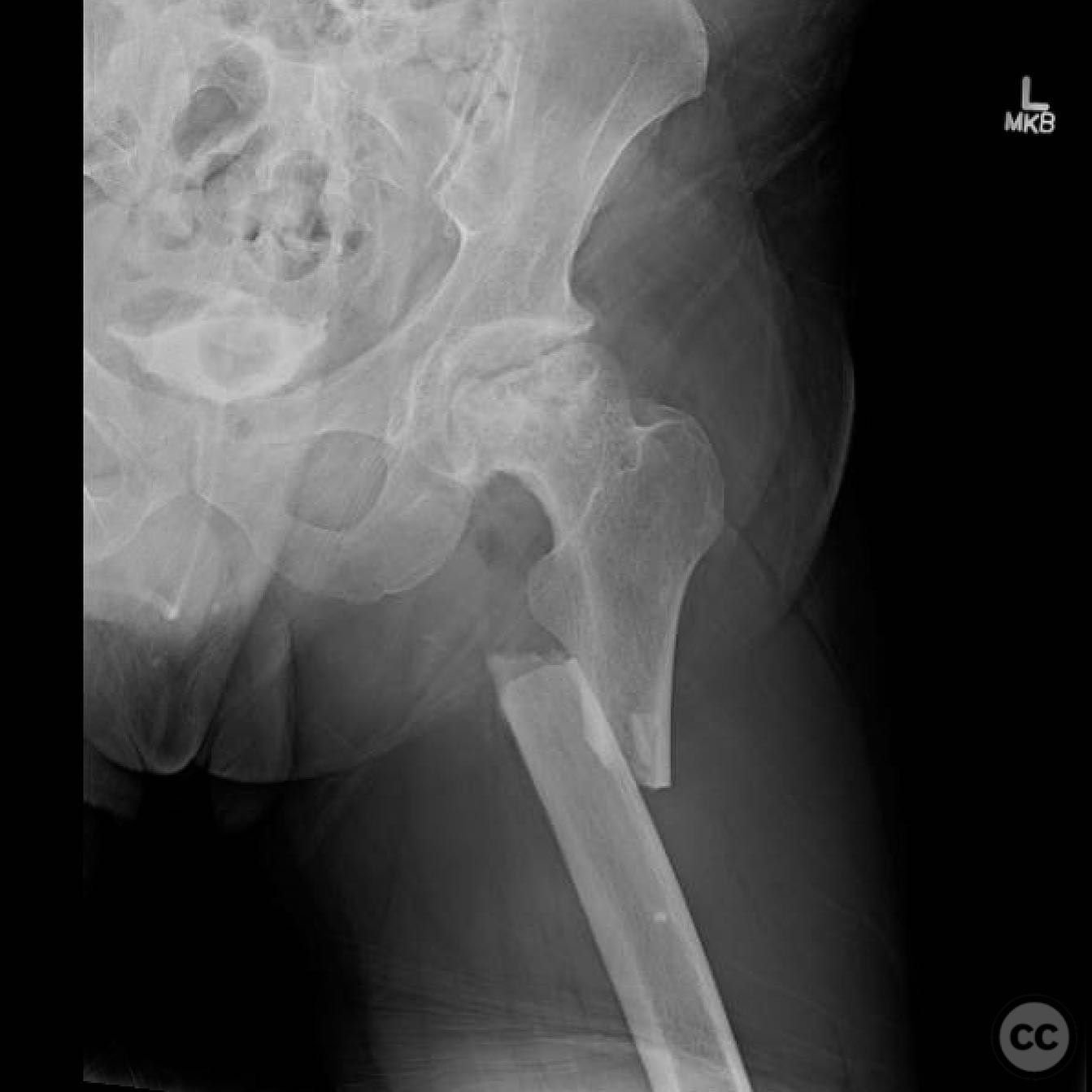
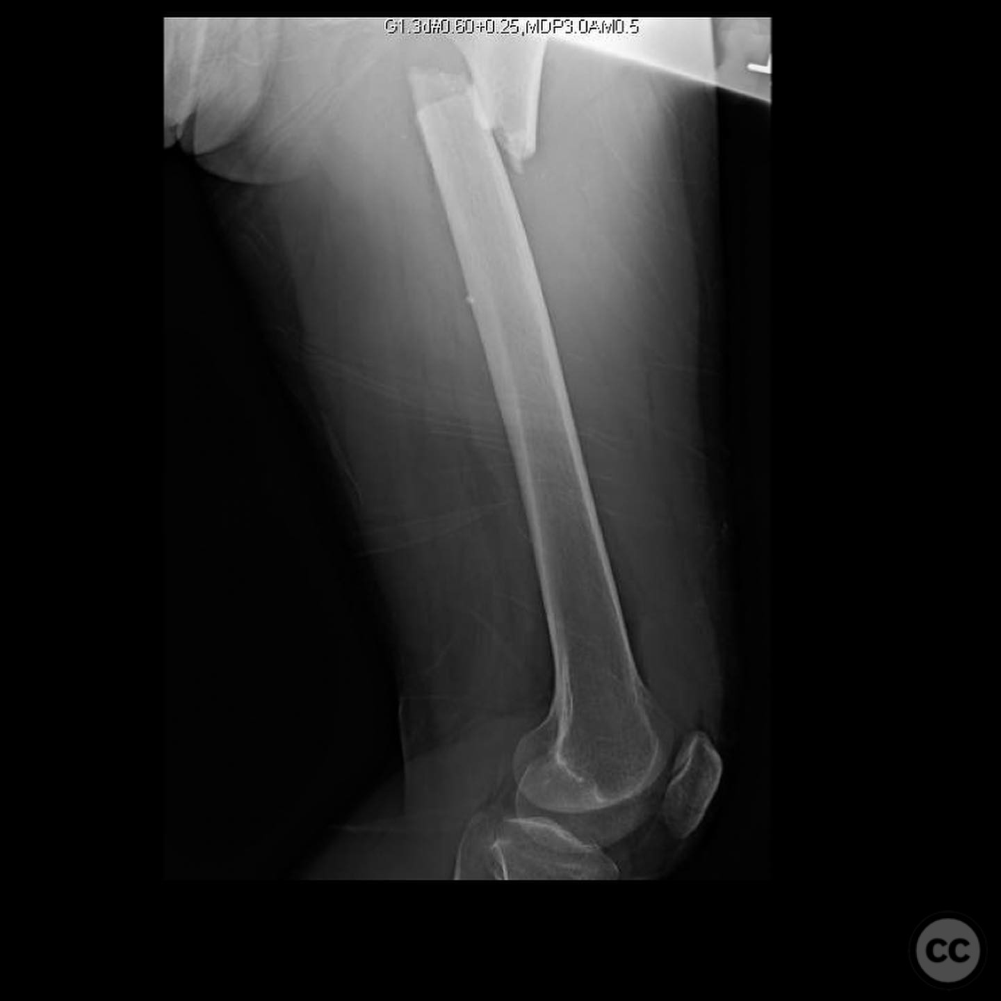
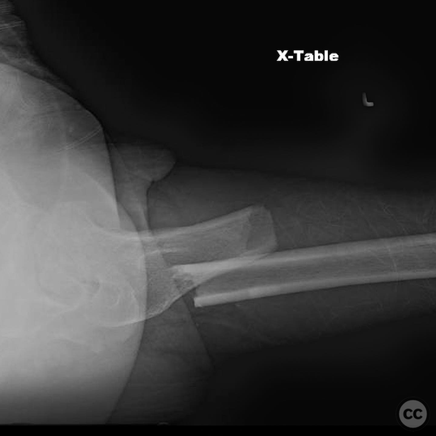
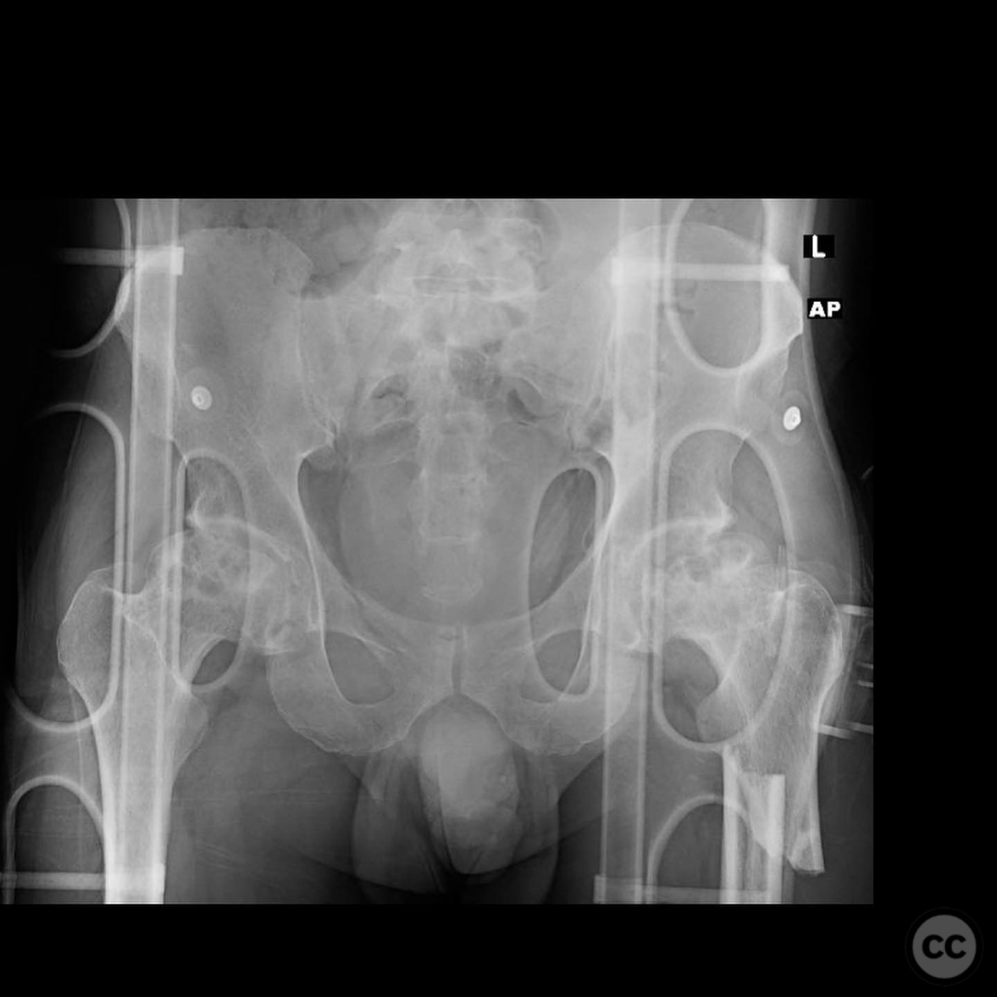
Clinical and radiological findings: A 45-year-old male presented with a high-energy subtrochanteric femur fracture following a motor vehicle accident. Initial radiographs demonstrated a comminuted subtrochanteric fracture with significant varus, flexion, and external rotation deformities of the proximal fragment. The fracture was classified as AO/OTA 32-A3. The patient had no other injuries and a normal neurovascular examination.
Preoperative Plan
Planning remarks: The preoperative plan involved a lateral approach for intramedullary nailing. Given the fracture pattern and deformity, an open reduction was anticipated if closed reduction techniques proved inadequate. Proper imaging in the lateral position was emphasized to ensure accurate reduction and nail placement.
Surgical Discussion
Patient positioning: The patient was positioned laterally on the operating table. The down leg was extended to prevent interference with imaging, while the operative leg was flexed to aid in neutralizing the deformity.
Anatomical surgical approach: A lateral approach was utilized, with an incision made over the greater trochanter. Subperiosteal dissection was performed to expose the proximal femur. Reduction was achieved using clamps placed laterally to tension the fracture site, ensuring they did not obstruct the nail jig.
Operative remarks:The surgeon noted that closed reduction attempts were insufficient due to the significant deforming forces. An open reduction was performed promptly, minimizing operative time and reducing the risk of malreduction. The importance of achieving proper alignment before nail insertion was emphasized to prevent varus malreduction and potential nonunion.
Postoperative protocol: Postoperatively, the patient was allowed partial weight-bearing with crutches for six weeks, progressing to full weight-bearing as tolerated by eight weeks. Physical therapy focused on range of motion and strengthening exercises.
Follow up: Not specified.
Orthopaedic implants used: Trochanteric entry intramedullary nail, reduction clamps.
Search for Related Literature

orthopaedic_trauma
- United States , Seattle
- Area of Specialty - General Trauma
- Position - Specialist Consultant

Industry Sponsership
contact us for advertising opportunities
Article viewed 360 times
16 Jul 2025
Add to Bookmarks
Full Citation
Cite this article:
Surname, Initial. (2025). Subtrochanteric Femur Fracture: Challenges and Solutions. Journal of Orthopaedic Surgery and Traumatology. Case Report 35458872 Published Online Jul 16 2025.