Lateral Epicondylar Osteotomy for Complex Distal Femur Fracture
Score and Comment on this Case
Clinical Details
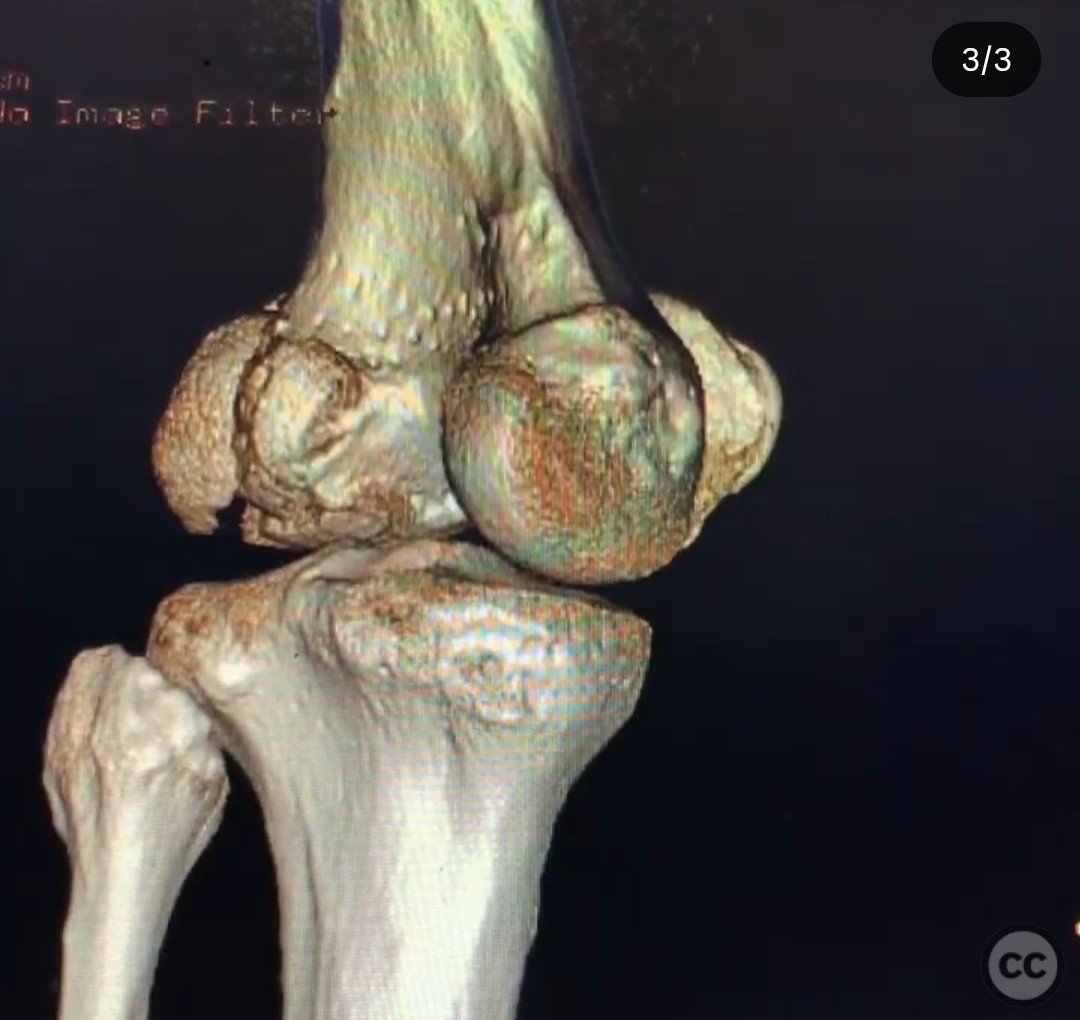
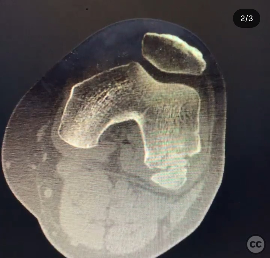
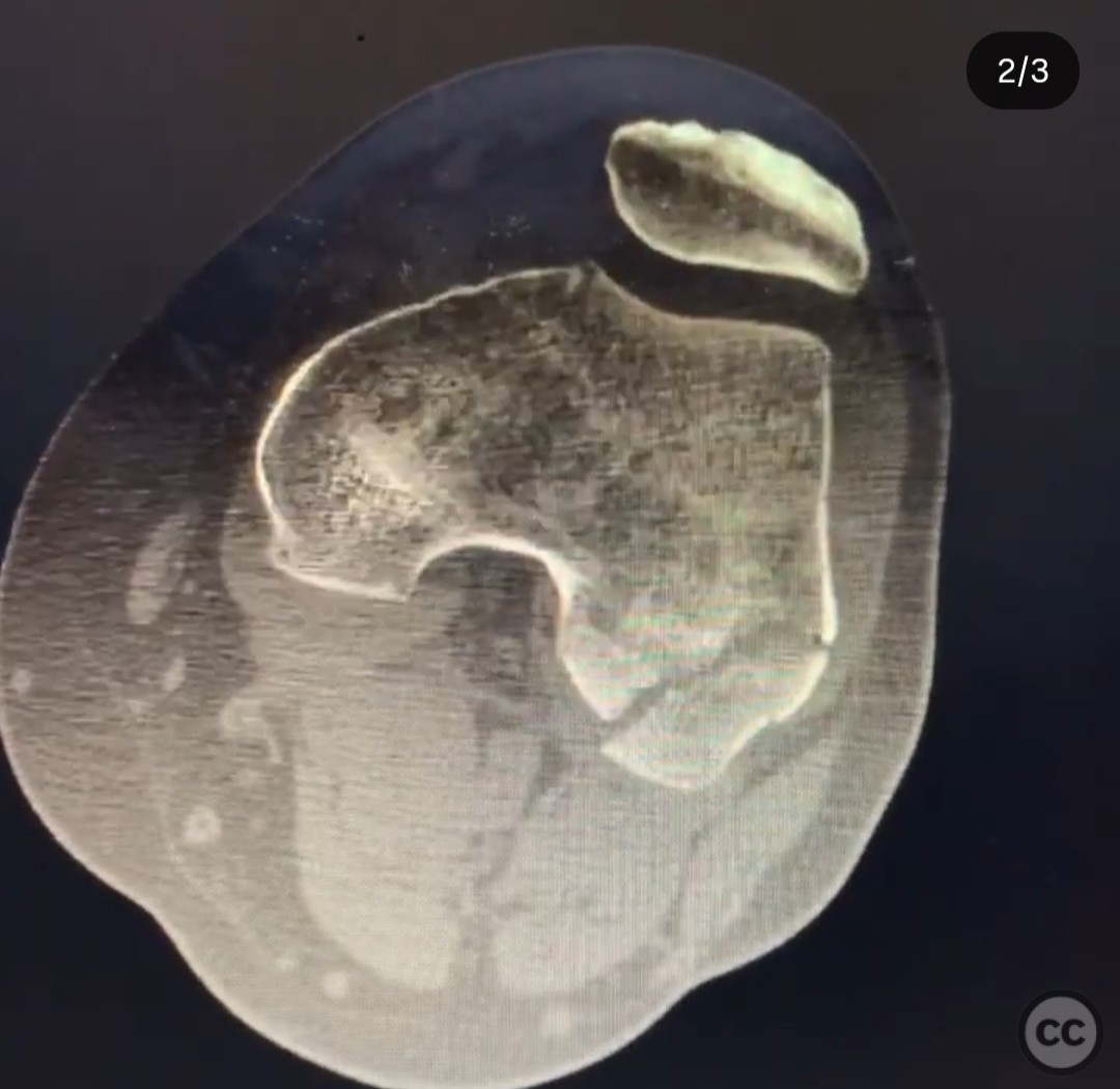
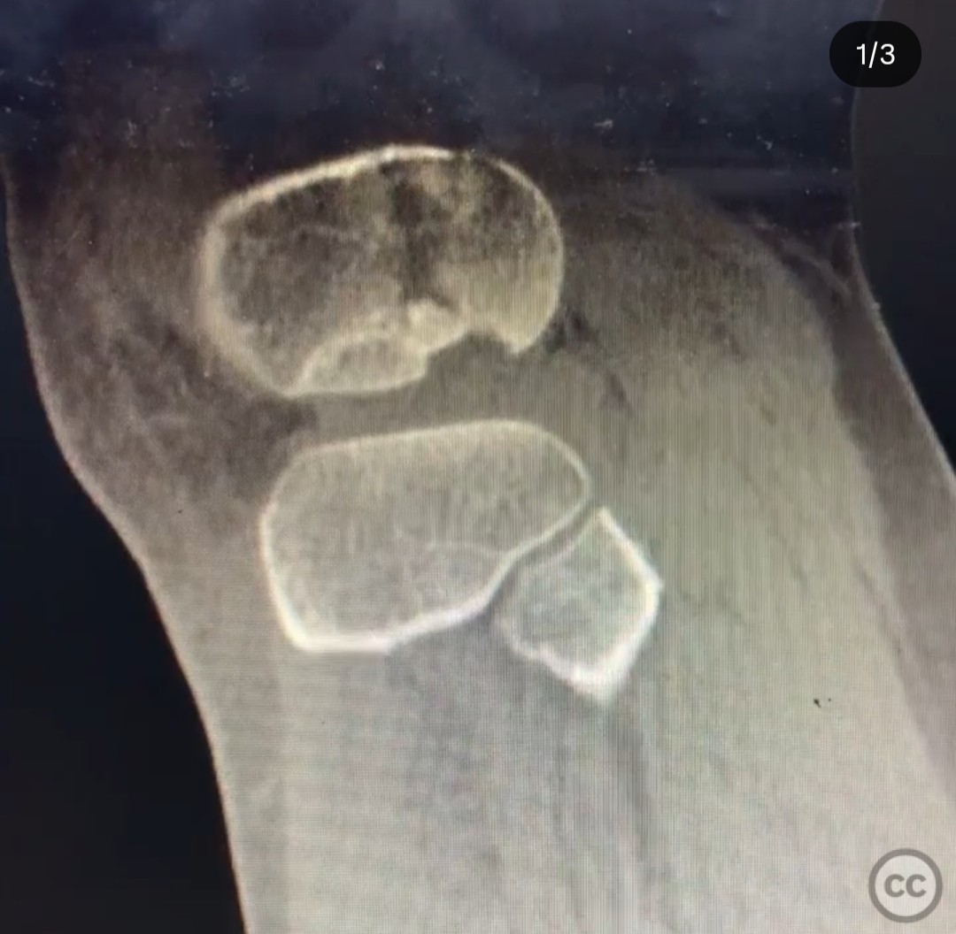
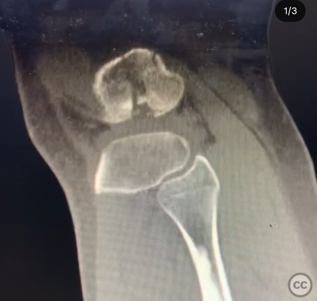
Clinical and radiological findings: A 35-year-old male experienced a high-energy motorcycle accident, resulting in a complex fracture of the distal femur. Initial imaging, including CT with sagittal cuts and 3D reconstruction, revealed a unique fracture pattern: a quarter-sized impacted fragment in the middle third of the condyle, a second free impacted fragment posterior to it, and a coronal plane split of the posterior condyle.
Preoperative Plan
Planning remarks: The preoperative plan involved a posterolateral corner approach with a lateral epicondylar osteotomy to access all fracture components and facilitate implant placement. This approach was chosen over standard lateral or prone direct posterior approaches due to its superior exposure and control of the fracture fragments.
Surgical Discussion
Patient positioning: The patient was positioned laterally to optimize access to the lateral distal femur and facilitate the surgical approach.
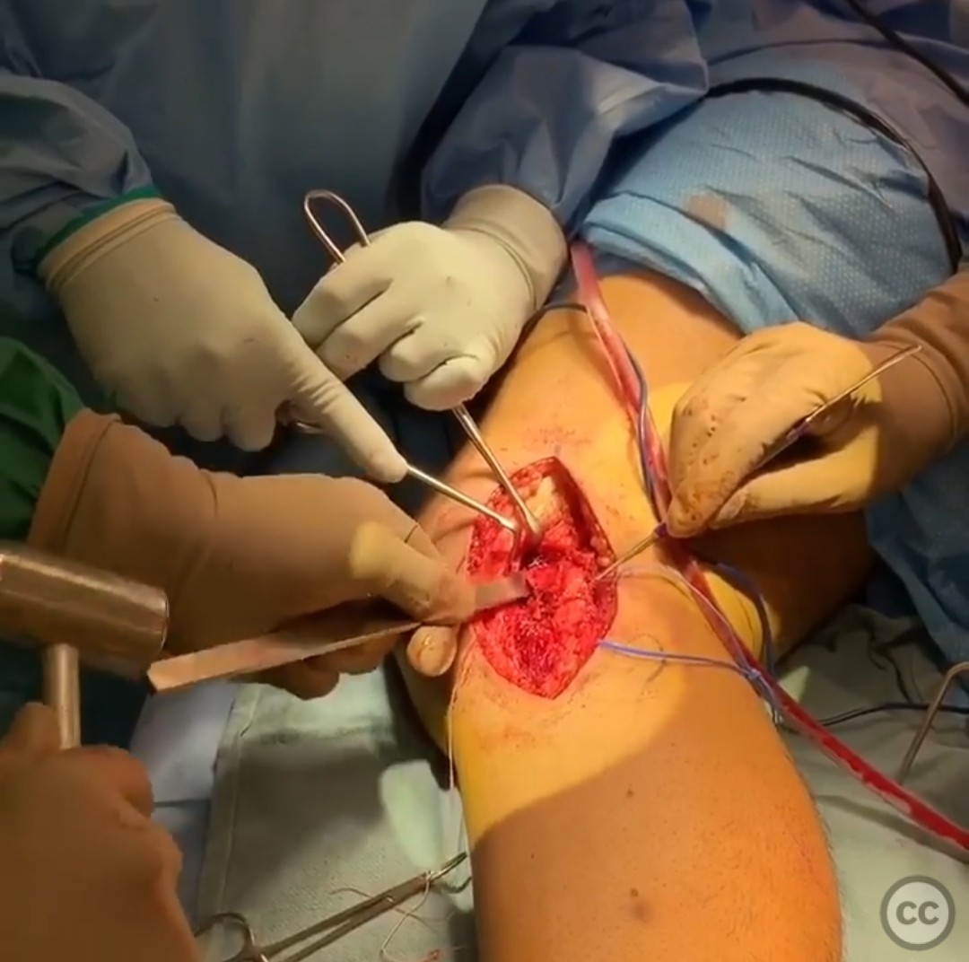
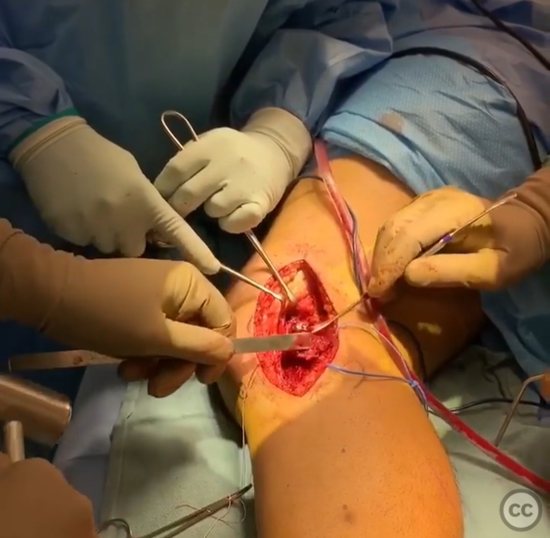
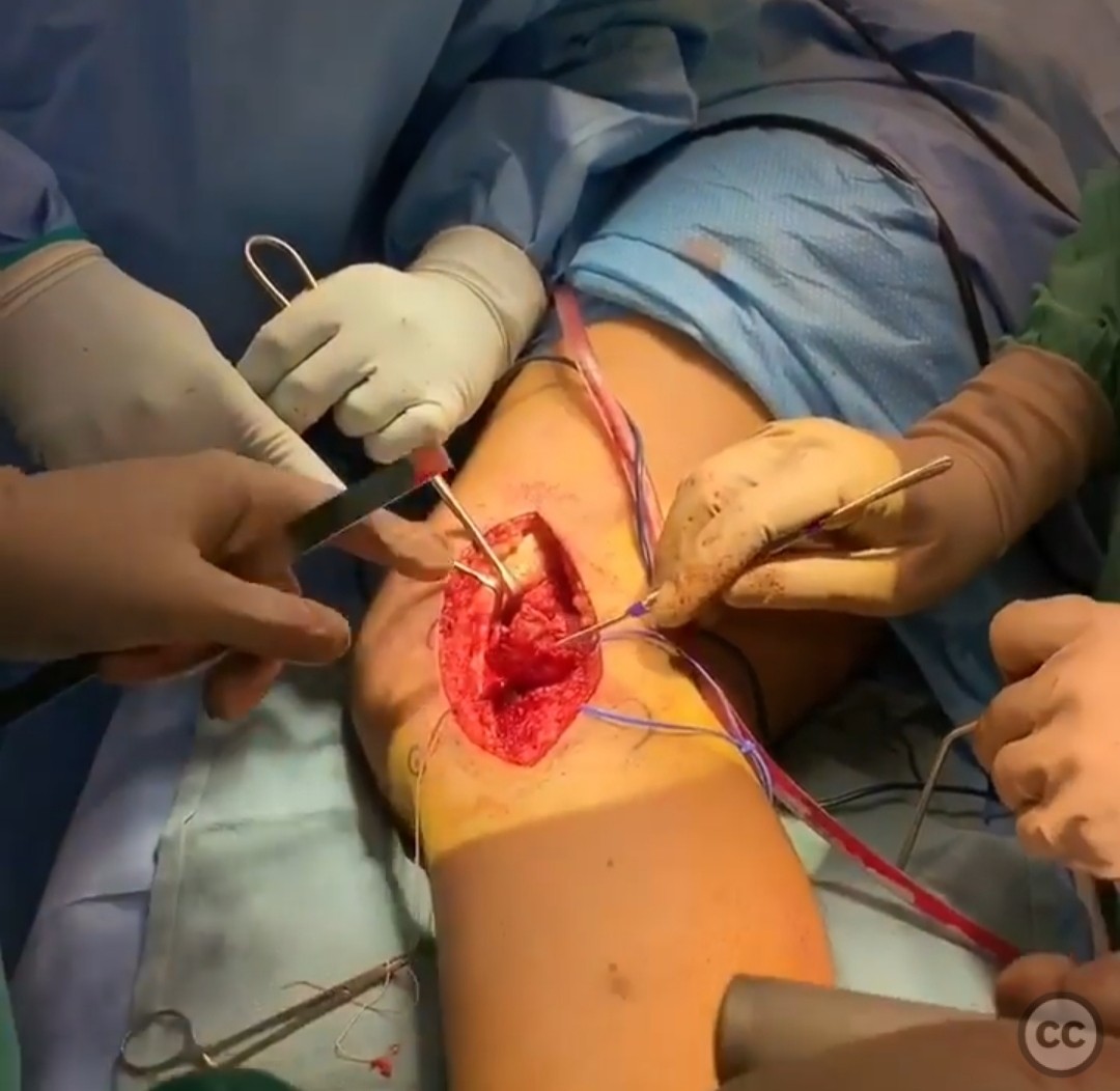
Anatomical surgical approach: The surgical approach was centered over the lateral epicondyle. The posterior border of the iliotibial band and biceps femoris were identified. The common peroneal nerve was mobilized and protected. The lateral collateral ligament (LCL) was clearly defined. A posterior capsulotomy was performed between the biceps femoris and LCL, as well as an anterior capsulotomy to the LCL. A lateral epicondylar osteotomy was executed, reflecting the posterolateral corner structures distally to expose the fracture site.
Operative remarks:The surgeon noted that the lateral epicondylar osteotomy provided excellent exposure to the posterior aspects of the lateral distal femur, allowing for direct reduction of the fracture fragments. Implants were placed behind the biceps femoris. The capsule and osteotomy were repaired following reduction and fixation.
Postoperative protocol: Postoperative rehabilitation included early range of motion exercises with weight-bearing as tolerated, progressing to full weight-bearing by 6 weeks post-surgery.
Follow up: Not specified
Orthopaedic implants used: Orthopaedic implants used included screws and plates for fracture fixation and osteotomy stabilization.
Search for Related Literature

orthopaedic_trauma
- United States , Seattle
- Area of Specialty - General Trauma
- Position - Specialist Consultant

Industry Sponsership
contact us for advertising opportunities
Article viewed 162 times
19 Jul 2025
Add to Bookmarks
Full Citation
Cite this article:
Surname, Initial. (2025). Lateral Epicondylar Osteotomy for Complex Distal Femur Fracture. Journal of Orthopaedic Surgery and Traumatology. Case Report 35021895 Published Online Jul 19 2025.