Open Pertrochanteric Femur Fracture with Critical Bone Loss
Score and Comment on this Case
Clinical Details
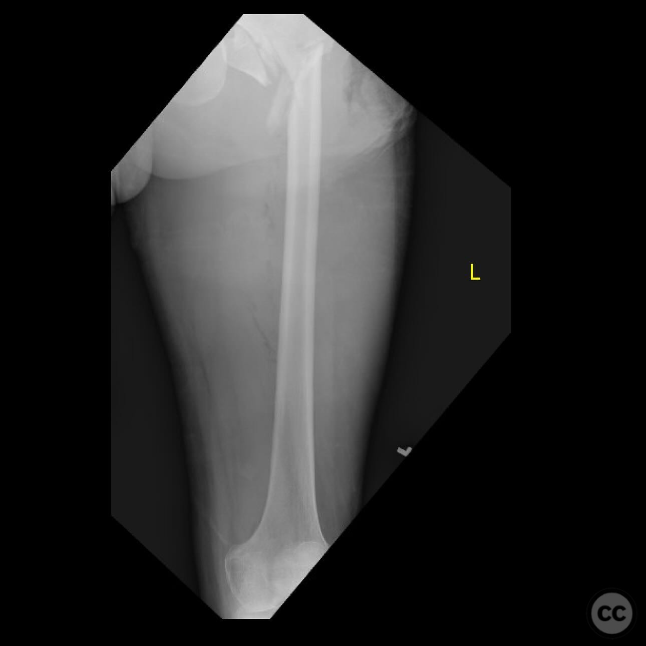
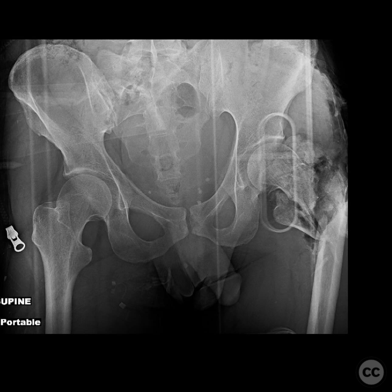
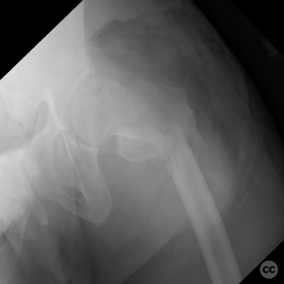
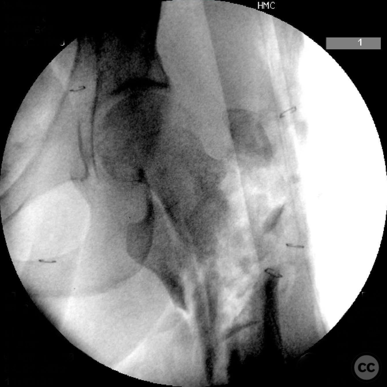
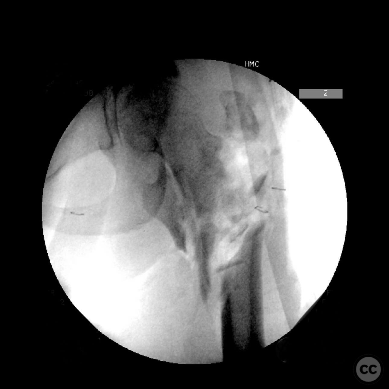
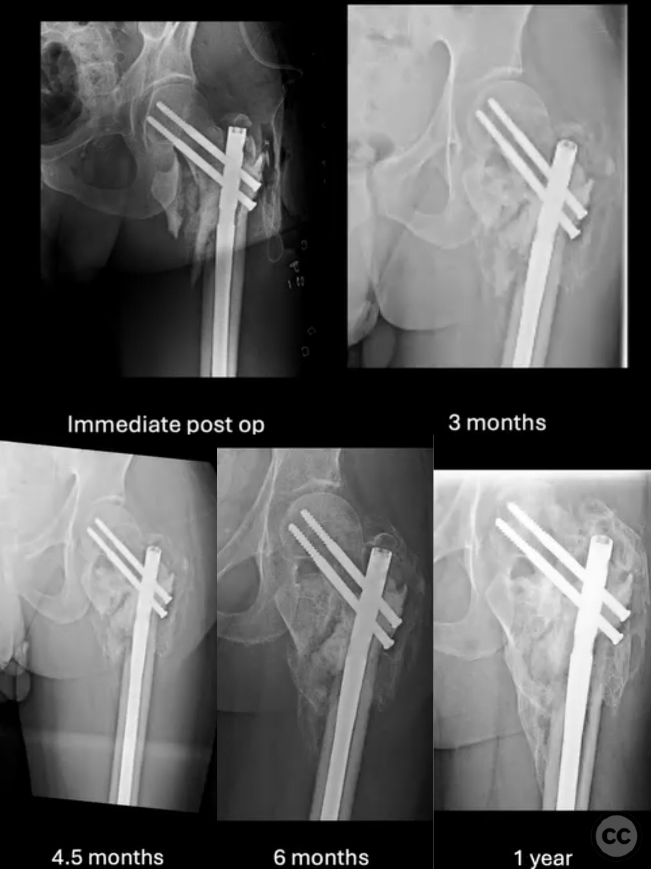
Clinical and radiological findings: A 33-year-old male involved in a high-speed motorcycle crash presented with multiple injuries, including a head bleed, hemopneumothorax, and splenic and liver lacerations. The left hip exhibited a 30 cm irregular open wound exposing a pertrochanteric femur fracture with significant contamination. The sciatic nerve was palpable and intact within the wound, and there was no vascular injury. Initial instability precluded immediate orthopedic intervention.
Preoperative Plan
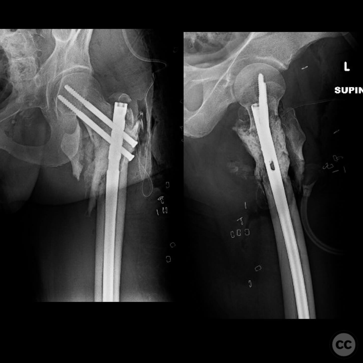
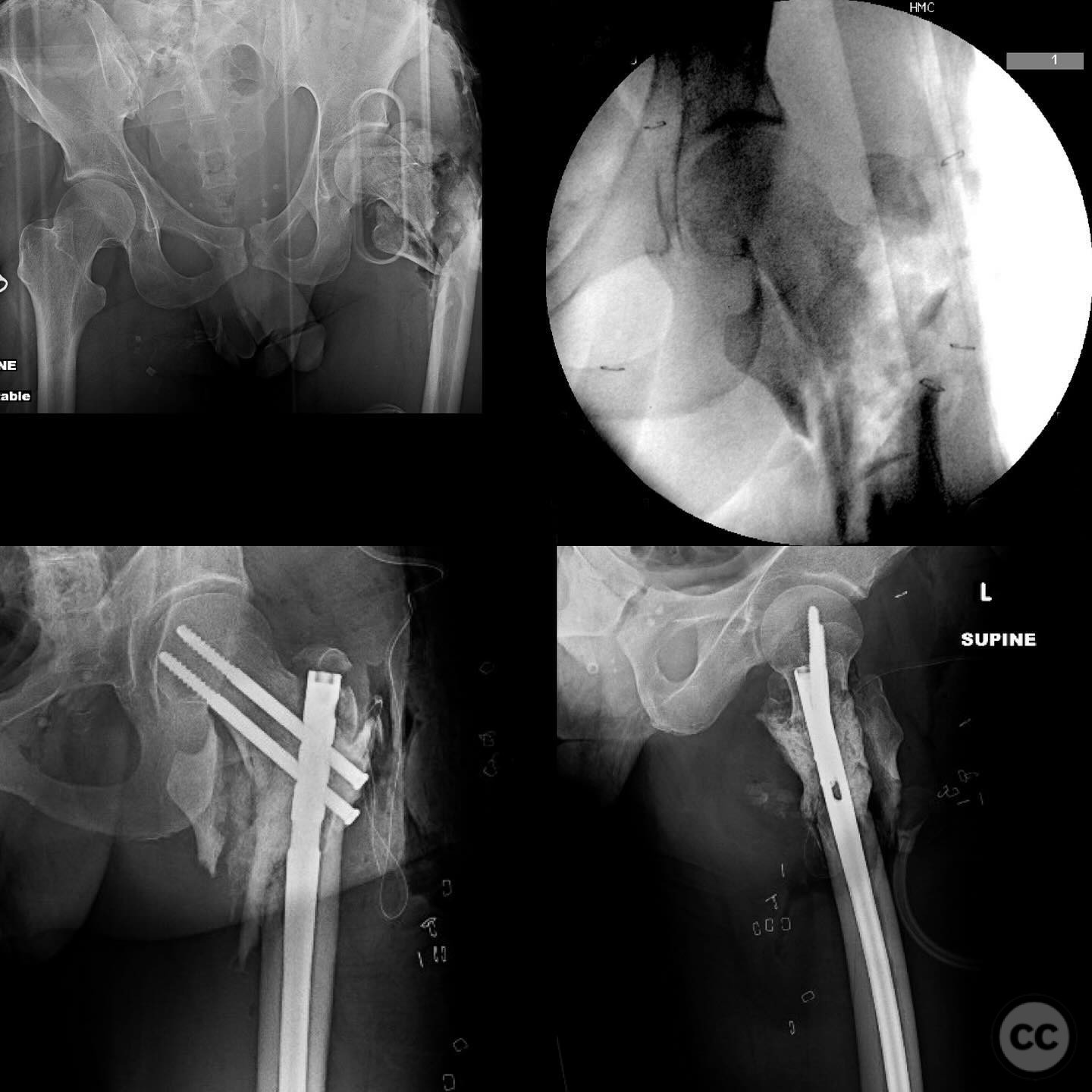
Planning remarks: The preoperative plan involved a single-stage thorough debridement and irrigation of the open wound, followed by definitive fixation using reconstruction intramedullary nailing. The approach was to manage the critical bone defect with the induced membrane technique (IMT), utilizing an antibiotic-loaded cement spacer.
Surgical Discussion
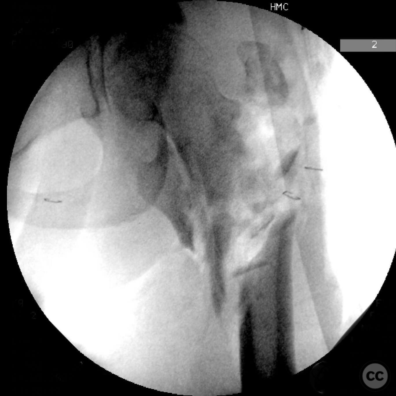
Patient positioning: The patient was positioned supine on a radiolucent operating table to facilitate access to the proximal femur and allow for intraoperative imaging.
Anatomical surgical approach: A lateral approach to the proximal femur was utilized, involving careful dissection to expose the fracture site. The wound was thoroughly debrided, and irrigation was performed to address contamination. Trochanteric fragments were stabilized using #5 sutures anchored into the gluteus medius tendon.
Operative remarks:The surgical team opted for a single-stage procedure due to the nature of the Gustilo-Anderson Type 3A open fracture, which was closeable despite moderate contamination. The critical bone defect was addressed using the Masquélet technique, with an antibiotic-loaded PMMA spacer containing vancomycin and tobramycin. This approach is supported by literature for managing critical-sized bone defects and allows for potential deferral of bone grafting if early callus formation is observed.
Postoperative protocol: Postoperatively, the patient followed a rehabilitation protocol emphasizing early mobilization within pain tolerance and weight-bearing as tolerated, with close monitoring for signs of infection or complications.
Follow up: Not specified
Orthopaedic implants used: Reconstruction intramedullary nail, #5 sutures, Antibiotic-loaded PMMA cement spacer (vancomycin and tobramycin).
Search for Related Literature

orthopaedic_trauma
- United States , Seattle
- Area of Specialty - General Trauma
- Position - Specialist Consultant

Industry Sponsership
contact us for advertising opportunities
Article viewed 247 times
08 Jul 2025
Add to Bookmarks
Full Citation
Cite this article:
Surname, Initial. (2025). Open Pertrochanteric Femur Fracture with Critical Bone Loss. Journal of Orthopaedic Surgery and Traumatology. Case Report 34810792 Published Online Jul 08 2025.