

Chopart's Fracture Dislocation with Lateral Column Crush.
Score and Comment on this Case
Clinical Details
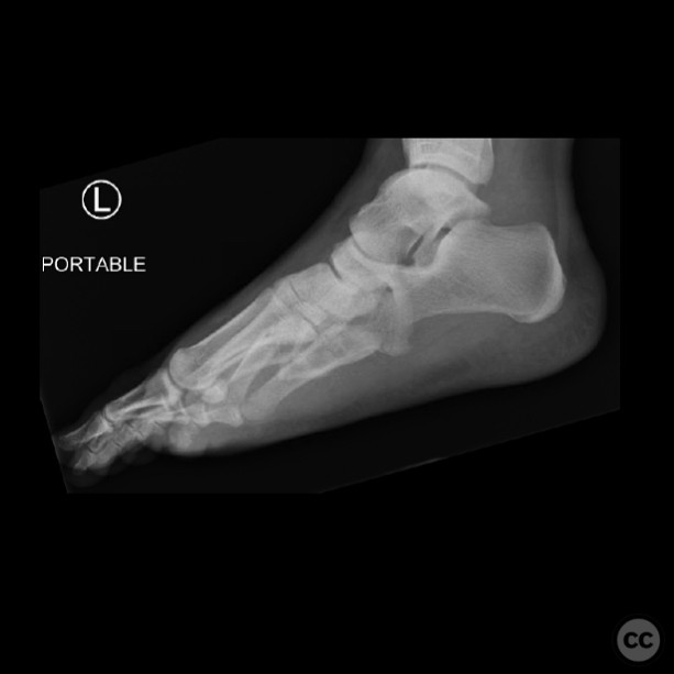
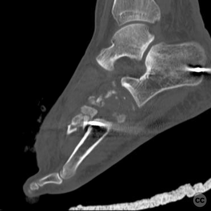
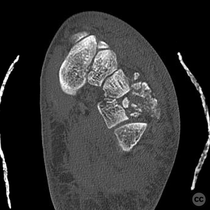
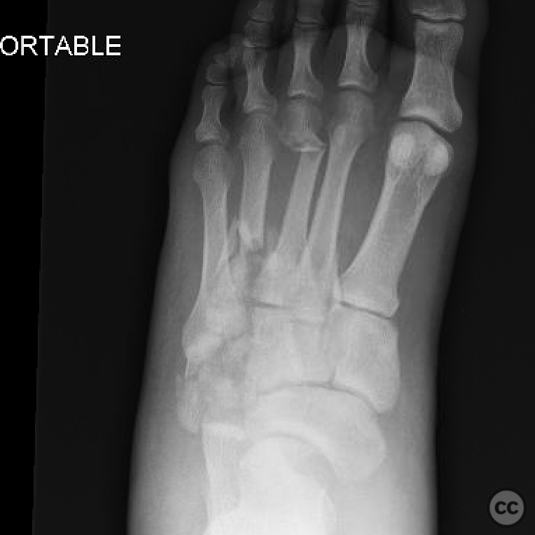
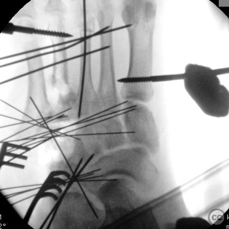
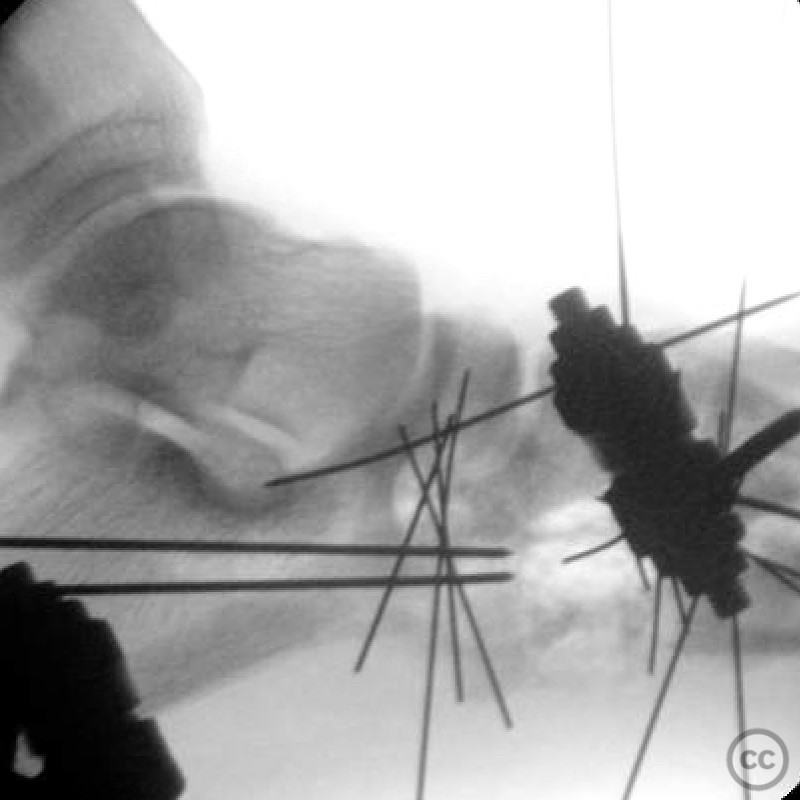
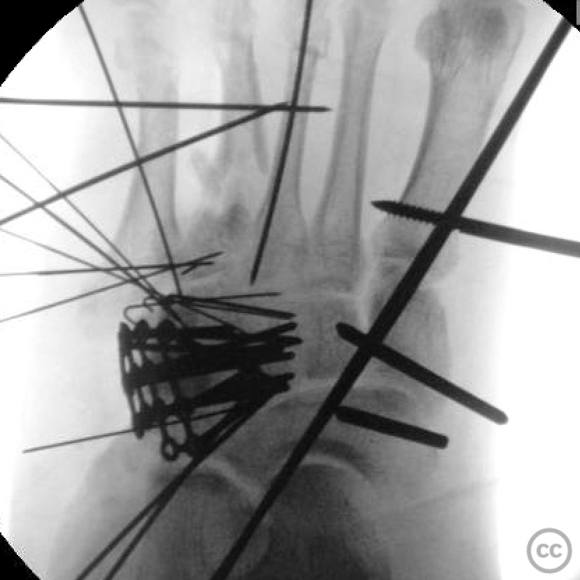
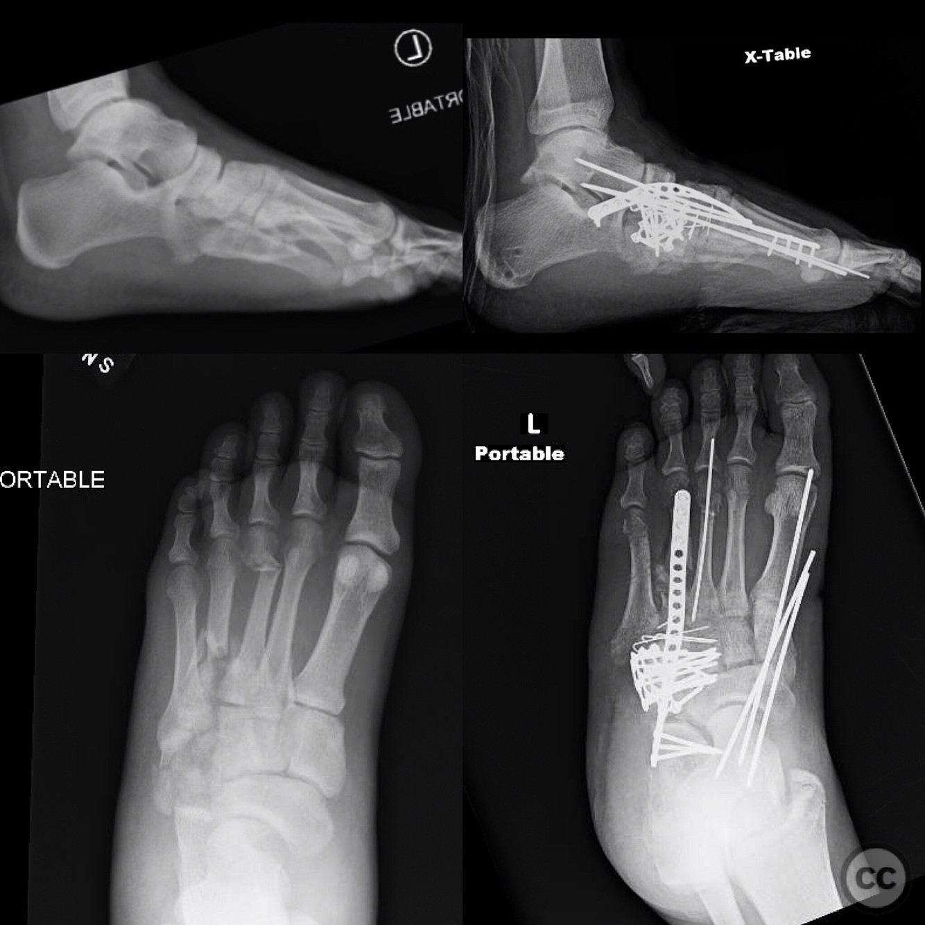
Clinical and radiological findings: A 27-year-old male presented following a motorcycle accident due to a stuck throttle. The patient sustained a closed Chopart's fracture dislocation, characterized by a lateral column crush involving the cuboid, talonavicular, and navicular-cuneiform joints. Radiographs revealed significant shortening of the lateral column and midfoot sagging, with no involvement of the Lisfranc joint. The cuboid was severely comminuted, and there was notable forefoot abduction and supination deformity.
Preoperative Plan
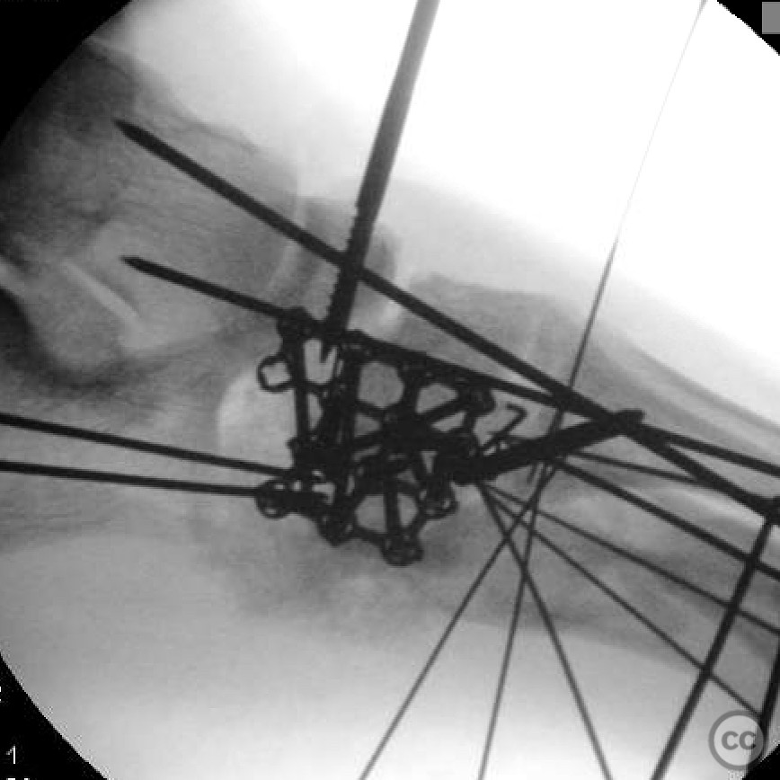
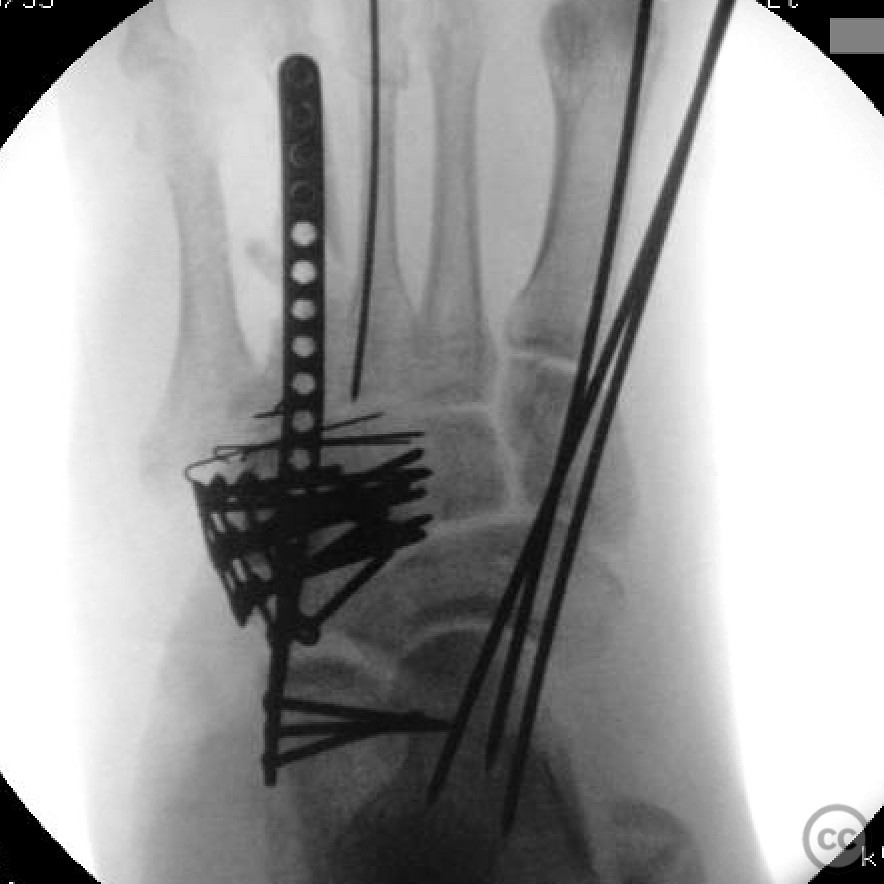
Planning remarks: The preoperative plan involved restoring the lateral column length and correcting the midfoot sag. Given the patient's previous 5-week period in an external fixator for soft tissue management, the decision was made to proceed with internal fixation by bridging the calcaneus to the fourth metatarsal.
Surgical Discussion
Patient positioning: Supine position with the affected foot elevated on a padded support to facilitate access to the lateral aspect of the foot.
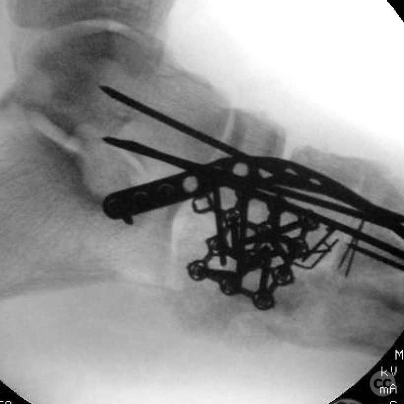
Anatomical surgical approach: A lateral approach to the foot was utilized, with an incision extending from the distal fibula to the base of the fourth metatarsal. Subperiosteal dissection was performed to expose the cuboid and adjacent structures. The calcaneocuboid joint was accessed, and attention was given to restoring the length of the lateral column by bridging fixation.
Operative remarks:The surgeon noted significant comminution of the cuboid, which necessitated meticulous reconstruction to restore the lateral column length. Due to the poor condition of the cuboid-metatarsal joints, achieving articular congruity was challenging. The decision to bridge from the calcaneus to the fourth metatarsal was made to provide stable fixation and maintain column length, as the external fixator pins were compromising the integrity of the fourth and fifth metatarsals.
Postoperative protocol: Postoperative rehabilitation included non-weight bearing for 8 weeks, followed by progressive weight bearing as tolerated. Range of motion exercises for the ankle and subtalar joint were initiated early to prevent stiffness.
Follow up: Not specified.
Orthopaedic implants used: Internal fixation with a bridging plate from calcaneus to fourth metatarsal.
Search for Related Literature

orthopaedic_trauma
- United States , Seattle
- Area of Specialty - General Trauma
- Position - Specialist Consultant

Industry Sponsership
contact us for advertising opportunities
Article viewed 304 times
26 Jul 2025
Add to Bookmarks
Full Citation
Cite this article:
Surname, Initial. (2025). Chopart's Fracture Dislocation with Lateral Column Crush.. Journal of Orthopaedic Surgery and Traumatology. Case Report 34317678 Published Online Jul 26 2025.