


Ipsilateral Talus and Calcaneus Fracture: Complex Hindfoot Reconstruction
Score and Comment on this Case
Clinical Details
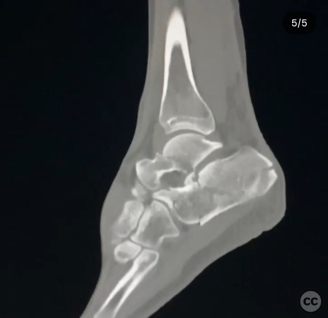
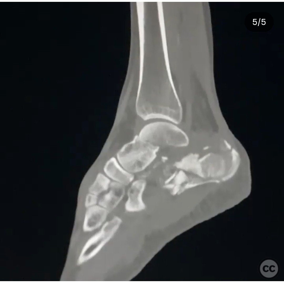
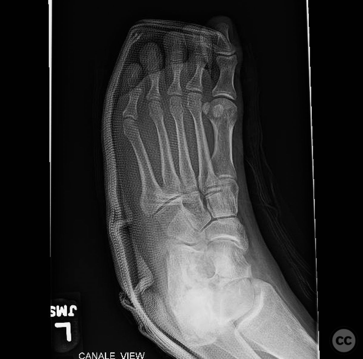
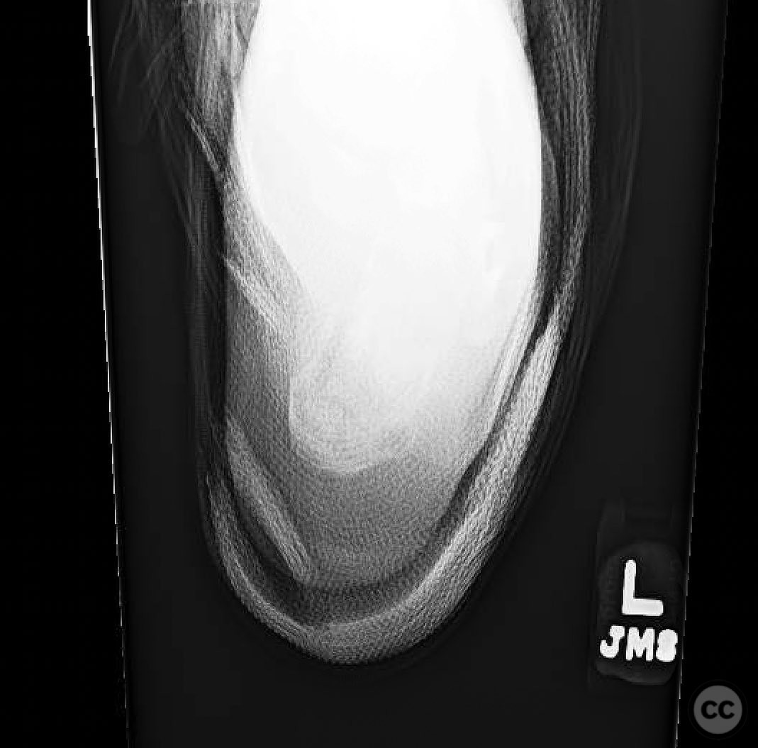
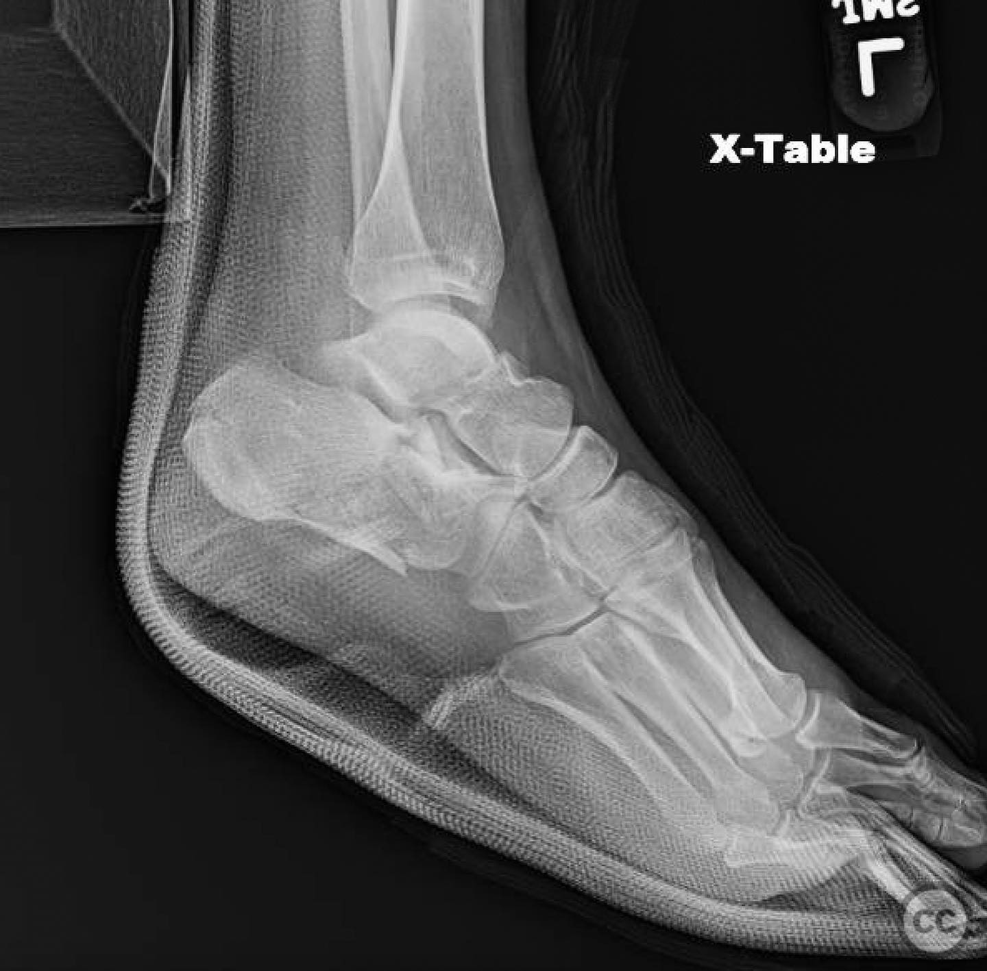
Clinical and radiological findings: A 26-year-old female patient presented with a closed ipsilateral talus and calcaneus fracture following a motor vehicle collision. Initial clinical examination revealed a fit and healthy individual with no significant comorbidities. Radiological assessment, including X-rays and CT scans, confirmed the presence of a talar neck fracture and a calcaneal fracture. The soft tissue envelope was intact, with no signs of neurovascular compromise.
Preoperative Plan
Planning remarks: The preoperative plan involved open reduction and internal fixation (ORIF) of the talar neck fracture using dorsolateral and dorsomedial approaches. Following a two-week interval to allow for soft tissue recovery, ORIF of the calcaneal fracture was planned through an extensile lateral approach. The sequence was chosen to optimize reduction and fixation of the talar neck prior to addressing the calcaneus.
Surgical Discussion
Patient positioning: The patient was positioned supine on the operating table with the affected limb elevated on a padded footrest to facilitate access to both the medial and lateral aspects of the hindfoot.
Anatomical surgical approach: The surgical approach for the talar neck involved dual incisions: a dorsolateral incision was made parallel to the fourth metatarsal, extending proximally towards the lateral malleolus, and a dorsomedial incision was made parallel to the first metatarsal, extending proximally towards the medial malleolus. Subperiosteal dissection was performed to expose the talar neck for reduction and fixation. For the calcaneal fracture, an extensile lateral approach was utilized, with a lateral incision extending from the posterior aspect of the lateral malleolus to the base of the fifth metatarsal, allowing for wide exposure of the calcaneus.
Operative remarks:The surgeon emphasized the importance of careful soft tissue handling and intraoperative assessment of vascularity using Doppler ultrasound to ensure flap viability. The sequence of addressing the talus prior to the calcaneus was critical to achieving optimal alignment and fixation. The use of dual approaches for the talar neck was necessary to achieve adequate reduction and fixation.
Postoperative protocol: Postoperatively, the patient was placed in a non-weight-bearing cast for six weeks, followed by progressive weight-bearing as tolerated. Physical therapy focused on range of motion exercises and gradual strengthening of the hindfoot and ankle.
Follow up: Not specified
Orthopaedic implants used: Orthopaedic implants used included cannulated screws for talar neck fixation and a locking plate system for calcaneal fracture stabilization.
Search for Related Literature

orthopaedic_trauma
- United States , Seattle
- Area of Specialty - General Trauma
- Position - Specialist Consultant

Industry Sponsership
contact us for advertising opportunities
Article viewed 328 times
17 Jul 2025
Add to Bookmarks
Full Citation
Cite this article:
Surname, Initial. (2025). Ipsilateral Talus and Calcaneus Fracture: Complex Hindfoot Reconstruction. Journal of Orthopaedic Surgery and Traumatology. Case Report 34022892 Published Online Jul 17 2025.