


Open B-Type Pilon Fracture with Fibular Nonunion and Suspected Infection.
Score and Comment on this Case
Clinical Details
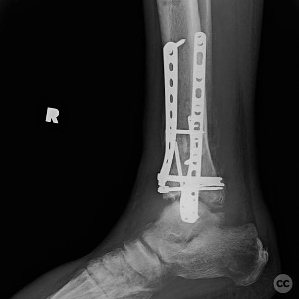
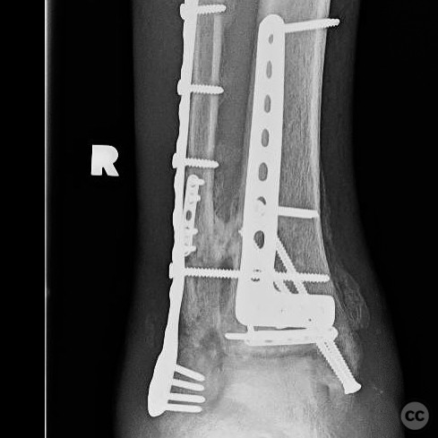
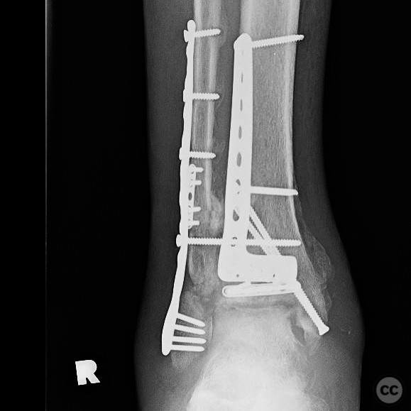
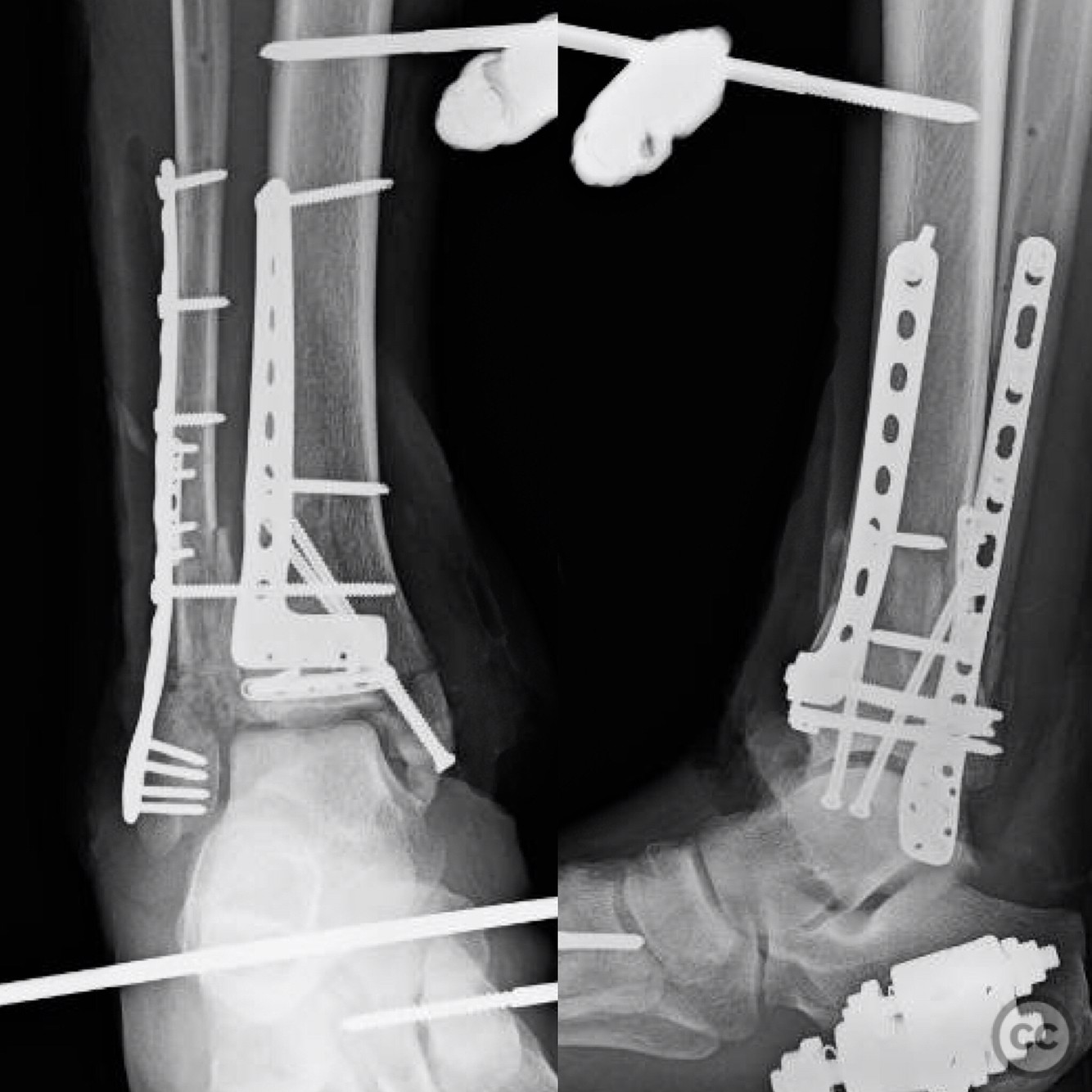
Clinical and radiological findings: A 46-year-old male sustained an open B-type pilon fracture after jumping off an overpass while evading police. Initial management included external fixation. Two weeks post-injury, the fibula was internally fixed, followed by open reduction and internal fixation (ORIF) of the pilon fracture another two weeks later. Immediate postoperative radiographs showed suboptimal articular reduction but satisfactory fixation. At three months, follow-up radiographs indicated potential deep infection or early Charcot arthropathy, with fibular nonunion and osteolysis.
Preoperative Plan
Planning remarks: The preoperative plan involved an anteromedial approach to the distal tibia for ORIF of the pilon fracture, as described by Benirschke. The fibula was addressed separately with internal fixation.
Surgical Discussion
Patient positioning: Supine positioning on a radiolucent table to facilitate fluoroscopic imaging during the procedure.
Anatomical surgical approach: Anteromedial approach to the distal tibia: A longitudinal incision was made along the anteromedial aspect of the distal tibia. Subcutaneous tissues were dissected to expose the fracture site. Careful handling of soft tissues was maintained to preserve vascular supply.
Operative remarks:The articular surface reduction was suboptimal, though fixation was deemed satisfactory at the time of surgery. The patient returned to incarceration postoperatively and missed follow-up appointments until three months post-surgery. Radiographs at that time revealed concerning signs of either deep infection or early Charcot changes, with fibular nonunion and progressive osteolysis. Despite these findings, the patient appeared clinically well with healed wounds and normal inflammatory markers.
Postoperative protocol: The patient was allowed to begin weight-bearing at three months postoperatively, given the clinical appearance of well-healed wounds and absence of systemic infection signs.
Follow up: Not specified.
Orthopaedic implants used: Orthopaedic implants used: Internal fixation devices for fibula and pilon fracture (specific implant details not provided).
Search for Related Literature

orthopaedic_trauma
- United States , Seattle
- Area of Specialty - General Trauma
- Position - Specialist Consultant

Industry Sponsership
contact us for advertising opportunities
Article viewed 318 times
26 Jul 2025
Add to Bookmarks
Full Citation
Cite this article:
Surname, Initial. (2025). Open B-Type Pilon Fracture with Fibular Nonunion and Suspected Infection.. Journal of Orthopaedic Surgery and Traumatology. Case Report 33550488 Published Online Jul 26 2025.