ORIF of Anterior Column/Posterior Hemi-Transverse Acetabular Fracture with Medial Dome Impaction in an Active Senior
Score and Comment on this Case
Clinical Details
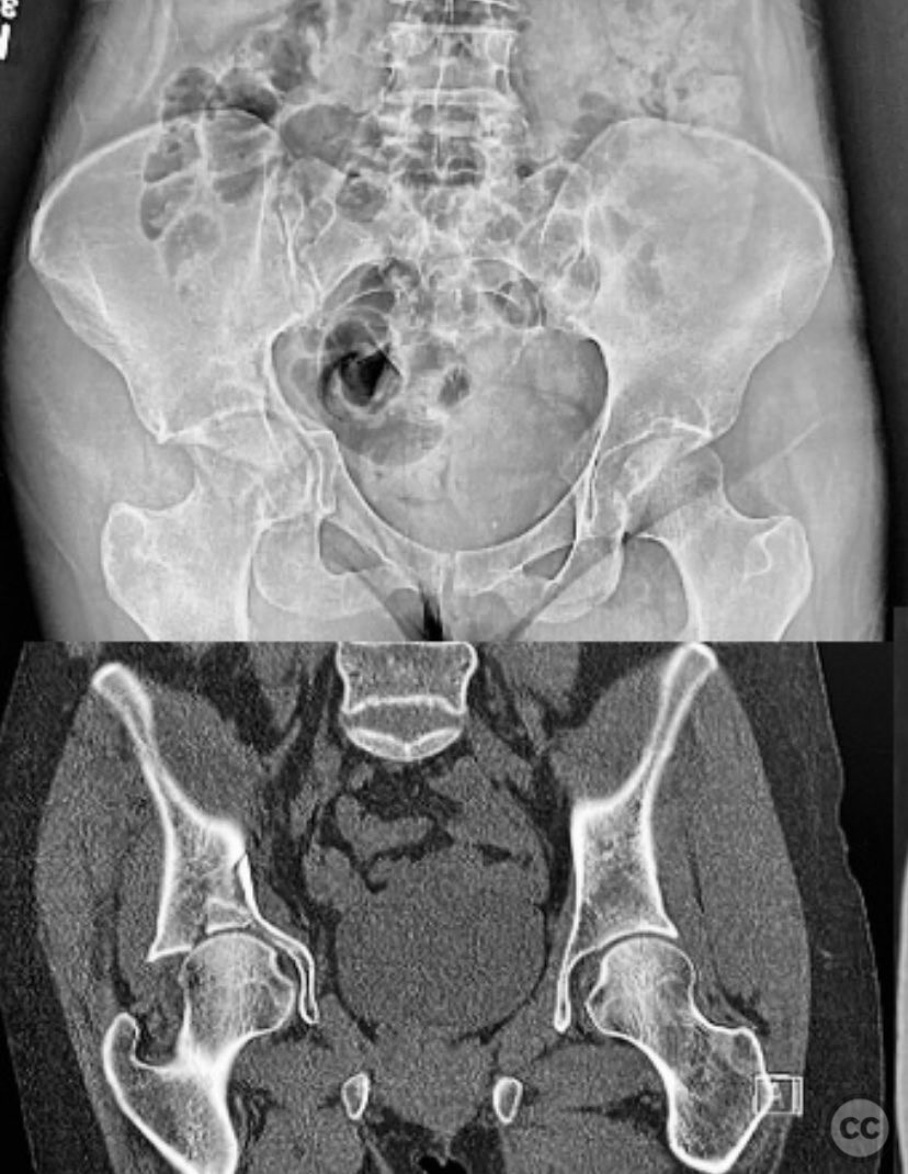
Clinical and radiological findings: An active senior patient sustained an intruded acetabular fracture, best classified as an anterior column/posterior hemi-transverse (AC/PHTr) pattern with associated medial dome impaction. Initial evaluation included clinical and radiological assessment, with CT imaging confirming the fracture configuration and impaction zone. Neurovascular examination was not specified. Skeletal traction of 10-15 pounds was applied during initial resuscitation and evaluation.
Preoperative Plan
Planning remarks: The preoperative plan involved open reduction and internal fixation (ORIF) via an ilioinguinal approach. The surgical objective was anatomical reduction of the anterior column and posterior column, buttressing the quadrilateral surface, and addressing the medial dome impaction with allograft bone support. Preoperative imaging was used to plan fragment-specific reduction and fixation.
Surgical Discussion
Patient positioning: The patient was positioned supine on the operating table.
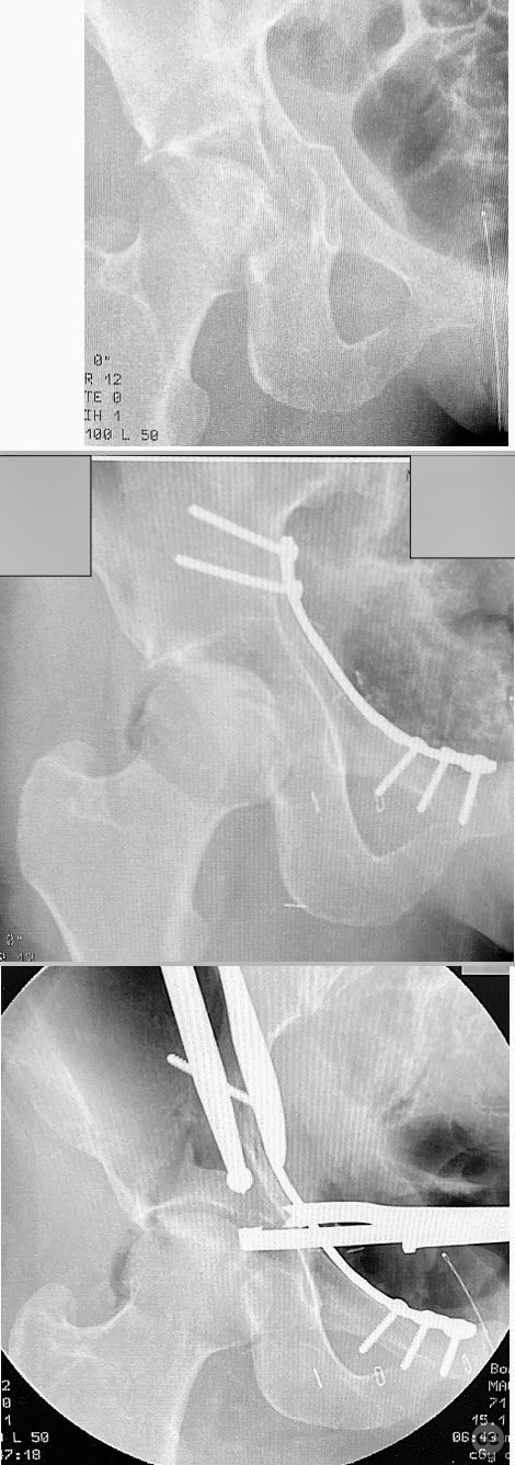
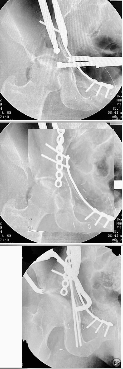
Anatomical surgical approach: A classical ilioinguinal approach was utilized. The incision was made parallel to the inguinal ligament, extending from just lateral to the anterior superior iliac spine (ASIS) towards the pubic tubercle. Dissection proceeded through the subcutaneous tissue and external oblique aponeurosis, with careful identification and protection of the neurovascular structures within the three windows of the ilioinguinal approach. The anterior column fragments were exposed and reduced using clamps. The quadrilateral surface and posterior column were buttressed with a contoured reconstruction plate. The medial dome impaction zone was identified under fluoroscopic guidance, reduced to the femoral head, and supported with allograft bone. The anterior column reduction was stabilized with a contoured reconstruction plate, and additional lag screws were placed as needed to secure the posterior column.
Operative remarks:Manual traction intraoperatively demonstrated the appropriate position of the femoral head. Reduction of the quadrilateral surface and posterior column was achieved prior to addressing the impaction zone. The impacted dome fragment was anatomically reduced and supported with allograft bone grafting. An offset clamp was utilized to improve posterior column reduction when necessary before definitive fixation with lag screws. Intraoperative fluoroscopy confirmed satisfactory reduction and implant placement.
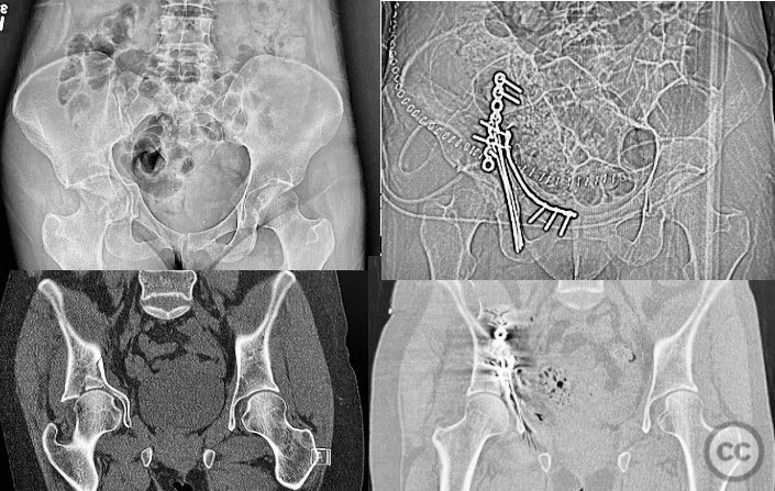
Postoperative protocol: Postoperatively, weight bearing was protected for 4-6 weeks using crutches or a walker as tolerated, though most patients weight bear as tolerated for various reasons.
Follow up: Not specified
Orthopaedic implants used: Contoured reconstruction plates, lag screws, allograft bone
Search for Related Literature


Industry Sponsership
contact us for advertising opportunities
Article viewed 226 times
11 Sep 2025
Add to Bookmarks
Full Citation
Cite this article:
Routt, ML. (2025). ORIF of Anterior Column/Posterior Hemi-Transverse Acetabular Fracture with Medial Dome Impaction in an Active Senior. Journal of Orthopaedic Surgery and Traumatology. Case Report 31042210 Published Online Sep 11 2025.