Associated Both Column Acetabular Fracture: Sequential ORIF via Ilioinguinal Windows and Percutaneous Posterior Column Fixation
Score and Comment on this Case
Clinical Details
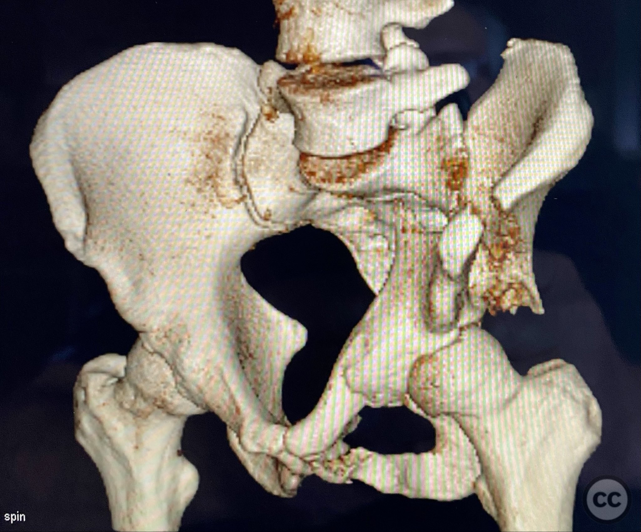
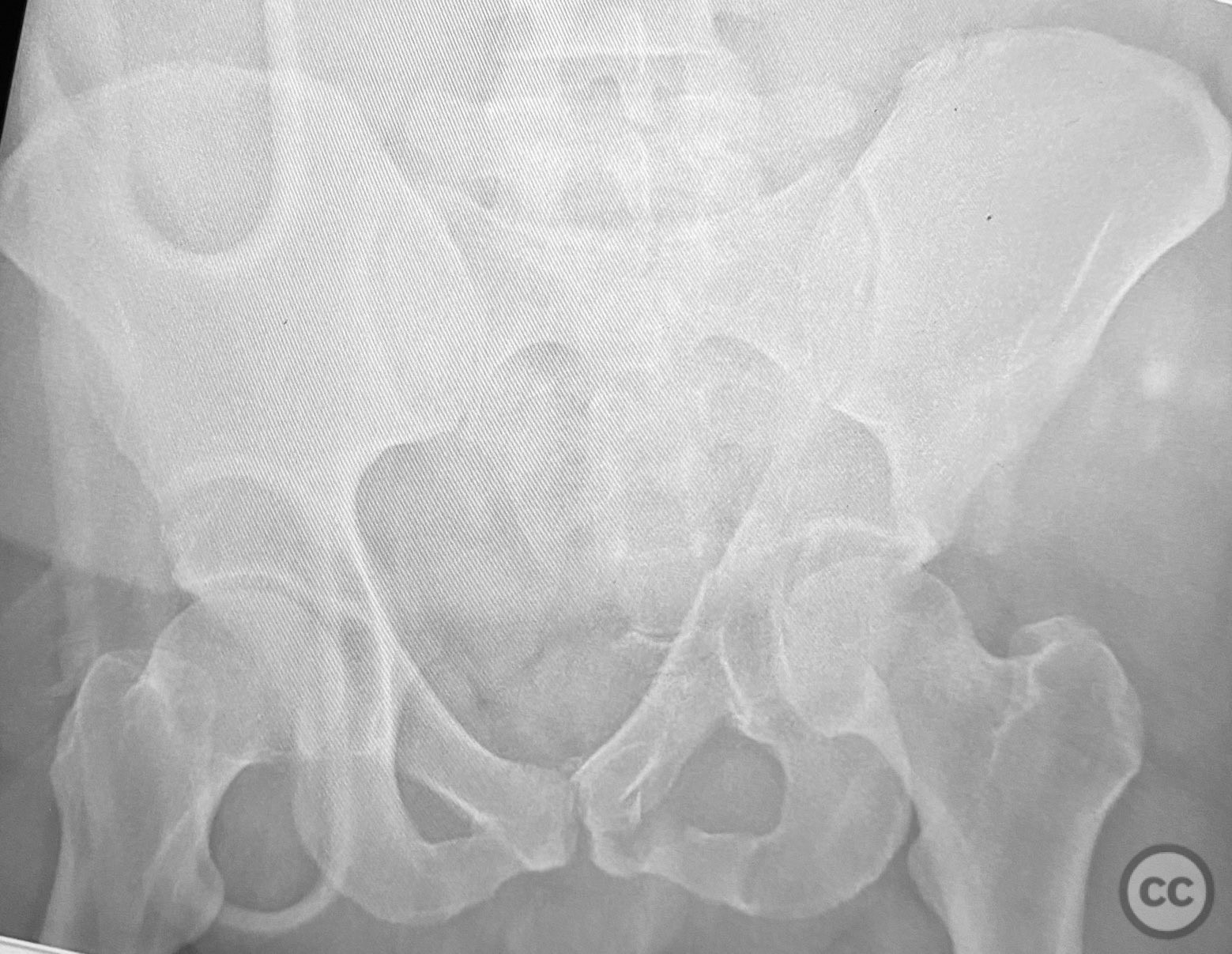
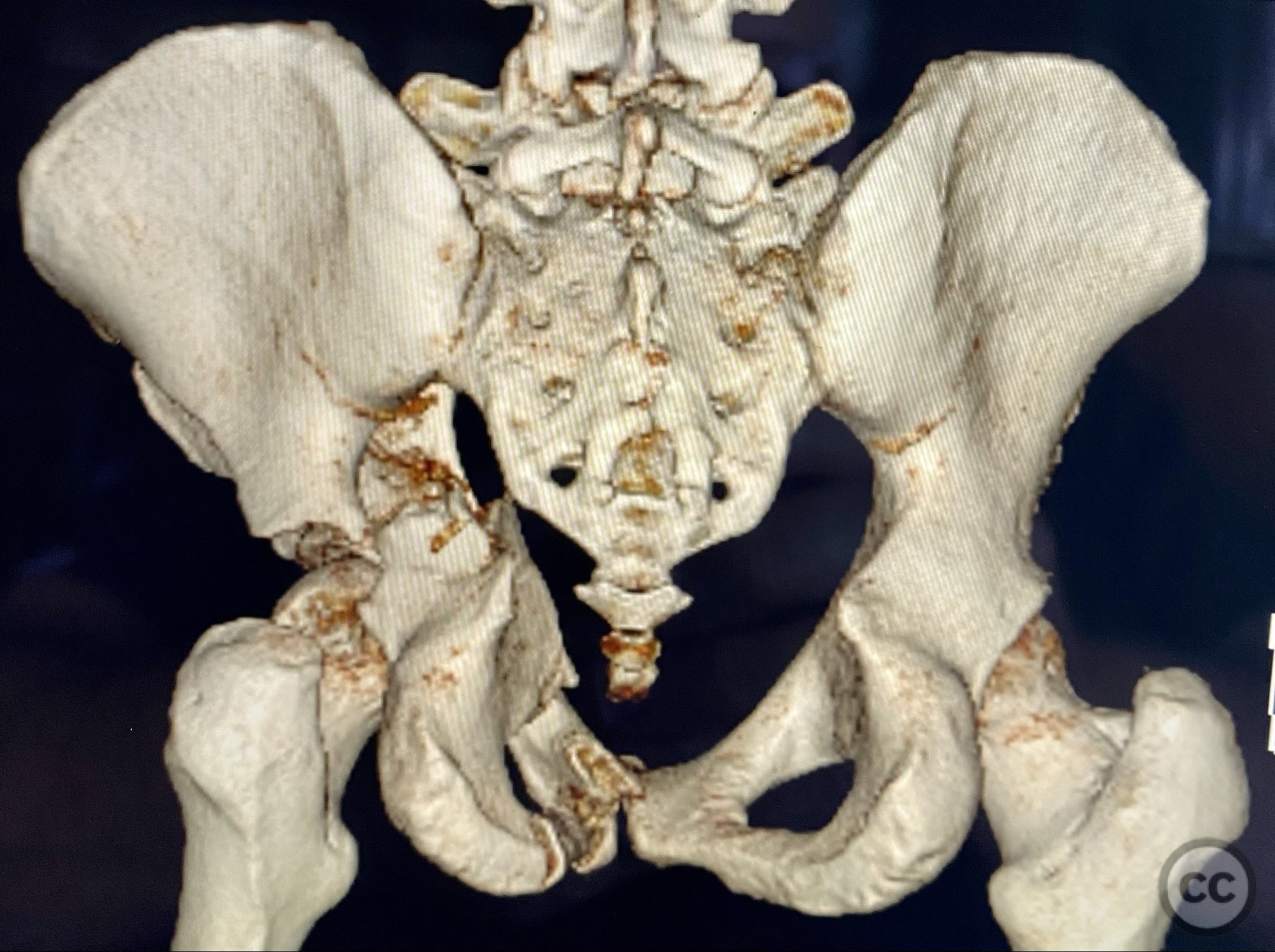
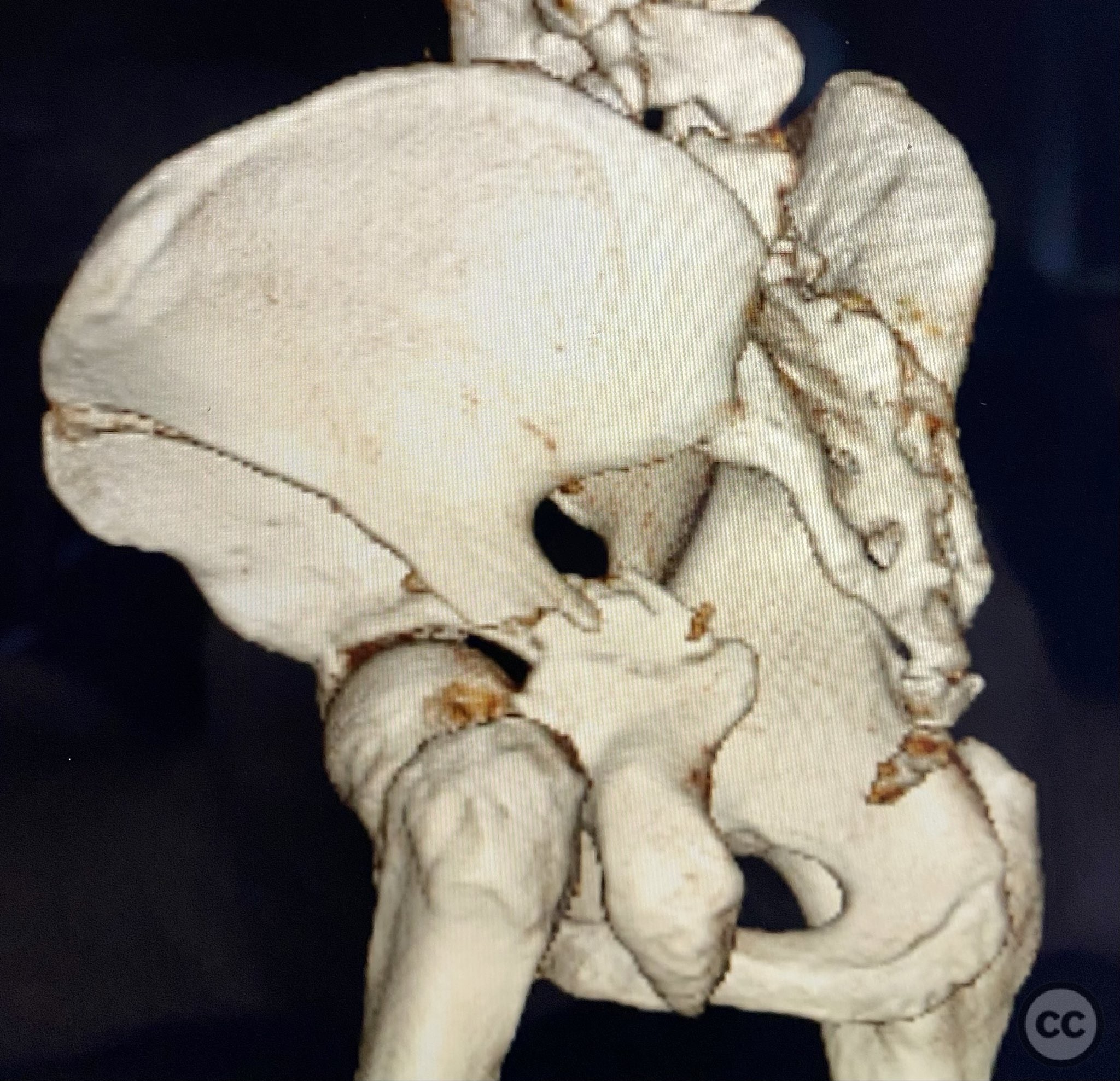
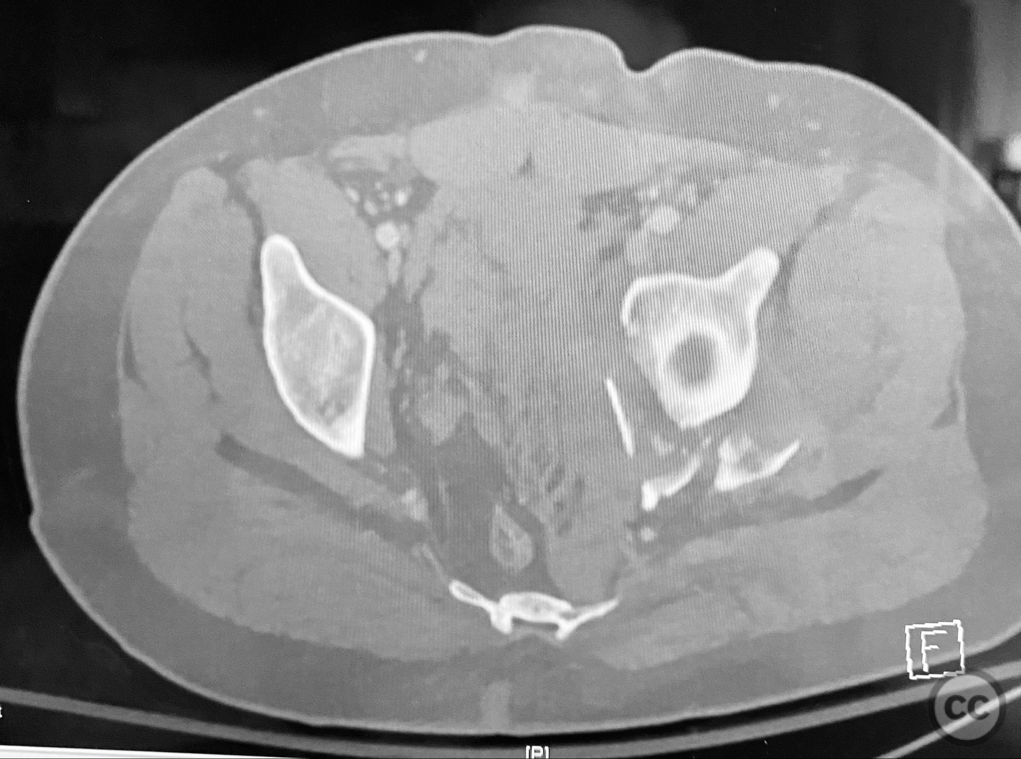
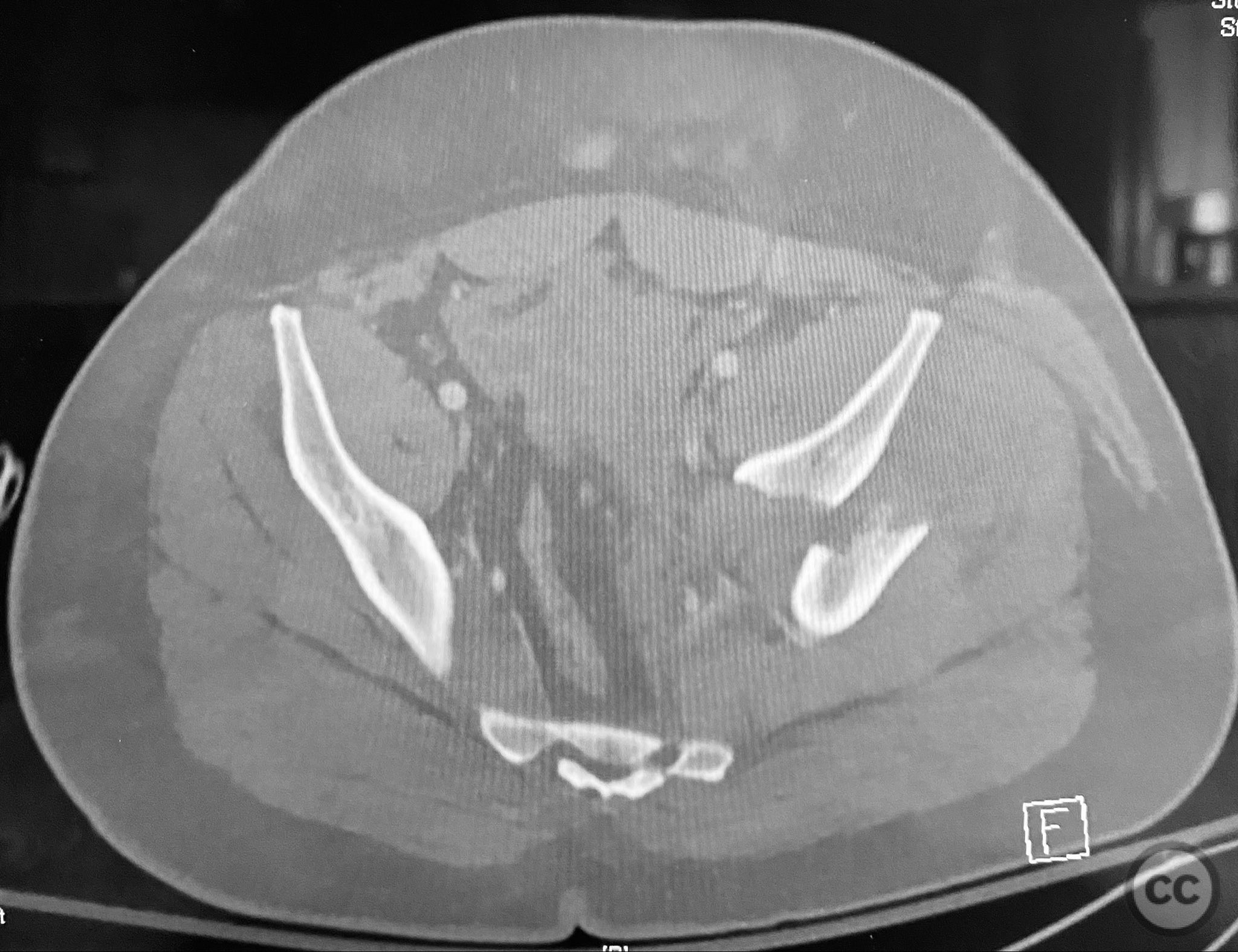
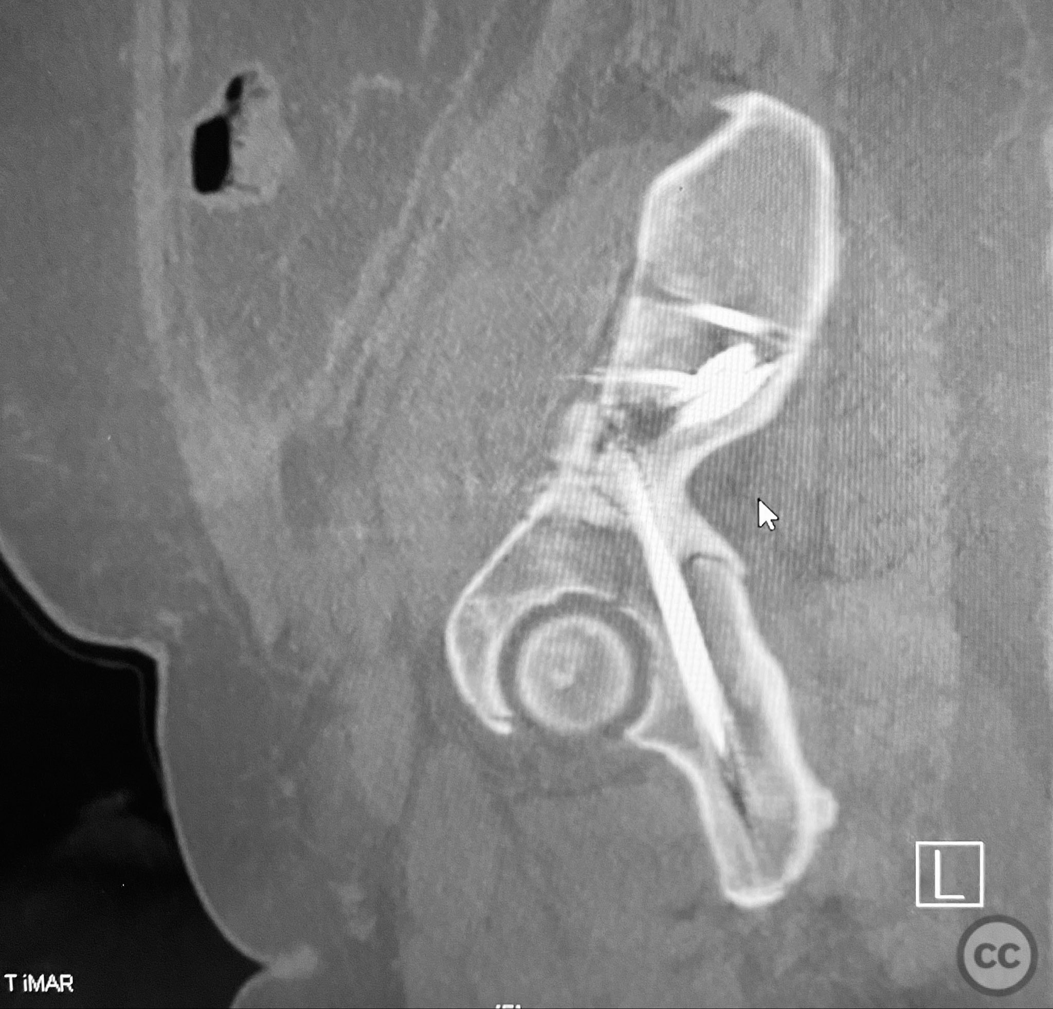
Clinical and radiological findings: A patient presented with a displaced associated both column acetabular fracture, characterized radiographically by the presence of the "spur sign" on the anteroposterior (AP) pelvis film, indicating the caudal aspect of the intact ilium exposed by medial displacement of the articular fragments. The spur was more clearly visualized on the obturator oblique rendered image. Axial computed tomography (CT) images demonstrated the intact ilium, medially displaced anterior column (AC) and posterior column (PC) fragments, and provided detailed visualization of surrounding soft tissues, including vascular structures enhanced by contrast. Multiplanar CT reconstructions (axial, sagittal, coronal) and 3D renderings were utilized for comprehensive fracture and soft tissue assessment. The patient’s overall clinical condition was a significant factor in surgical planning.
Preoperative Plan
Planning remarks: The preoperative plan involved open reduction and internal fixation (ORIF) of the anterior column via the ilioinguinal approach, utilizing two separate windows for exposure and reduction. Given the patient’s clinical status, a staged approach was selected: initial AC reduction and fixation, with planned subsequent percutaneous reduction and fixation of the PC. Implant positioning during the first operation was planned to avoid obstructing subsequent percutaneous PC fixation.
Surgical Discussion
Patient positioning: The patient was positioned supine on a radiolucent operating table to facilitate anterior pelvic access and intraoperative fluoroscopic imaging.
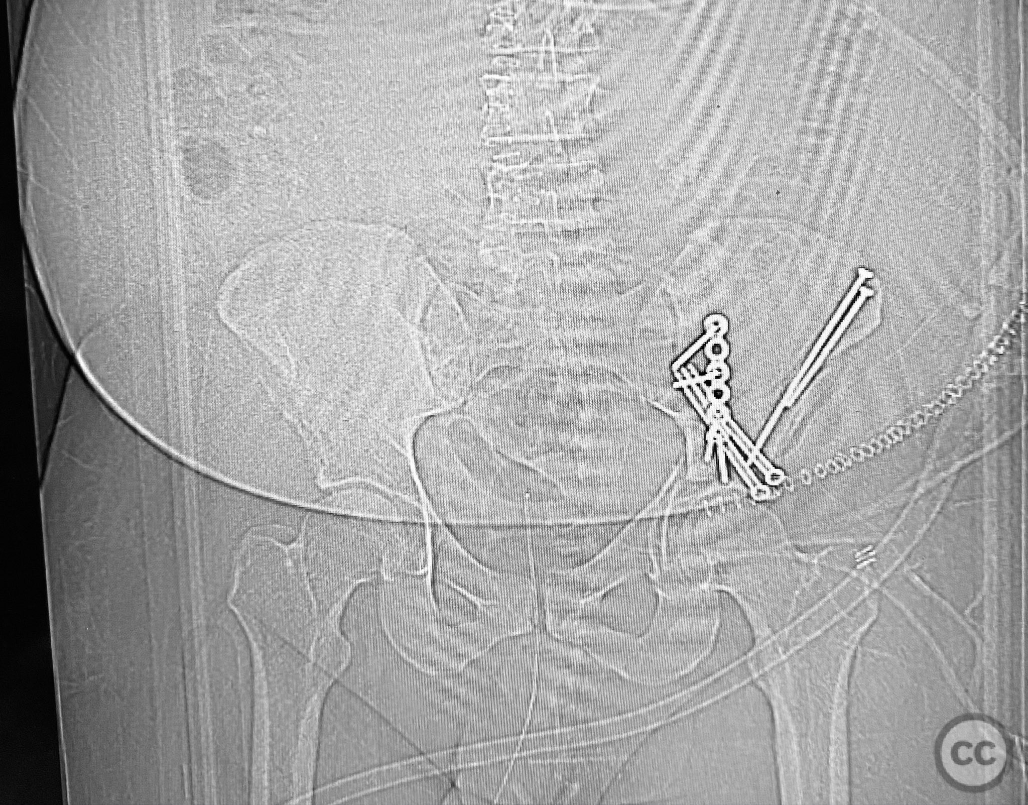
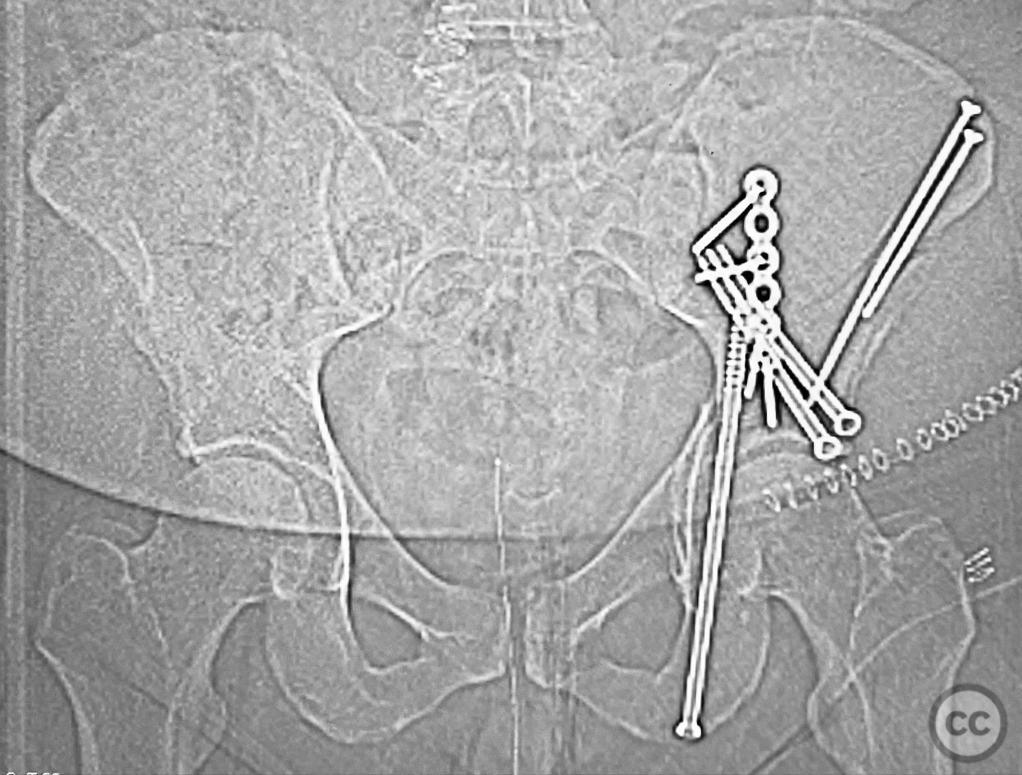
Anatomical surgical approach: A classical ilioinguinal approach was performed, with two windows developed: the lateral window between the musculus sartorius and musculus tensor fasciae latae, and the middle window between the musculus iliopsoas and external iliac vessels. Subcutaneous dissection was performed to create two opposing slabs of adipose tissue ("walls of fat"), minimizing random subcutaneous incisions. The anterior column fragment was exposed, reduced anatomically, and fixed under direct visualization. At a subsequent stage, percutaneous reduction and fixation of the posterior column were performed under fluoroscopic guidance, with careful trajectory planning to avoid interference from previously placed AC implants.
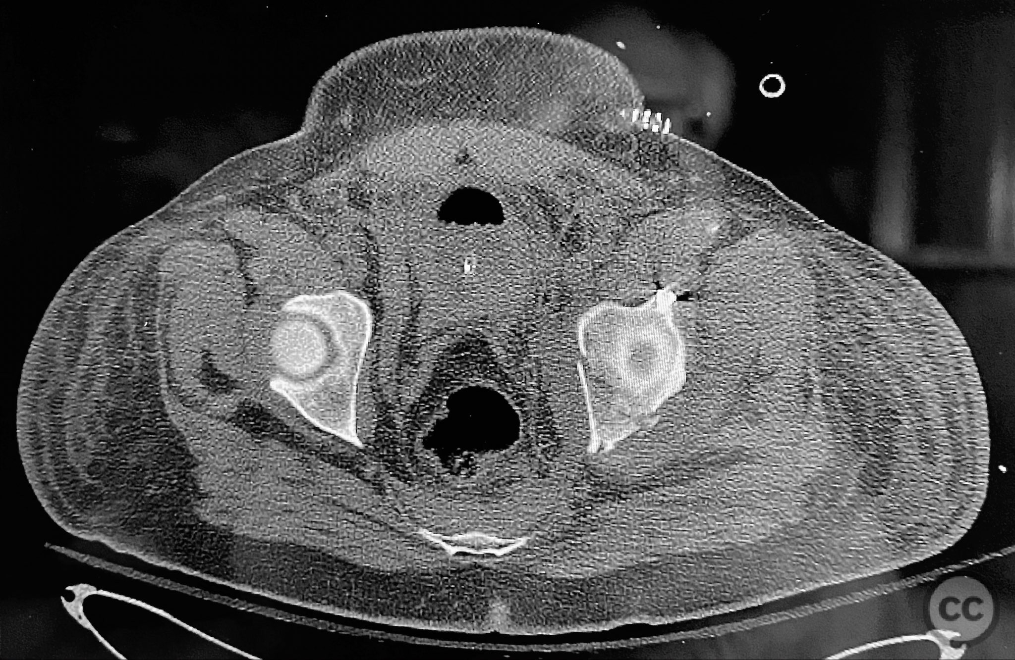
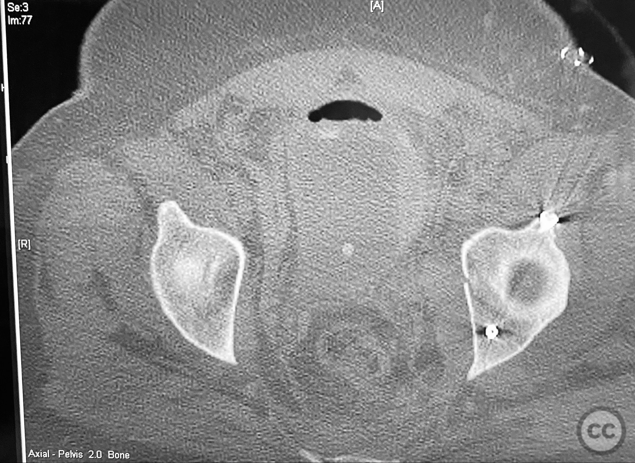
Operative remarks:The operative sequence was dictated by the patient’s overall clinical status, necessitating staged management. Intraoperative attention was paid to implant placement during AC fixation to preserve access for subsequent percutaneous PC fixation. Postoperative CT confirmed satisfactory reduction and fixation of both columns, with appropriate implant positioning and no evidence of neurovascular compromise or significant soft tissue complications.
Postoperative protocol: Early passive range of motion exercises were initiated postoperatively. Weight bearing was restricted according to fracture stability and healing progression, with toe-touch weight bearing for 6–8 weeks, followed by gradual advancement to partial and then full weight bearing as radiographic union permitted.
Follow up: Not specified
Orthopaedic implants used: 3.5 mm reconstruction plates (anterior column), cannulated screws (posterior column, percutaneous technique)
Search for Related Literature


Industry Sponsership
contact us for advertising opportunities
Article viewed 203 times
12 Sep 2025
Add to Bookmarks
Full Citation
Cite this article:
Routt, ML. (2025). Associated Both Column Acetabular Fracture: Sequential ORIF via Ilioinguinal Windows and Percutaneous Posterior Column Fixation. Journal of Orthopaedic Surgery and Traumatology. Case Report 30132427 Published Online Sep 12 2025.