 for discussion wrapped into one case_ ._1.jpg)
 for discussion wrapped into one case_ ._1.jpg)
 for discussion wrapped into one case_ ._3.jpg)
 for discussion wrapped into one case_ ._2.jpg)
 for discussion wrapped into one case_ ._(.jpg)
 for discussion wrapped into one case_ ._4.jpg)
 for discussion wrapped into one case_ ._5.jpg)
AO/OTA 43-C3 Open Pilon Fracture with Medial Tension Failure
Score and Comment on this Case
Clinical Details
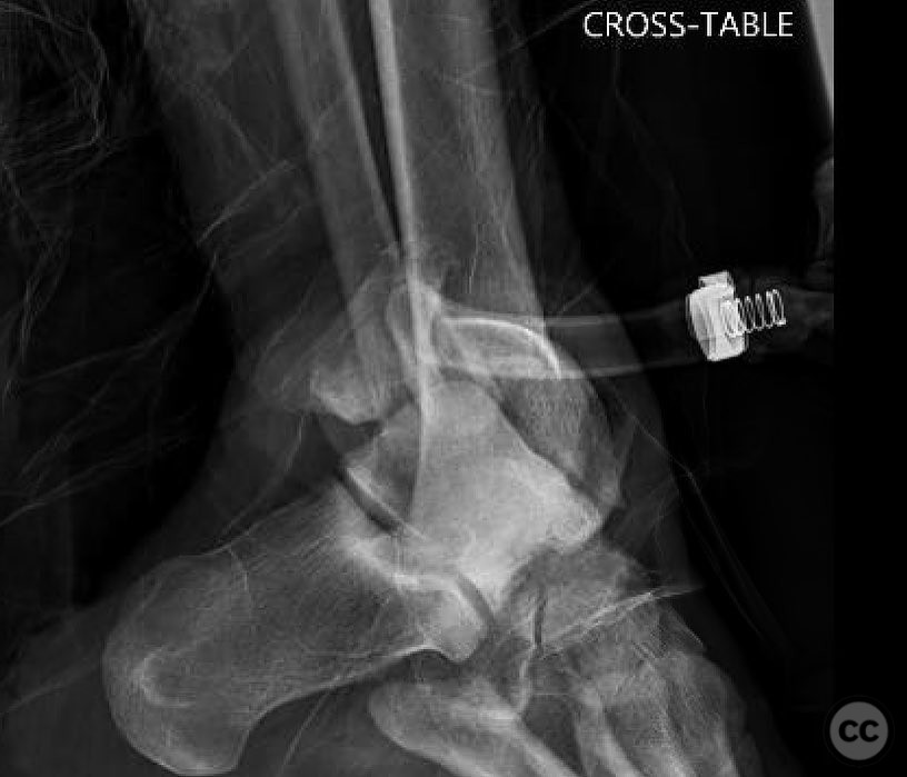
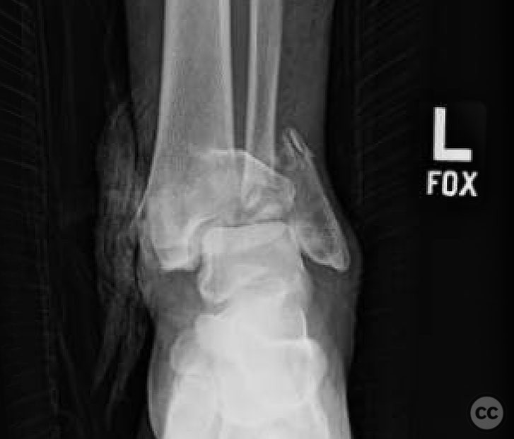
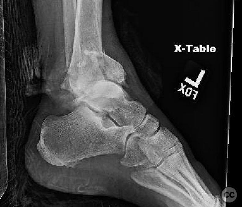
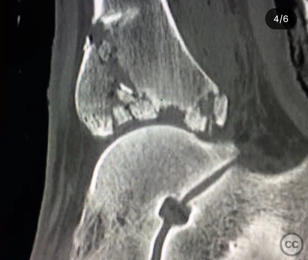
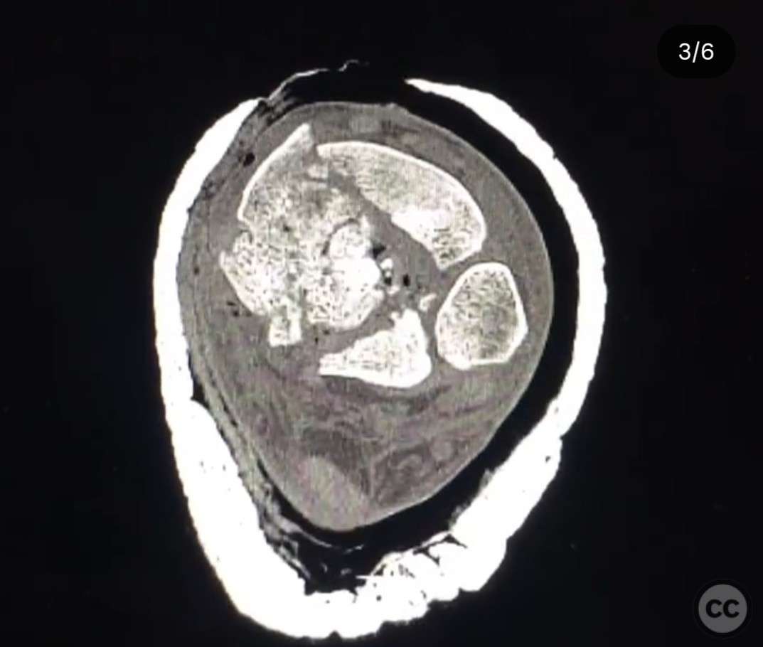
Clinical and radiological findings: A 23-year-old female involved in a motor vehicle accident, ejected from the vehicle, sustained a 3a open C-type pilon fracture. The injury presented with a 10cm transverse medial tension failure wound. Initial imaging included a CT scan performed prior to transfer, revealing a complex fracture pattern.
Preoperative Plan
Planning remarks: The preoperative plan involved initial debridement and external fixation through the traumatic medial wound, addressing the medial fracture components. Definitive fixation was planned in stages: first, a posterolateral approach for Volkmann fragment and fibula fixation; second, a small posteromedial approach for posteromedial fragments; and finally, an anterolateral approach for central joint injury and Chaput fragment fixation.
Surgical Discussion
Patient positioning: Prone positioning was utilized for the posterolateral and posteromedial approaches, allowing optimal access to the posterior and medial fracture components.
Anatomical surgical approach: The initial surgical approach involved using the traumatic medial wound for debridement and external fixation. The posterolateral approach was employed to address the Volkmann fragment and fibula, ensuring distal screws were contained within the Volkmann fragment. A small posteromedial incision facilitated fixation of the posteromedial components. The final stage utilized an anterolateral approach to address the central joint injury and Chaput fragment.
Operative remarks:The surgeon emphasized the importance of avoiding an anteromedial approach due to the transverse open wound location. Fixation of the medial side was completed during initial debridement to prevent further intervention in that area. Care was taken to ensure distal screws in the Volkmann fragment did not interfere with subsequent anterior reduction.
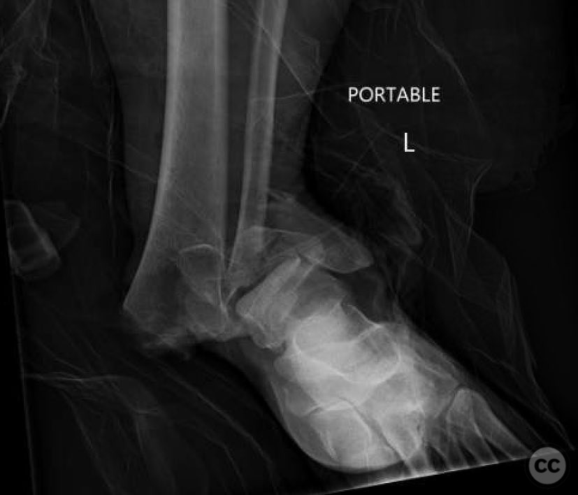
Postoperative protocol: Postoperative rehabilitation included non-weight bearing initially, progressing to weight bearing as tolerated over several months. At four months postoperatively, weight-bearing lateral radiographs demonstrated maintained fracture alignment.
Follow up: Not specified
Orthopaedic implants used: External fixator, low-profile plates for Volkmann and fibula fixation, screws for Chaput and central joint stabilization.
Search for Related Literature

orthopaedic_trauma
- United States , Seattle
- Area of Specialty - General Trauma
- Position - Specialist Consultant

Industry Sponsership
contact us for advertising opportunities
Article viewed 277 times
25 Jul 2025
Add to Bookmarks
Full Citation
Cite this article:
Surname, Initial. (2025). AO/OTA 43-C3 Open Pilon Fracture with Medial Tension Failure. Journal of Orthopaedic Surgery and Traumatology. Case Report 29611077 Published Online Jul 25 2025.