Subtrochanteric Femur Fracture with LC1 Pelvic Ring Injury in a Cyclist
Score and Comment on this Case
Clinical Details
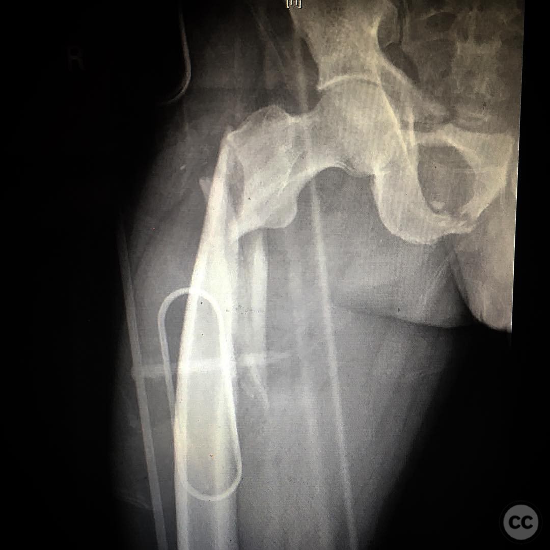
Clinical and radiological findings: A 36-year-old male cyclist was involved in a collision with a car, resulting in a subtrochanteric femur fracture and an LC1 pelvic ring injury. The patient presented with significant pain and inability to bear weight on the affected limb. Radiographic evaluation confirmed the presence of a subtrochanteric femur fracture, classified as AO/OTA 32-A3, and an LC1 pelvic ring injury, characterized by unilateral sacral compression without disruption of the posterior arch.
Preoperative Plan
Planning remarks: The preoperative plan involved a lateral approach to the femur, utilizing a flattop table for optimal access and visualization. The surgical strategy included attempting a percutaneous reduction of the subtrochanteric fracture, with the option to perform an open reduction if necessary. A reconstruction type intramedullary nail was selected for fixation.
Surgical Discussion
Patient positioning: The patient was positioned laterally on a flattop operating table to facilitate access to the proximal femur and allow for accurate reduction and fixation.
Anatomical surgical approach: A lateral approach to the femur was employed, involving a small incision over the greater trochanter. The vastus lateralis muscle was retracted anteriorly to expose the proximal femur. Percutaneous techniques were initially attempted for fracture reduction, with open reduction performed as needed.
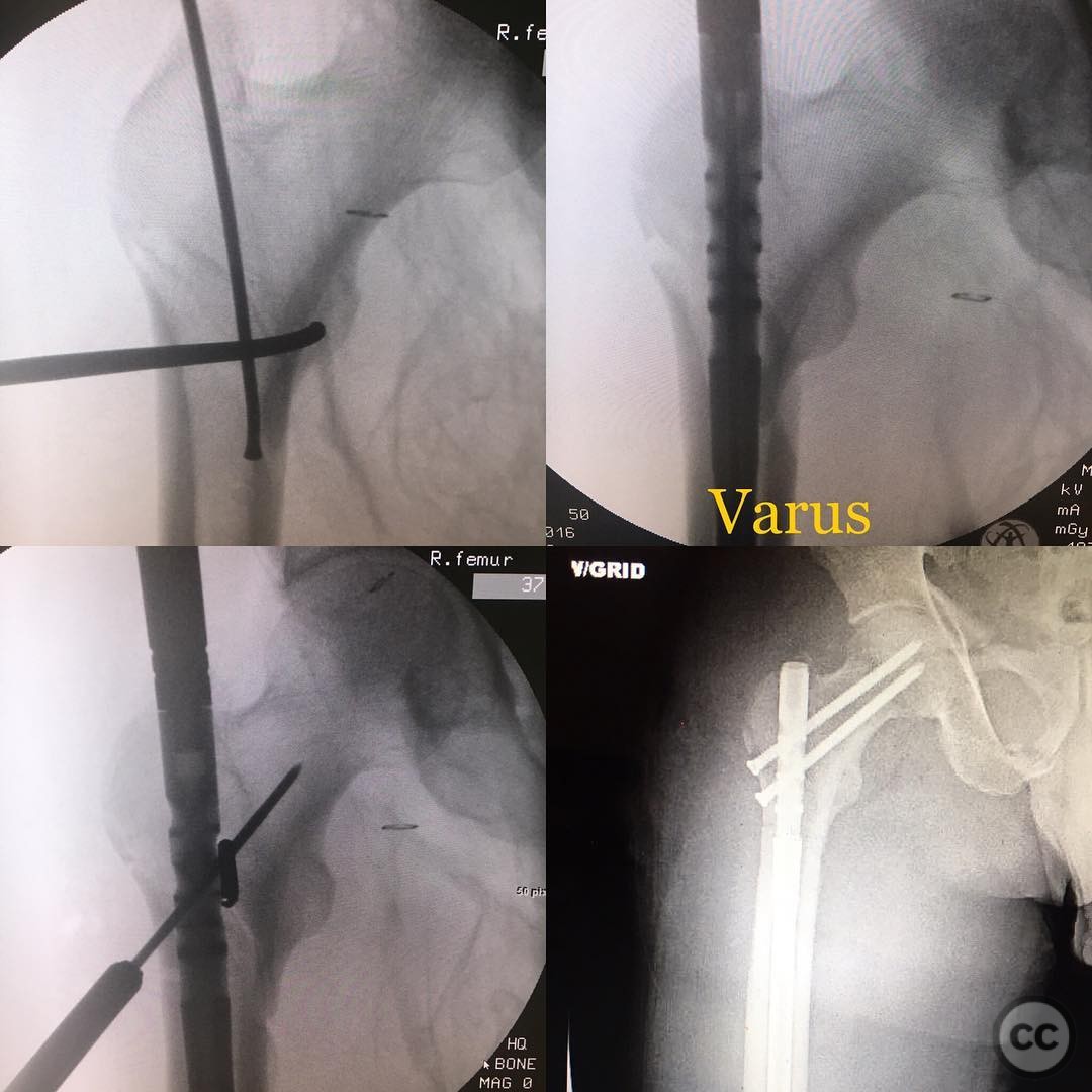
Operative remarks:During the procedure, an initial attempt at nail insertion resulted in a varus deformity due to a too lateral start site. To address this, the intramedullary nail was removed, and a blocking bit was placed to guide proper alignment. This adjustment allowed for maintenance of a high-quality reduction during subsequent nail placement and screw fixation.
Postoperative protocol: Postoperatively, the patient was advised to adhere to partial weight-bearing on the affected limb for six weeks, followed by progressive weight-bearing as tolerated. Physical therapy focused on range of motion exercises and strengthening of the surrounding musculature.
Follow up: Not specified.
Orthopaedic implants used: Reconstruction type intramedullary nail, blocking bit.
Search for Related Literature

orthopaedic_trauma
- United States , Seattle
- Area of Specialty - General Trauma
- Position - Specialist Consultant

Industry Sponsership
contact us for advertising opportunities
Article viewed 429 times
05 Aug 2025
Add to Bookmarks
Full Citation
Cite this article:
Surname, Initial. (2025). Subtrochanteric Femur Fracture with LC1 Pelvic Ring Injury in a Cyclist. Journal of Orthopaedic Surgery and Traumatology. Case Report 28799240 Published Online Aug 05 2025.