 _2.jpg)
 _1.jpg)
 _3.jpg)
 _4.jpg)
 _5.jpg)
 _6.jpg)
 _7.jpg)
 _8.jpg)
Posterior Wall Acetabular Fracture Dislocation with Cranial Extension
Score and Comment on this Case
Clinical Details
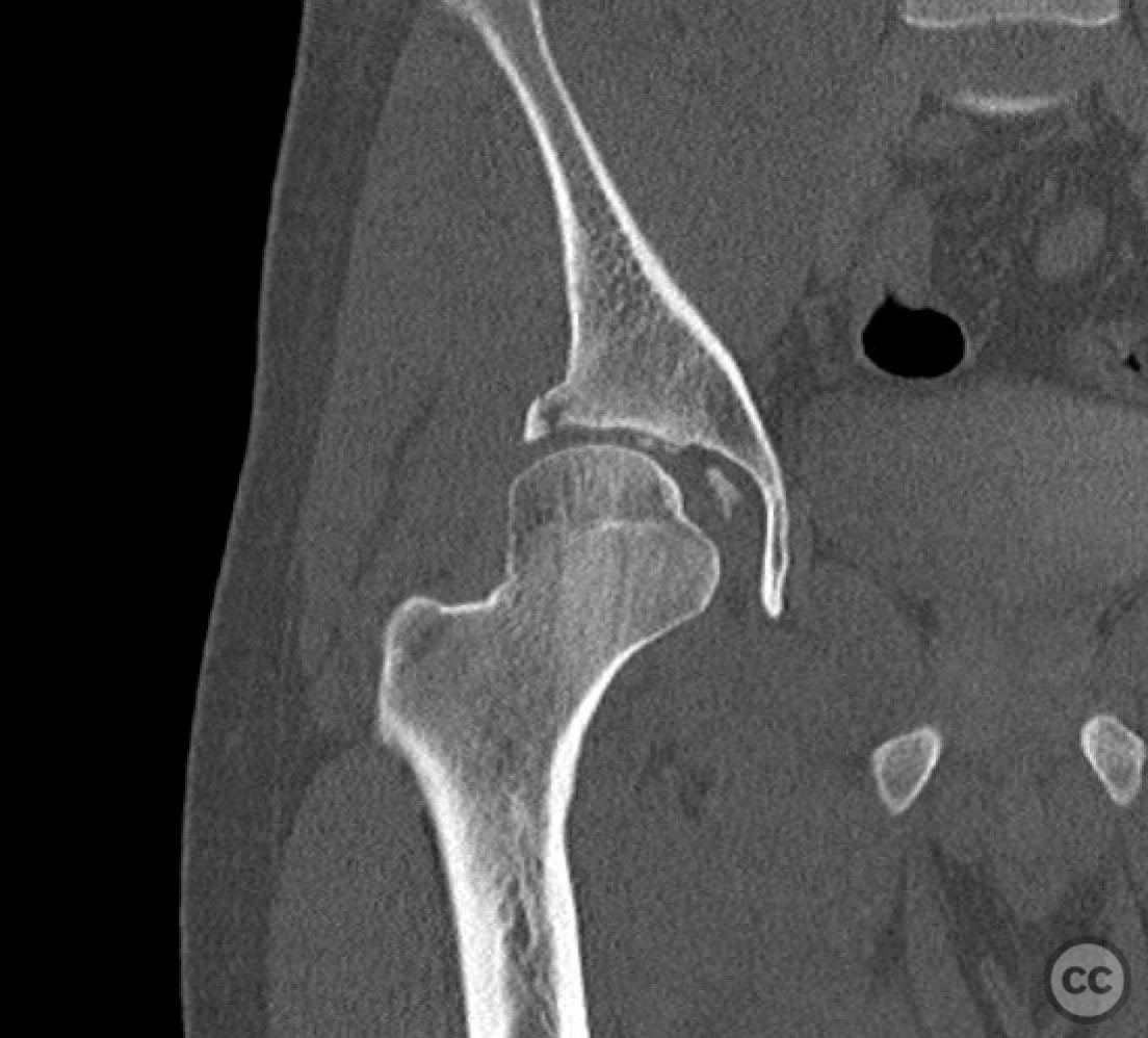
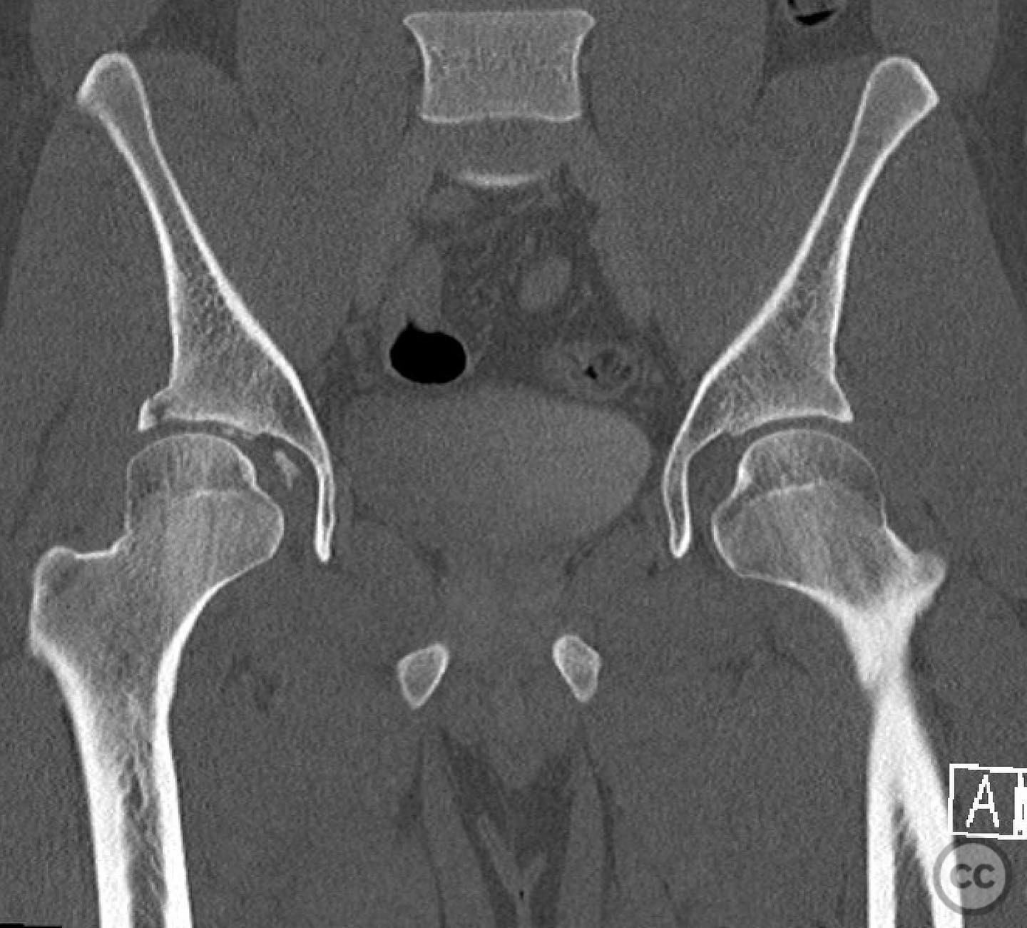
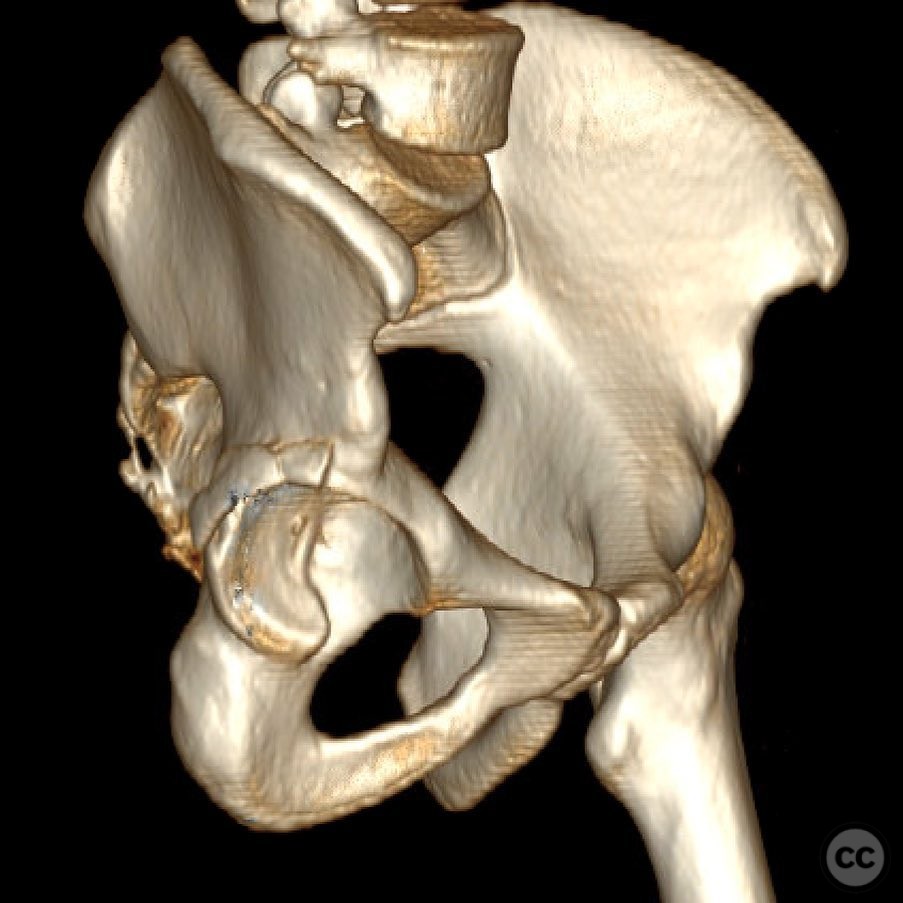
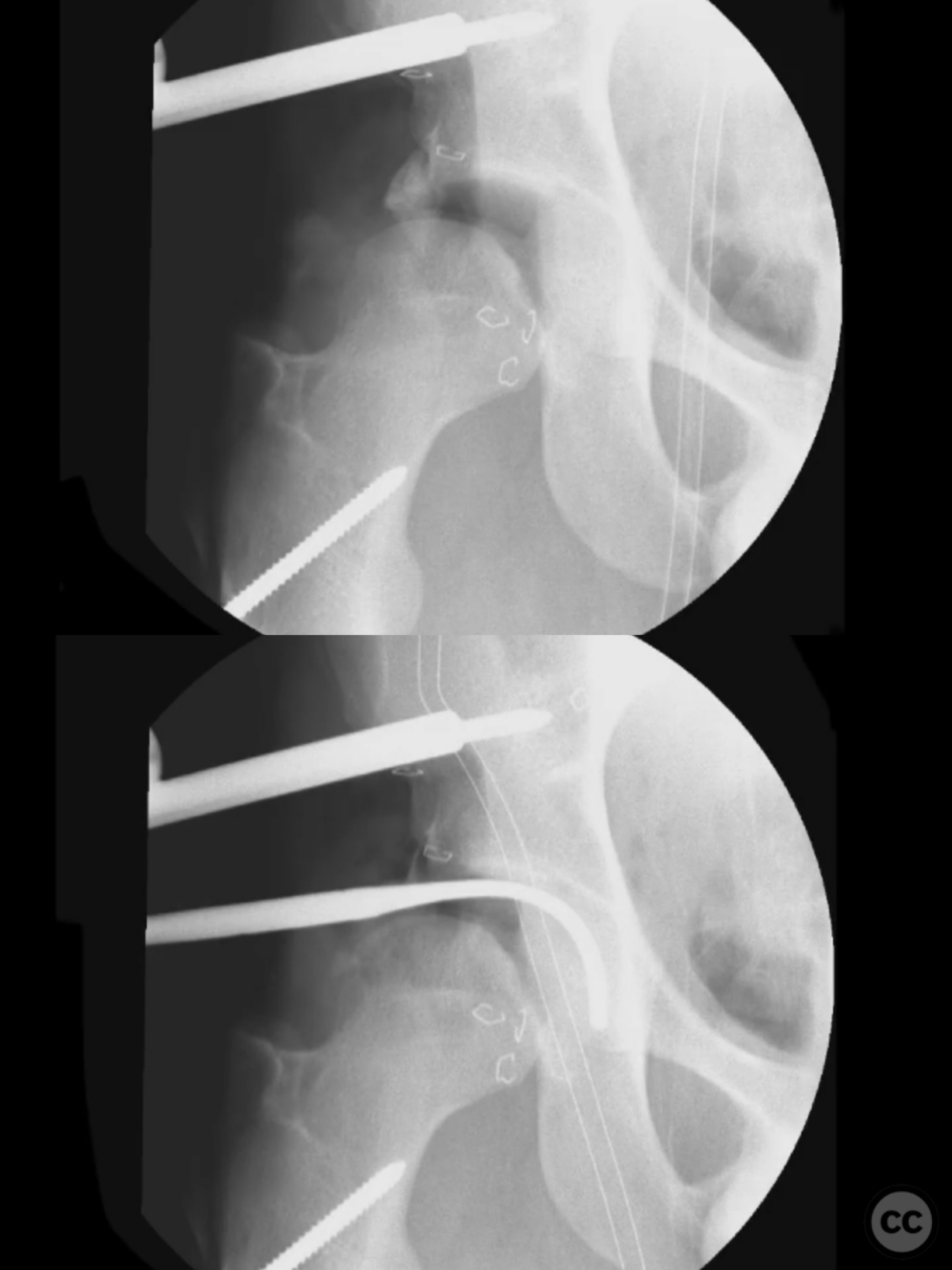
Clinical and radiological findings: A 22-year-old patient presented with a posterior wall acetabular fracture dislocation characterized by a very cranial extension with anterior involvement. The hip was noted to be slightly dysplastic, which may have contributed to the peripheral and cranial nature of the fracture pattern. Radiological assessment revealed cranial marginal impaction and free osteochondral fragments within the joint space.
Preoperative Plan
Planning remarks: The preoperative plan involved a trochanteric slide osteotomy to enhance anterior supra-acetabular visualization and minimize trauma to the gluteus medius and superior gluteal neurovascular structures. A Gibson approach was selected for optimal access, given the fracture's anterior extension, in conjunction with the osteotomy.
Surgical Discussion
Patient positioning: The patient was positioned laterally to facilitate the Gibson approach and trochanteric slide osteotomy.
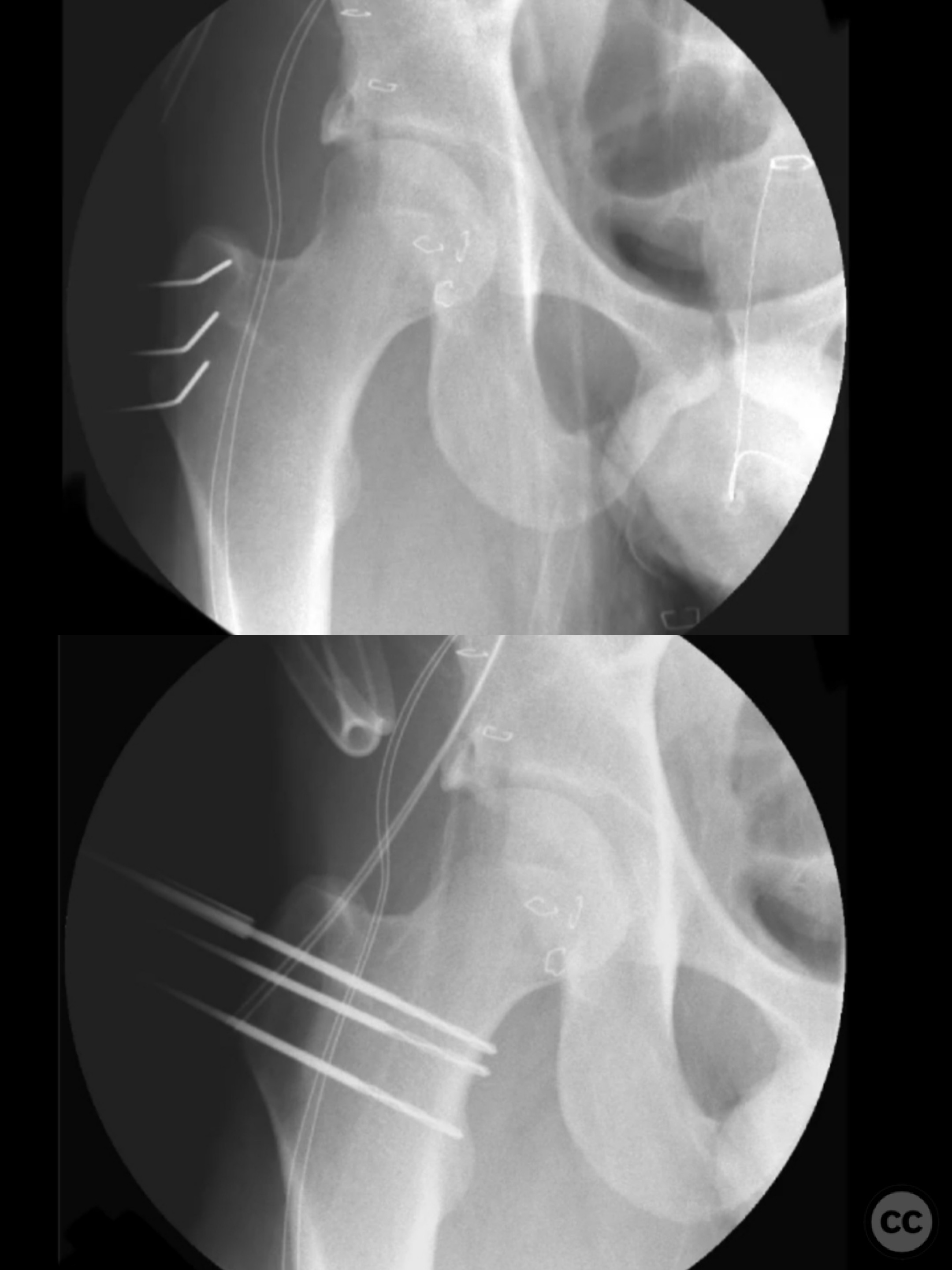
Anatomical surgical approach: The surgical approach involved a Gibson incision, with a trochanteric slide osteotomy performed to improve visualization and access to the anterior supra-acetabular region. This was followed by the use of a distractor and a Satinsky clamp to extract intra-articular fragments and address the caudal capsulolabral complex.
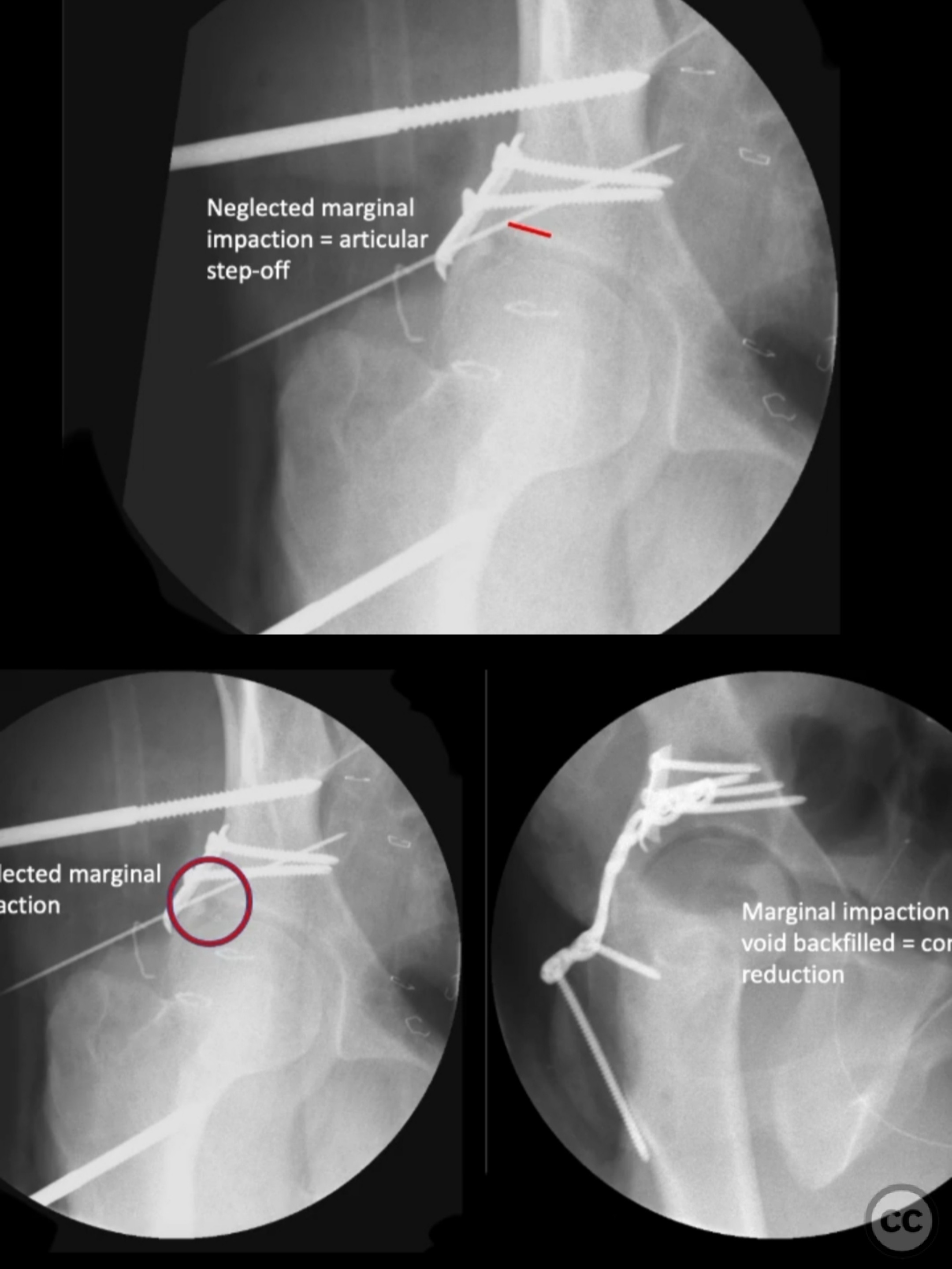
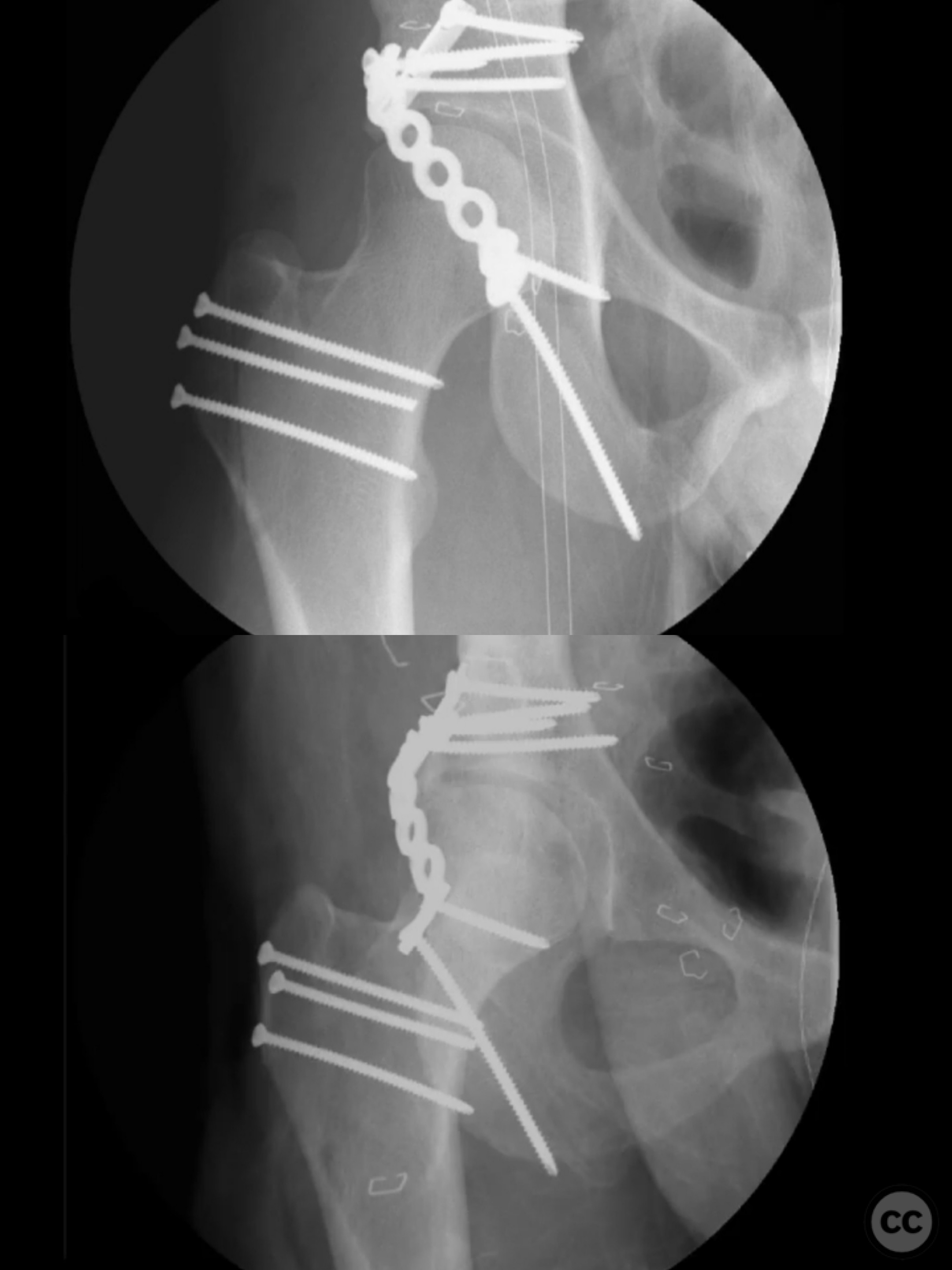
Operative remarks:During the procedure, marginal impaction was addressed to restore articular congruity. Initial fluoroscopic assessment revealed an articular step-off due to missed impaction, necessitating plate removal, identification, disimpaction, backfilling, and refixation of the impacted area. The capsulolabral complex was repaired using soft tissue anchors caudally, and a buttress plate was applied over a spring plate for reinforcement. For the osteotomy, k-wires were placed in the planned cut plane for fragment size control, and pre-drilling was performed for ease of repair.
Postoperative protocol: Not specified
Follow up: Not specified
Orthopaedic implants used: Buttress plate, spring plate, soft tissue anchors, k-wires
Search for Related Literature

orthopaedic_trauma
- United States , Seattle
- Area of Specialty - General Trauma
- Position - Specialist Consultant

Industry Sponsership
contact us for advertising opportunities
Article viewed 318 times
12 Jul 2025
Add to Bookmarks
Full Citation
Cite this article:
Surname, Initial. (2025). Posterior Wall Acetabular Fracture Dislocation with Cranial Extension. Journal of Orthopaedic Surgery and Traumatology. Case Report 28543731 Published Online Jul 12 2025.