Peri-Implant Failure in Subtrochanteric Femur Fracture with Diaphyseal Extension
Score and Comment on this Case
Clinical Details
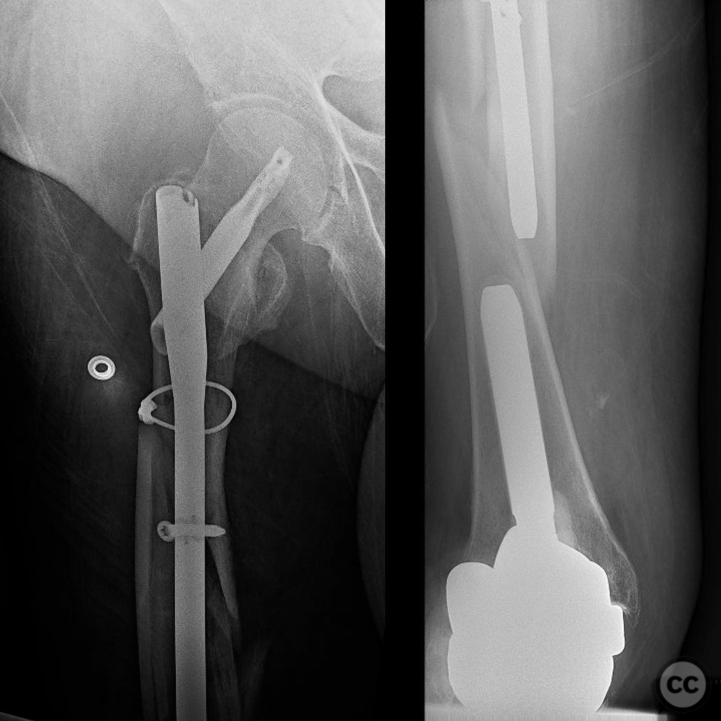
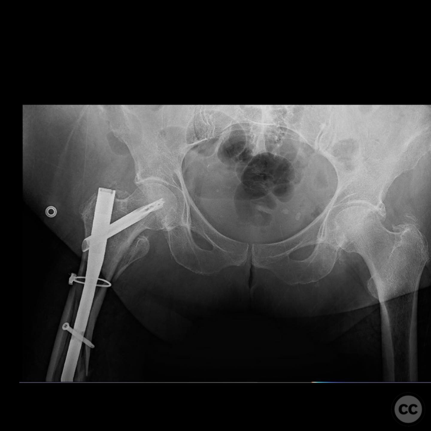
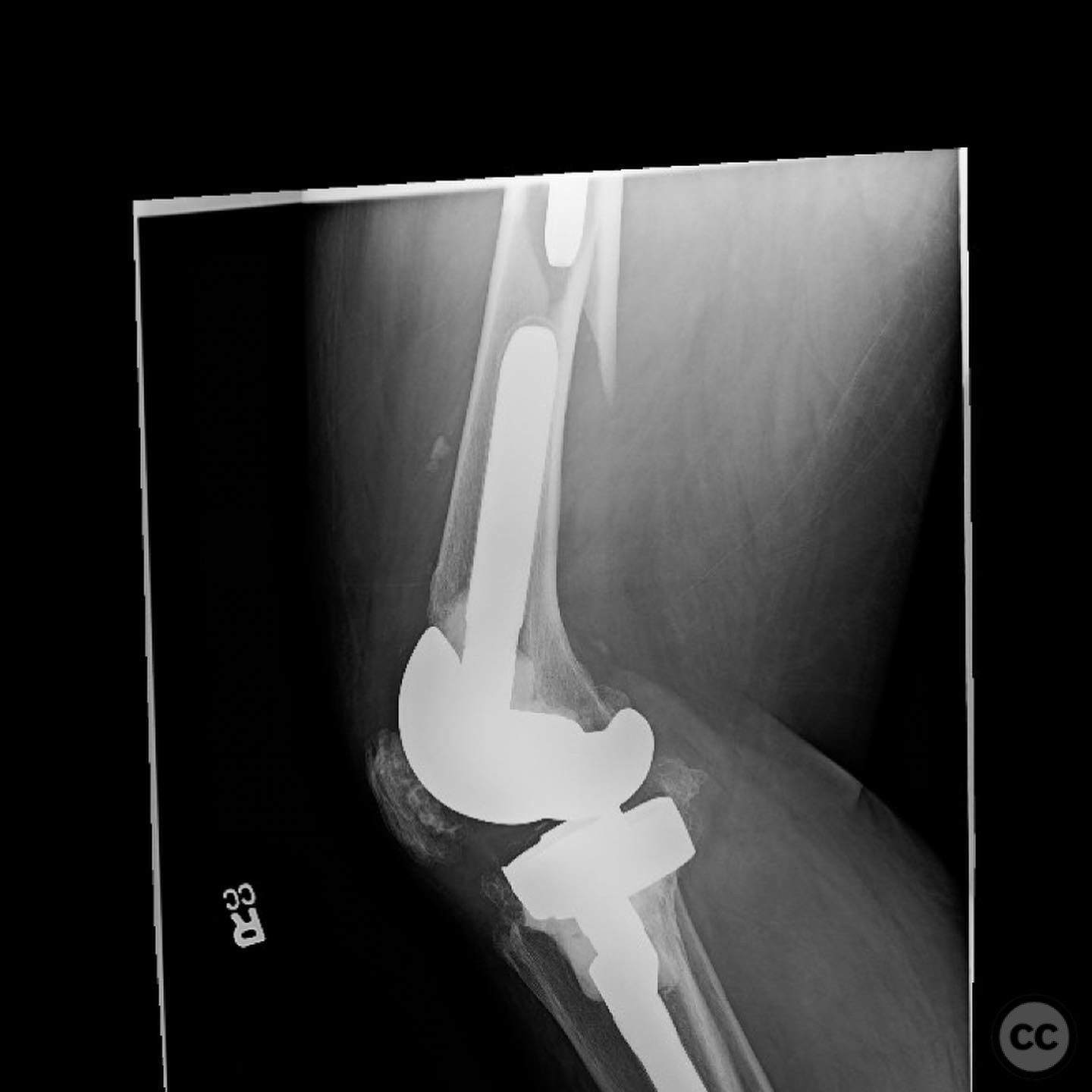
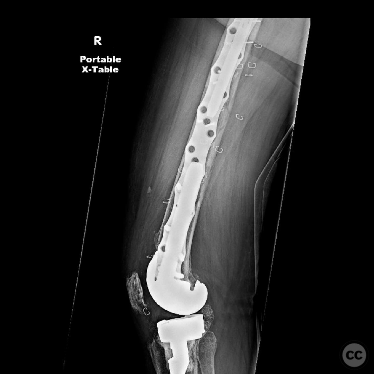
Clinical and radiological findings: A 68-year-old female presented to the emergency department following a twisting fall four weeks post-operatively from an initial open reduction and internal fixation of a subtrochanteric femur fracture. The initial injury, sustained two months prior, was a subtrochanteric fracture with diaphyseal extension above a stemmed femoral component. No initial injury films were available. The previous fixation involved a short intramedullary nail, which inadequately spanned the fracture site, with a single interlocking bolt traversing the fracture line. This led to mechanical failure and subsequent peri-implant fracture during early mobilization.
Preoperative Plan
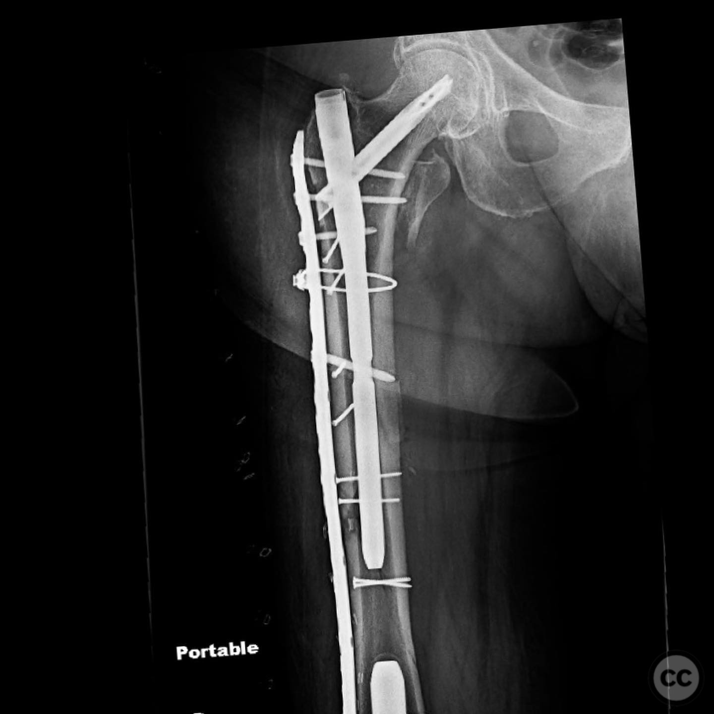
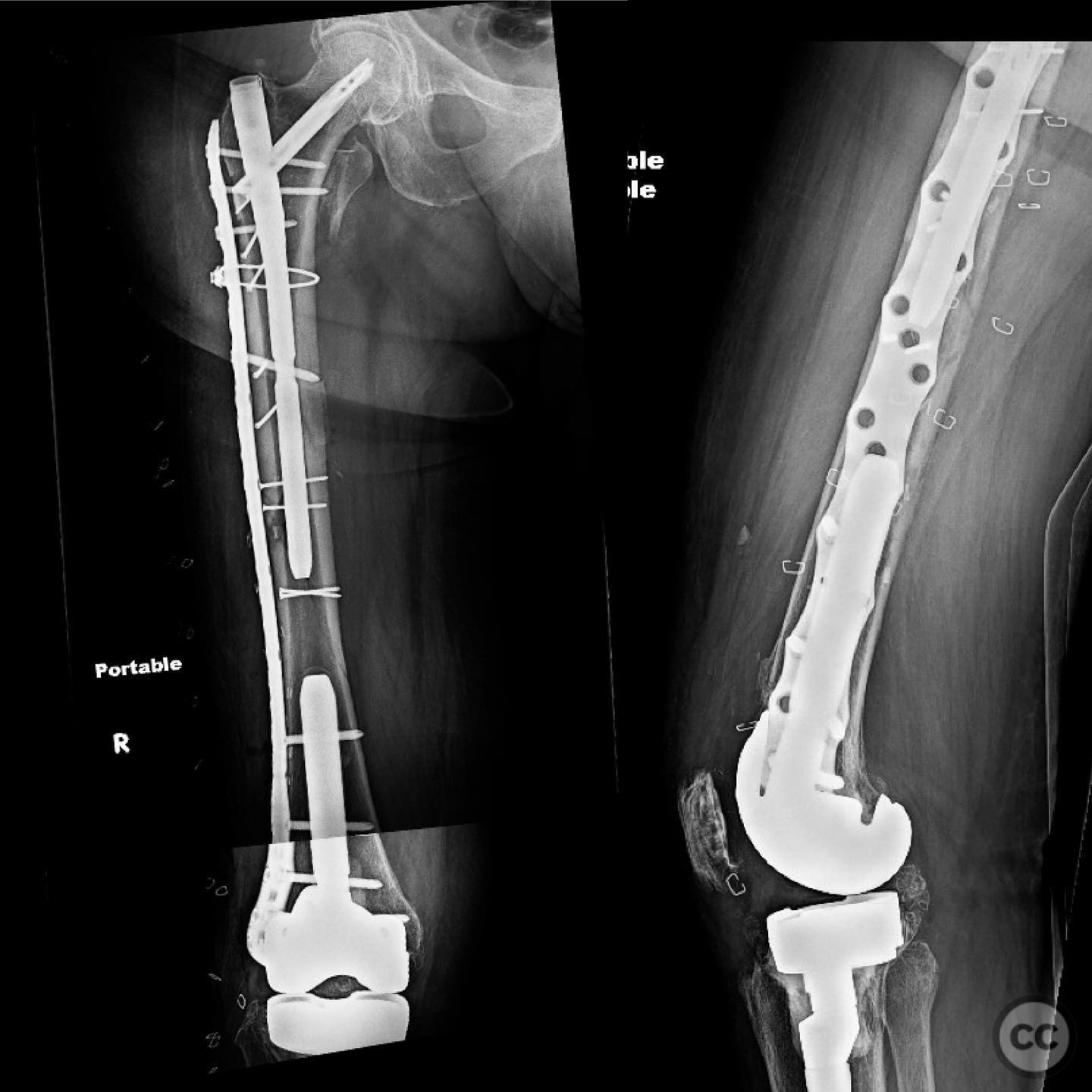
Planning remarks: The preoperative plan for revision surgery involved maintaining the existing intramedullary nail and augmenting fixation with a lateral plate. The strategy was to provide adequate stabilization by linking the plate to the nail using a 5mm locking screw for proximal fixation.
Surgical Discussion
Patient positioning: The patient was positioned laterally on the operating table to facilitate access to the lateral aspect of the femur for plating.
Anatomical surgical approach: A lateral approach to the femur was utilized. An incision was made along the lateral aspect of the thigh, extending from the greater trochanter distally. Subcutaneous tissues were dissected, and the fascia lata was incised to expose the lateral surface of the femur. Careful dissection around the existing nail was performed to allow for placement of the lateral plate.
Operative remarks:The surgeon noted that proximal fixation was challenging due to the presence of the stemmed femoral component and existing nail. The decision to link the plate and nail with a 5mm locking screw was critical in achieving stable fixation. This approach allowed for effective load sharing between the plate and nail, addressing the mechanical insufficiency of the initial construct.
Postoperative protocol: Postoperative rehabilitation included protected weight-bearing with crutches for six weeks, followed by gradual progression to full weight-bearing as tolerated. Range of motion exercises were initiated early to prevent joint stiffness.
Follow up: Not specified
Orthopaedic implants used: Long intramedullary nail, lateral femoral locking plate, 5mm locking screw.
Search for Related Literature

orthopaedic_trauma
- United States , Seattle
- Area of Specialty - General Trauma
- Position - Specialist Consultant

Industry Sponsership
contact us for advertising opportunities
Article viewed 181 times
11 Jul 2025
Add to Bookmarks
Full Citation
Cite this article:
Surname, Initial. (2025). Peri-Implant Failure in Subtrochanteric Femur Fracture with Diaphyseal Extension. Journal of Orthopaedic Surgery and Traumatology. Case Report 28414683 Published Online Jul 11 2025.