Complex Pediatric Upper Extremity Trauma with Supracondylar Humerus Fracture and Vascular Compromise
Score and Comment on this Case
Clinical Details
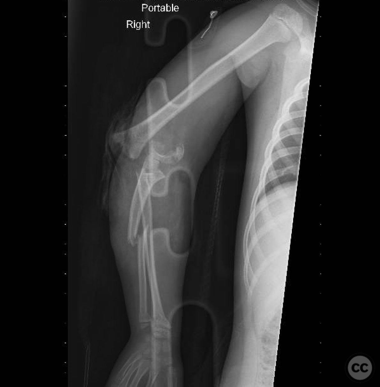
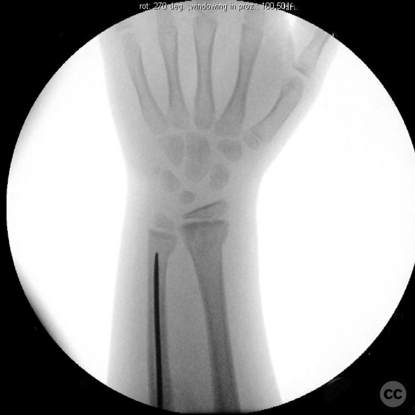
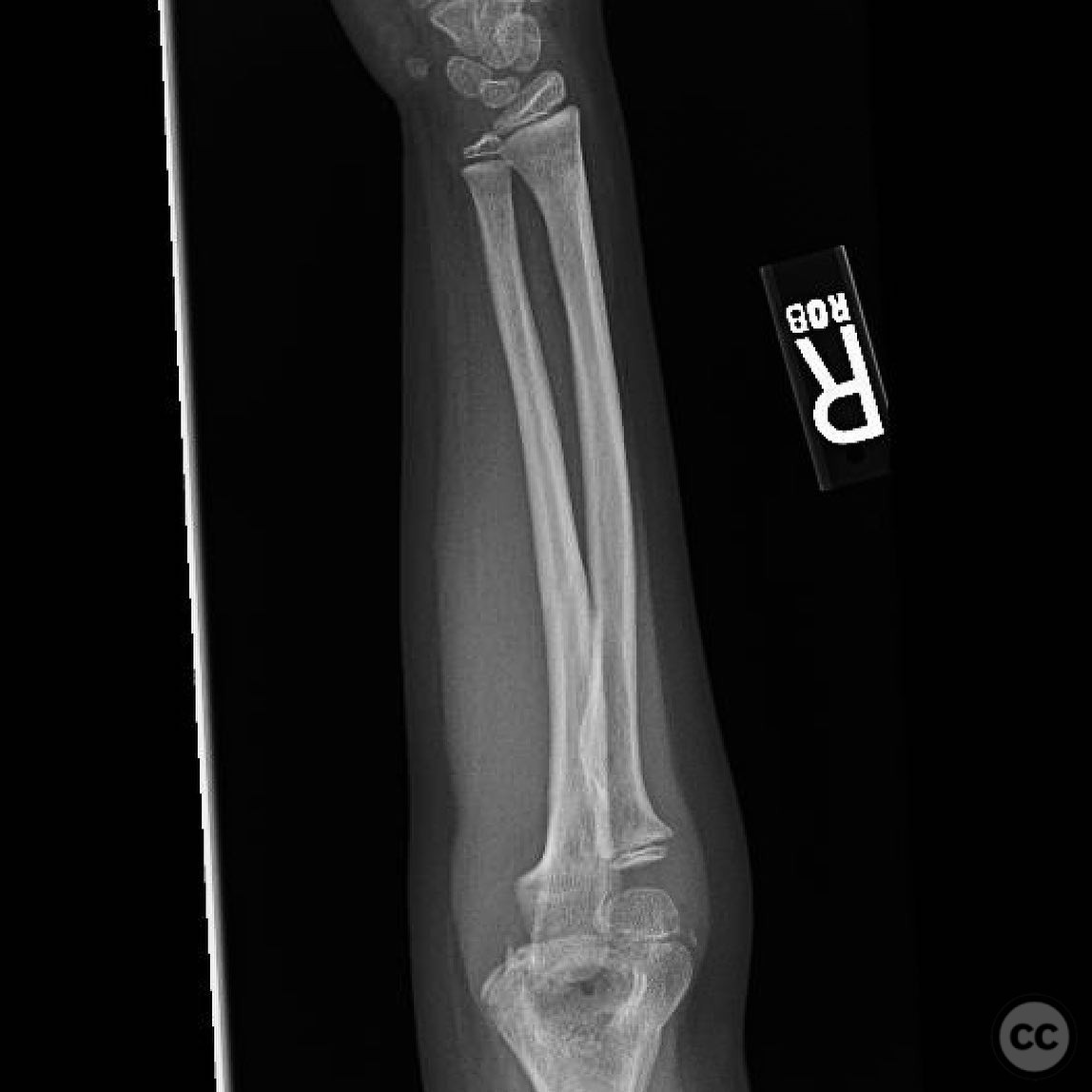
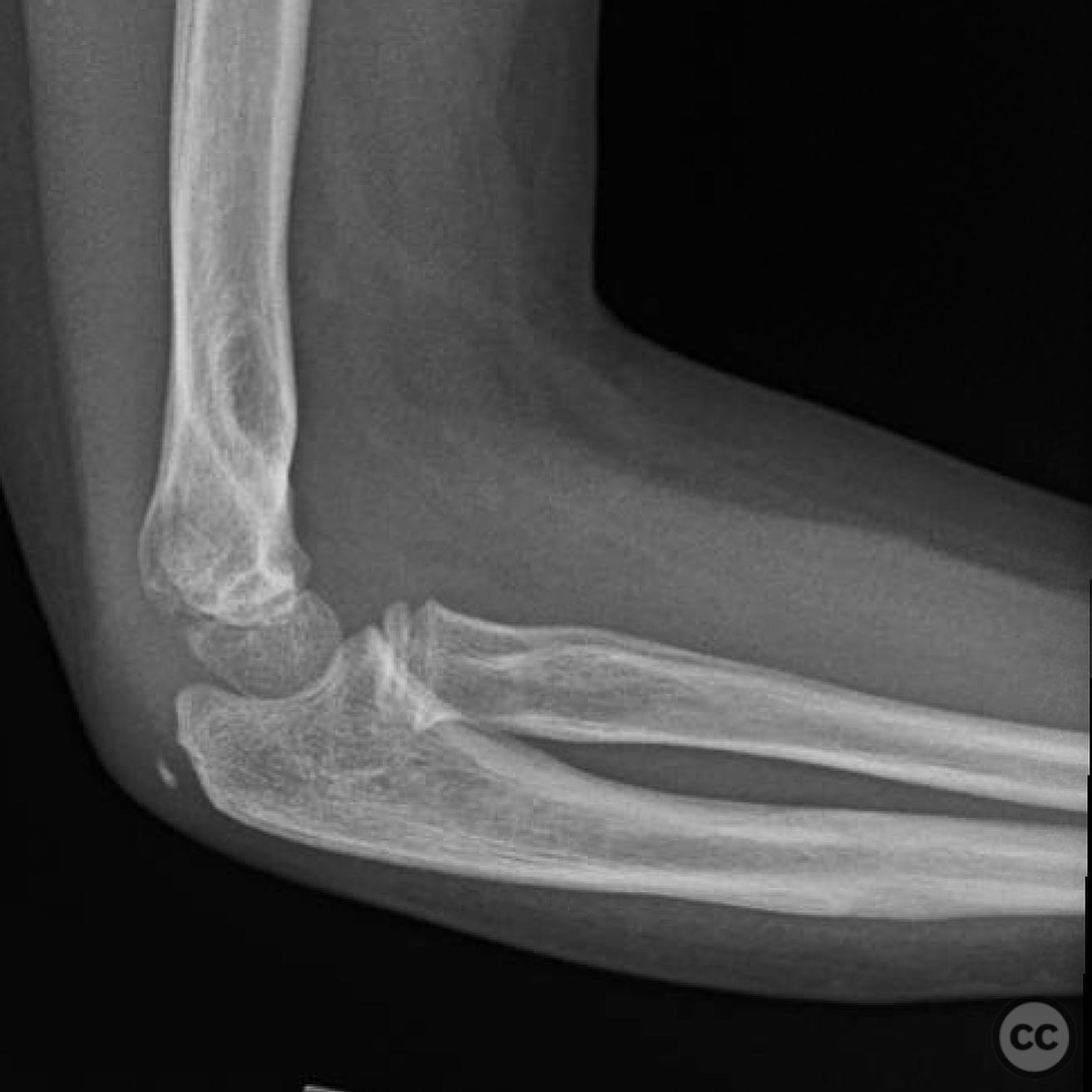
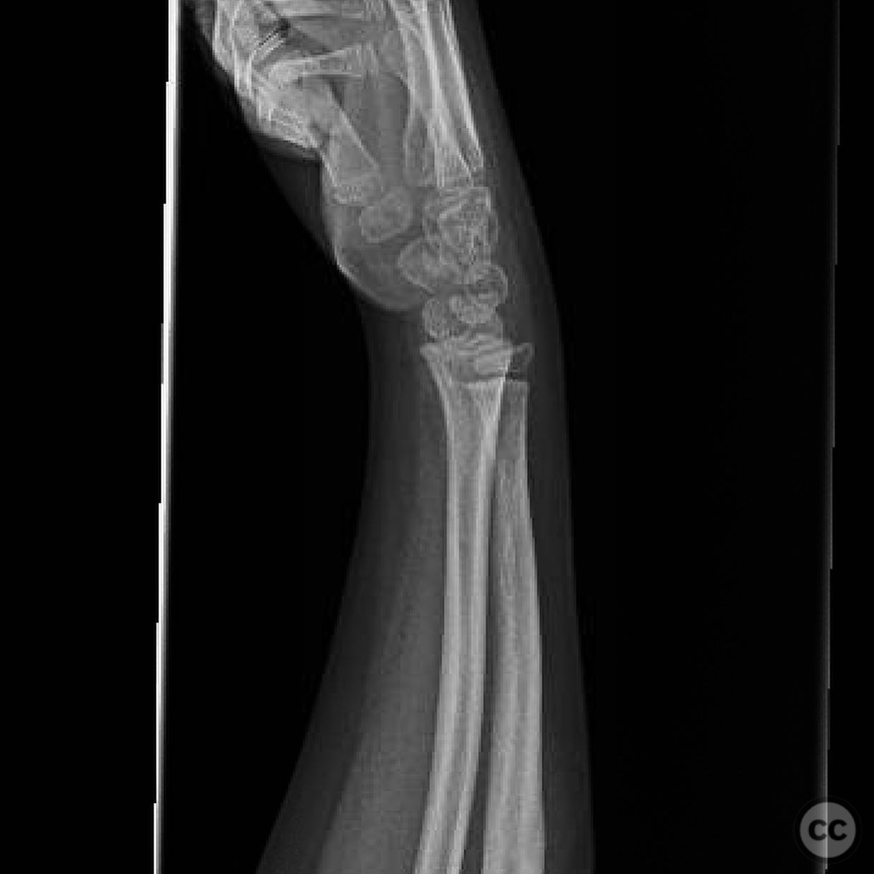
Clinical and radiological findings: A 5-year-old female sustained a fall from a two-story height onto concrete, resulting in an isolated upper extremity injury. Initial examination revealed an anterolateral open oblique wound measuring 4 cm and a 1 cm open dorsal ulnar wound at the level of the ulnar fracture. The arm was soft, and the hand appeared pinkish with a capillary refill time of 2-3 seconds, but no palpable pulse was detected. Motor and sensory examination was unreliable. Radiological assessment identified a Gartland type IV supracondylar humerus fracture, a radial neck fracture, a type I open ulna fracture, and closed distal radius and ulna fractures.
Preoperative Plan
Planning remarks: The preoperative plan involved urgent exploration of the open wounds to assess and catalog injured structures, followed by irrigation and debridement. The surgical strategy included stabilization of the ulna fracture with intramedullary nailing to provide a stable distal segment for subsequent reduction of the supracondylar humerus fracture. The radial neck fracture was to be addressed through manipulation via the traumatic wound.
Surgical Discussion
Patient positioning: The patient was positioned supine on the operating table with the affected arm extended on an arm board to facilitate access to the upper extremity.
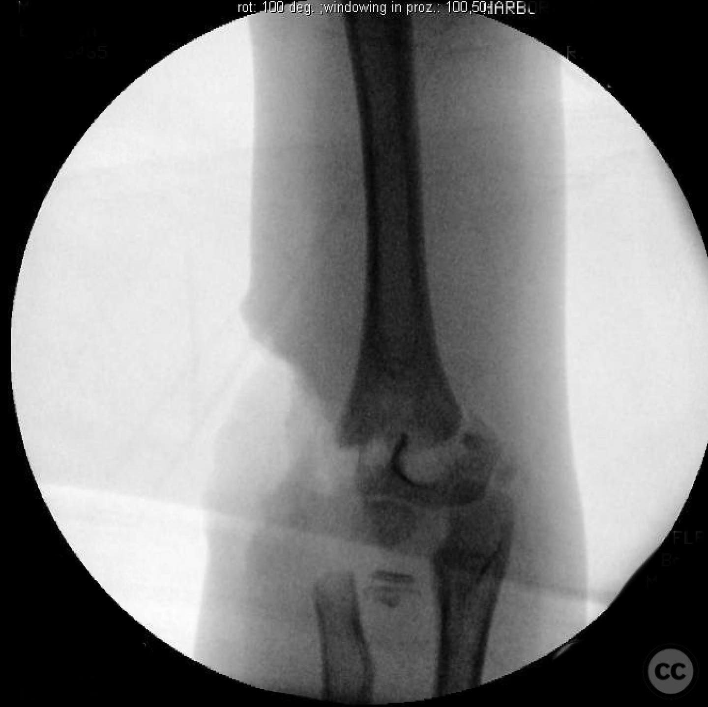
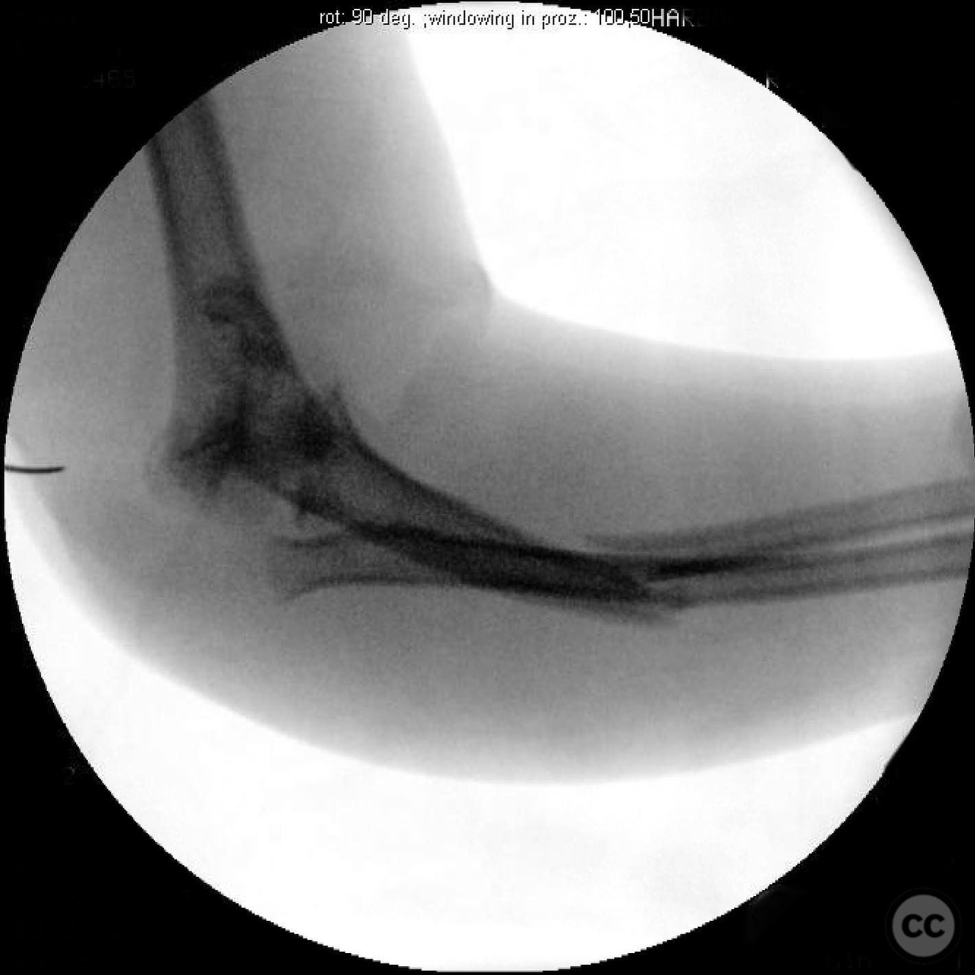
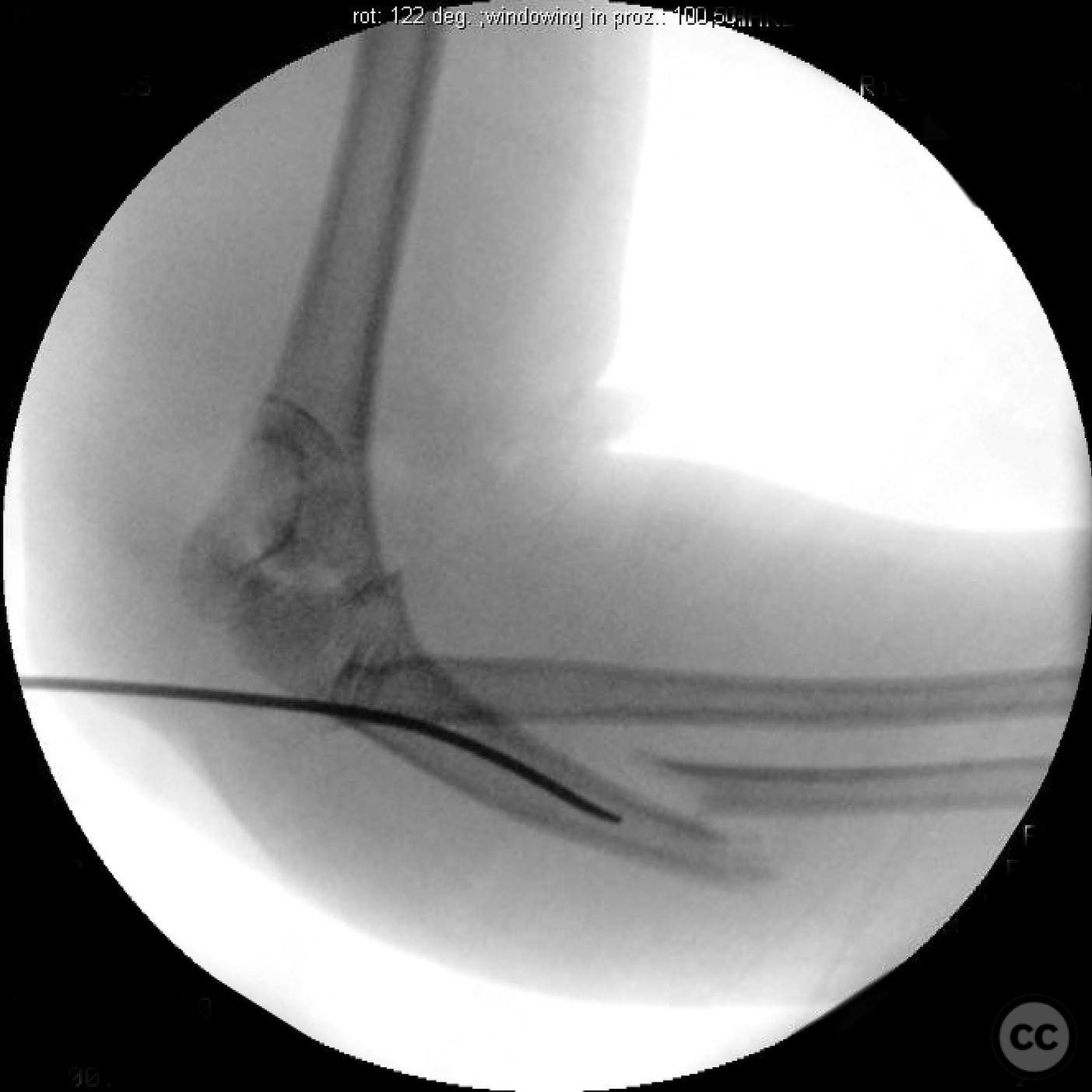
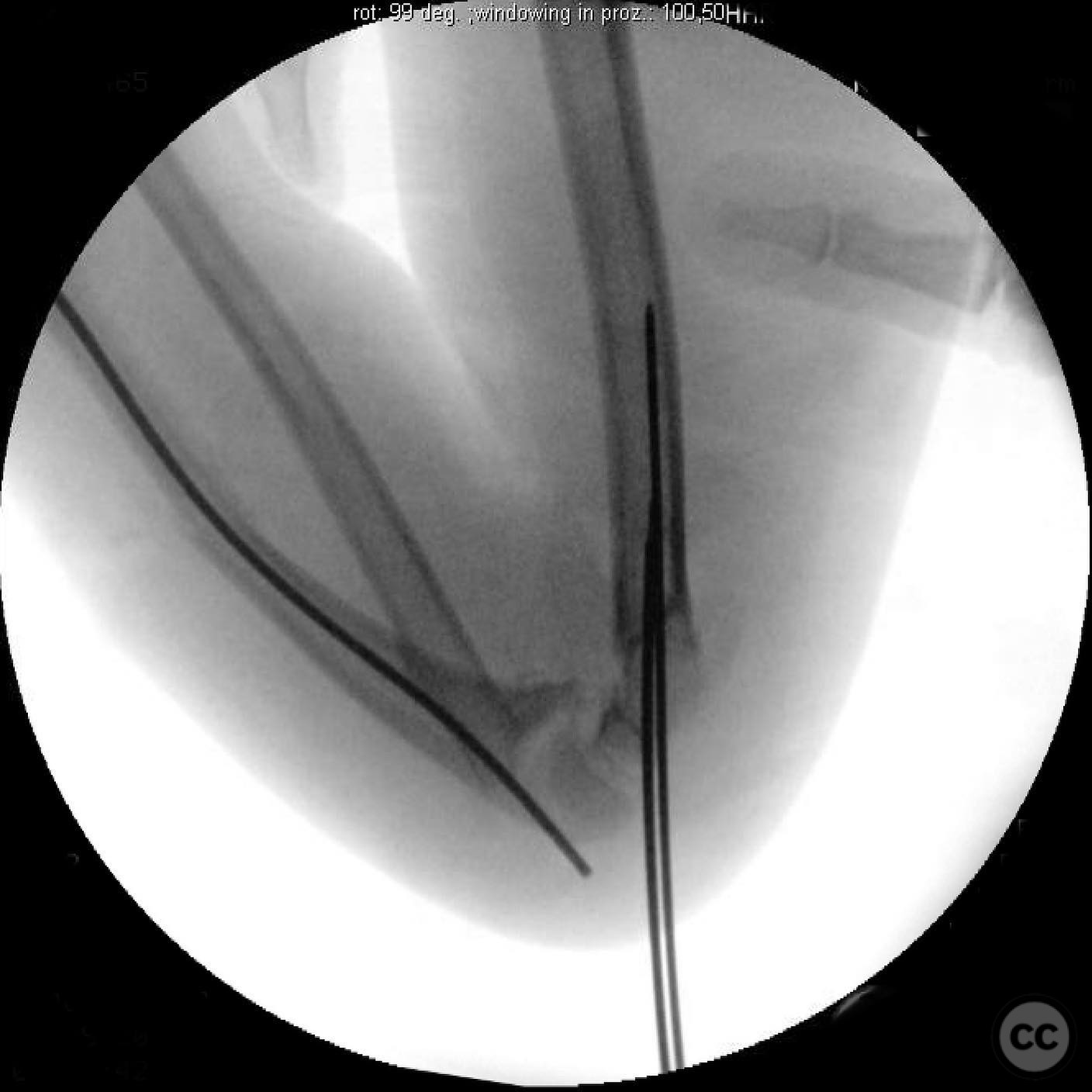
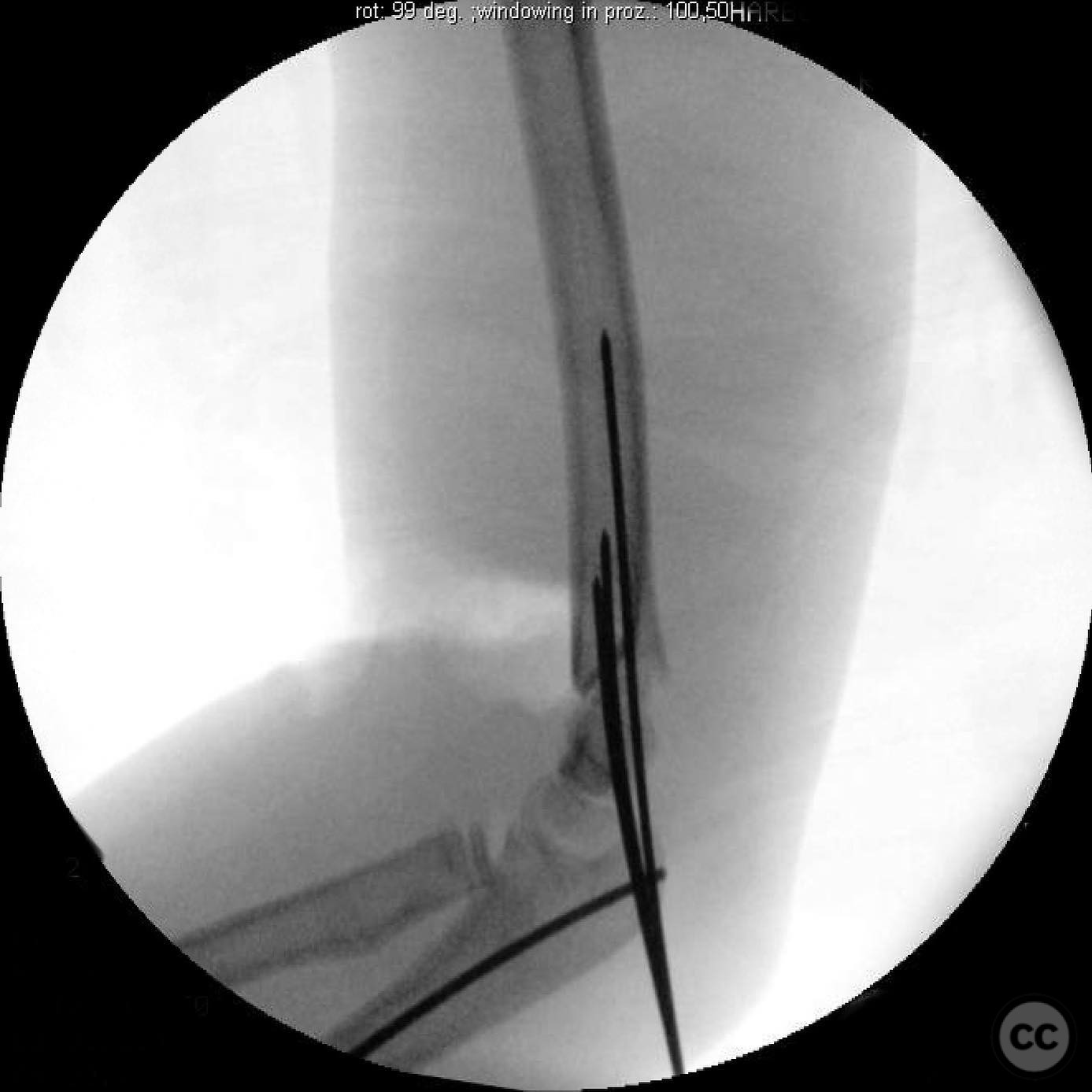
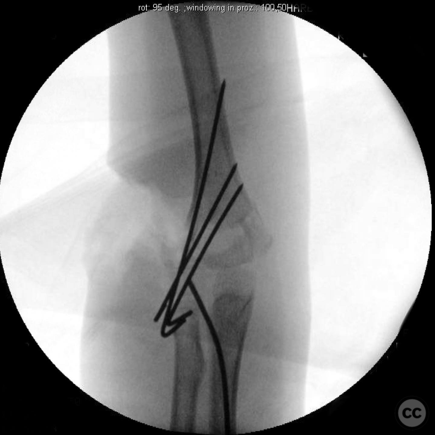
Anatomical surgical approach: A direct approach to the open wounds was utilized for exploration and debridement. The brachial artery was visualized and confirmed intact with Doppler signals. Both the radial and median nerves were found intact. The ulna was approached through a mini-open incision for reduction and intramedullary nailing. The supracondylar humerus fracture was reduced and stabilized with three lateral Kirschner wires. The radial neck was manipulated and reduced through the existing traumatic wound.
Operative remarks:Intraoperative findings confirmed that the brachial artery was intact, allowing restoration of a palpable radial pulse post-reduction. The compartments remained soft throughout the procedure, negating the need for fasciotomy. Post-reduction, the elbow and radial neck were stable through a range of motion. A well-molded splint was applied after wound closure. Postoperative monitoring focused on compartment checks and pain management due to high risk of compartment syndrome. The patient experienced a radial nerve motor neuropraxia, which improved over several months.
Postoperative protocol: Postoperative care included vigilant monitoring for signs of compartment syndrome, with emphasis on escalating pain medication requirements as an indicator. Rehabilitation involved maintaining immobilization in a splint, with gradual progression to active range of motion exercises as healing permitted.
Follow up: Not specified
Orthopaedic implants used: Intramedullary nail for ulna fixation, Kirschner wires for supracondylar humerus fracture stabilization.
Search for Related Literature

orthopaedic_trauma
- United States , Seattle
- Area of Specialty - General Trauma
- Position - Specialist Consultant

Industry Sponsership
contact us for advertising opportunities
Article viewed 288 times
16 Jul 2025
Add to Bookmarks
Full Citation
Cite this article:
Surname, Initial. (2025). Complex Pediatric Upper Extremity Trauma with Supracondylar Humerus Fracture and Vascular Compromise. Journal of Orthopaedic Surgery and Traumatology. Case Report 28175562 Published Online Jul 16 2025.