Ipsilateral Tibial Shaft and Trimalleolar Ankle Fracture: Case Report
Score and Comment on this Case
Clinical Details
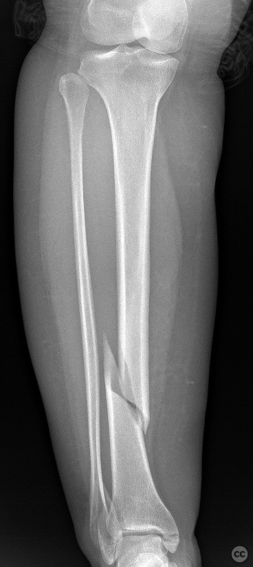
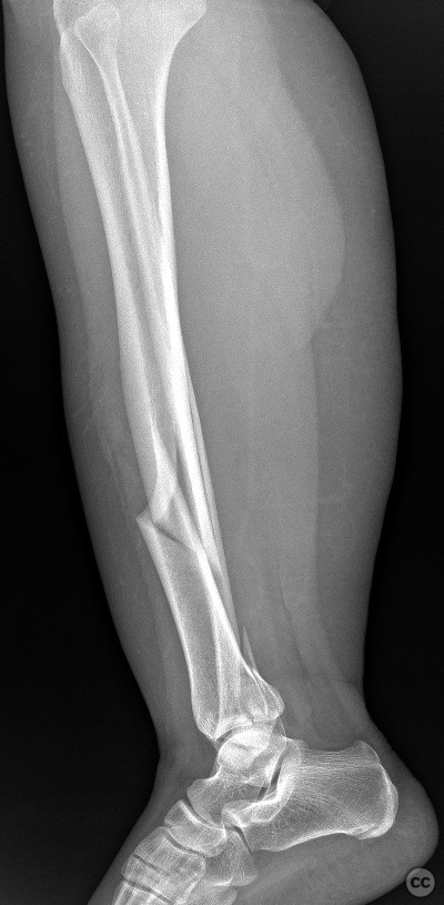
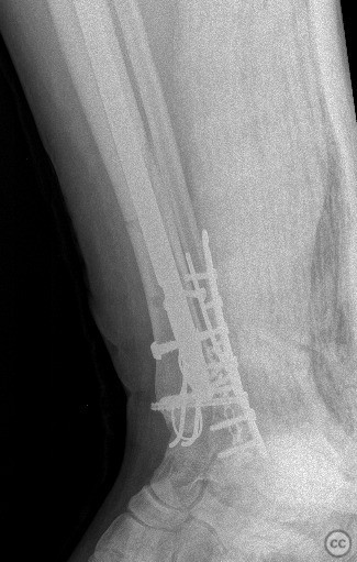
Clinical and radiological findings: A 45-year-old male sustained a high-energy trauma resulting in an ipsilateral closed tibial shaft fracture and a trimalleolar fracture of the ankle. Initial radiographs demonstrated a displaced mid-diaphyseal tibial fracture (AO/OTA 42-A3) and a trimalleolar ankle fracture (AO/OTA 44-C1), with involvement of the medial malleolus, lateral malleolus, and posterior malleolus. No evidence of open injury or neurovascular compromise was present on examination.
Preoperative Plan
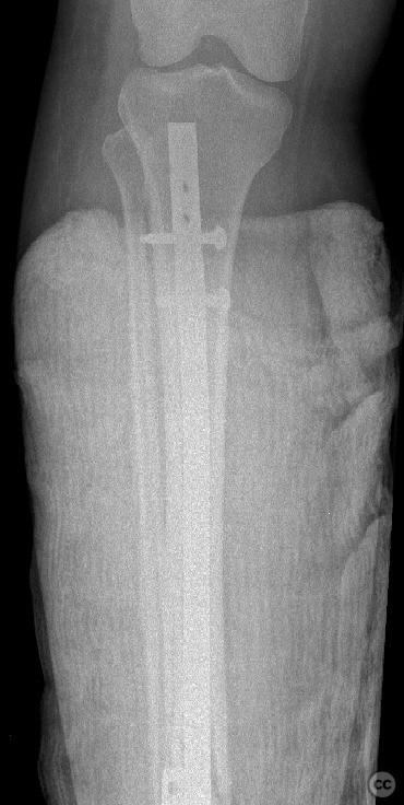
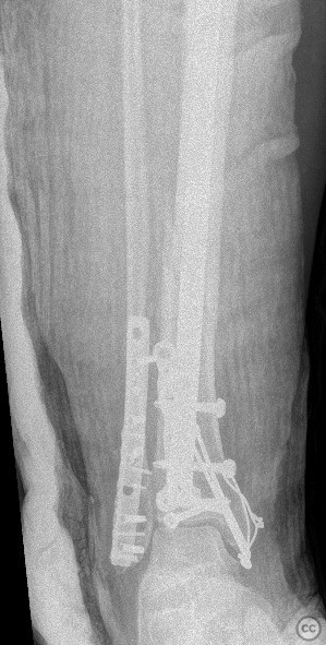
Planning remarks: The preoperative plan included a staged approach: initial stabilization of the tibial shaft fracture with an intramedullary nail via an infrapatellar approach, followed by open reduction and internal fixation of the trimalleolar ankle fracture through separate medial and posterolateral incisions.
Surgical Discussion
Patient positioning: tibia nail and medial malleol----supin///// lateral and posterior malleol------prone position
Operative remarks:The combination of ipsilateral tibial shaft and trimalleolar ankle fractures presents technical challenges in maintaining alignment and stability during fixation. Sequential fixation allowed for restoration of tibial length and rotation prior to addressing the articular surface of the ankle. Careful soft tissue handling was required due to the proximity of multiple incisions. Stable fixation of all malleolar fragments was achieved with lag screws and neutralization plating as indicated.
Postoperative protocol: Postoperatively, the limb was immobilized in a well-padded posterior splint with strict non-weight bearing for 6 weeks. Early active range of motion exercises for the knee and ankle were initiated at 2 weeks postoperatively, progressing to partial weight bearing at 6 weeks and full weight bearing by 10-12 weeks, contingent on radiographic evidence of healing.
Follow up: Not specified
Orthopaedic implants used: tibial intramedullary nail; 3.5mm tubular plate; 4.0mm cannulated screws; mini plate, k wire, cerclage
Search for Related Literature


Industry Sponsership
contact us for advertising opportunities
Article viewed 733 times
08 Dec 2025
Add to Bookmarks
Full Citation
Cite this article:
Seçkin Doğan. (2025). Ipsilateral Tibial Shaft and Trimalleolar Ankle Fracture: Case Report. Journal of Orthopaedic Surgery and Traumatology. Case Report 27959886 Published Online Dec 08 2025.