. See the previous 4 posts for injury films and CT scan. I treated thi(.jpg)
Sanders 3ab Calcaneal Fracture with Staged ORIF.
Score and Comment on this Case
Clinical Details
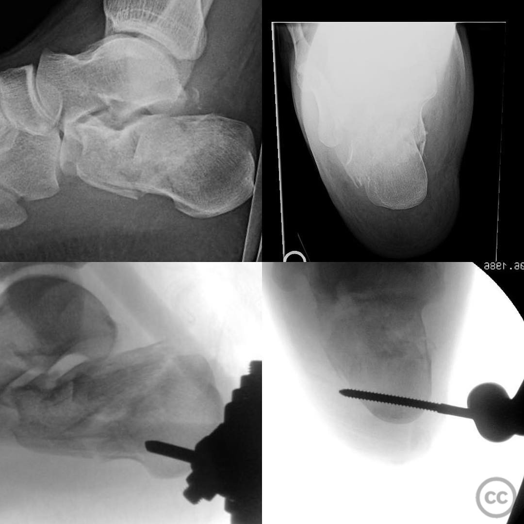
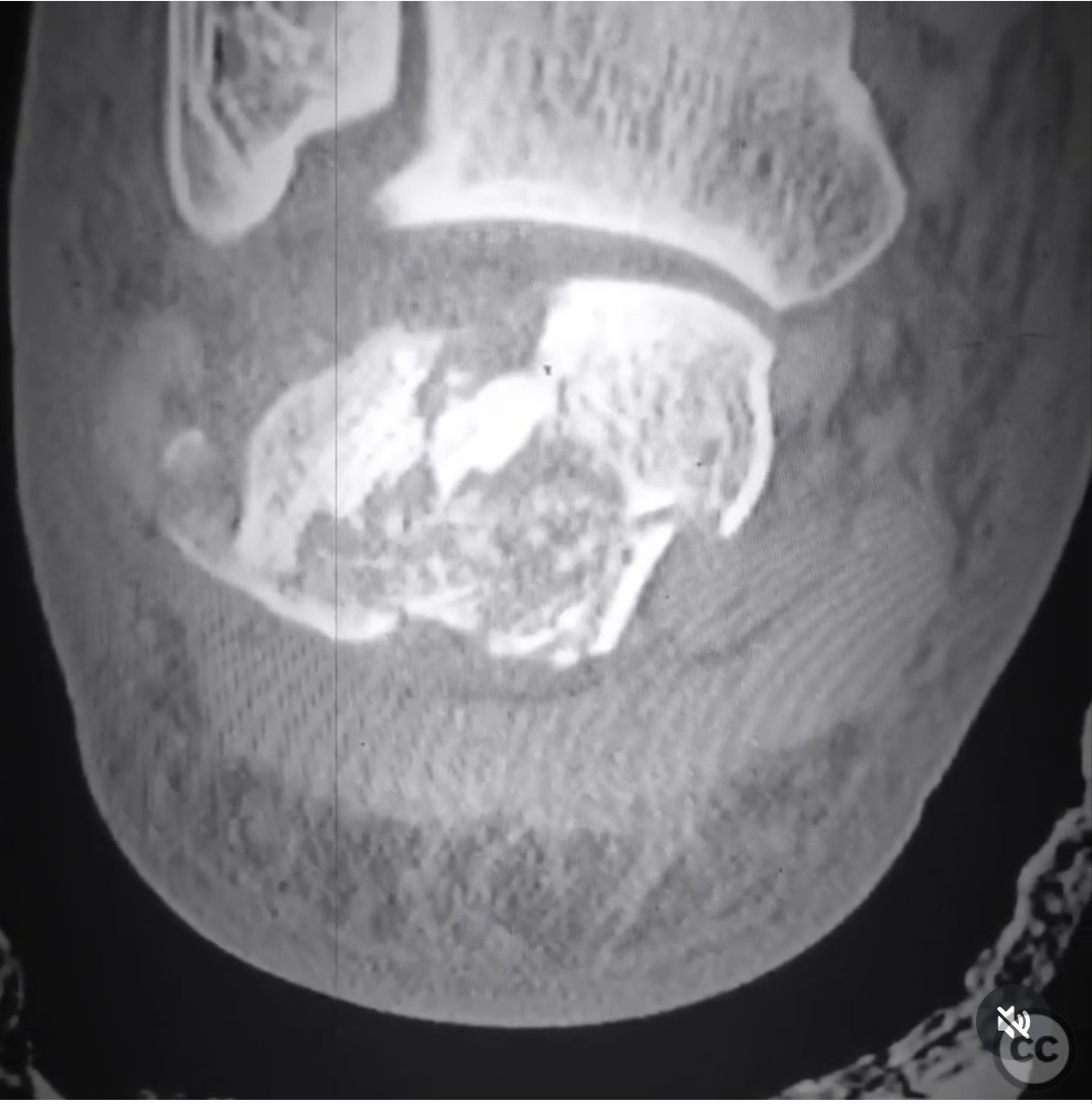
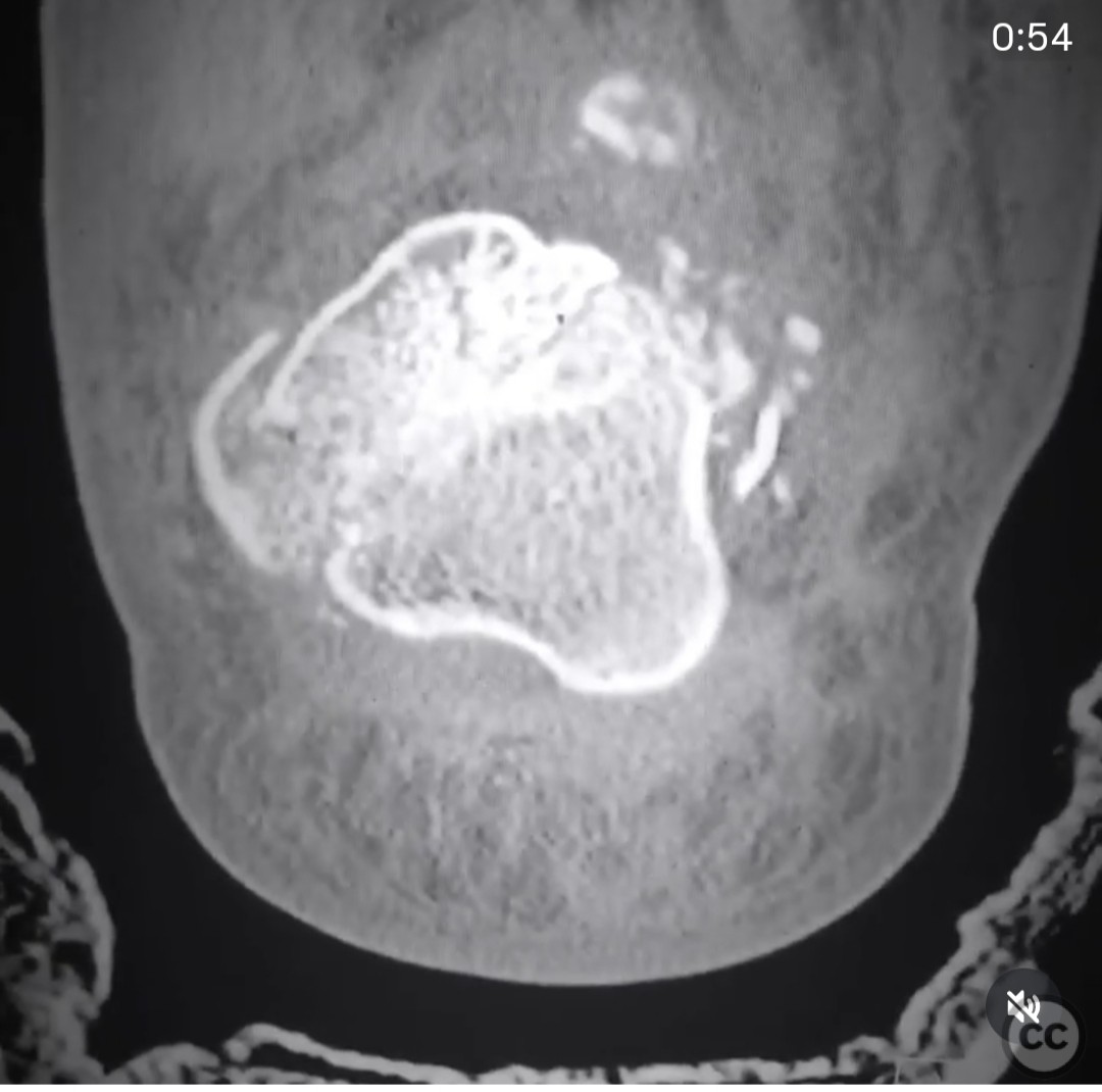
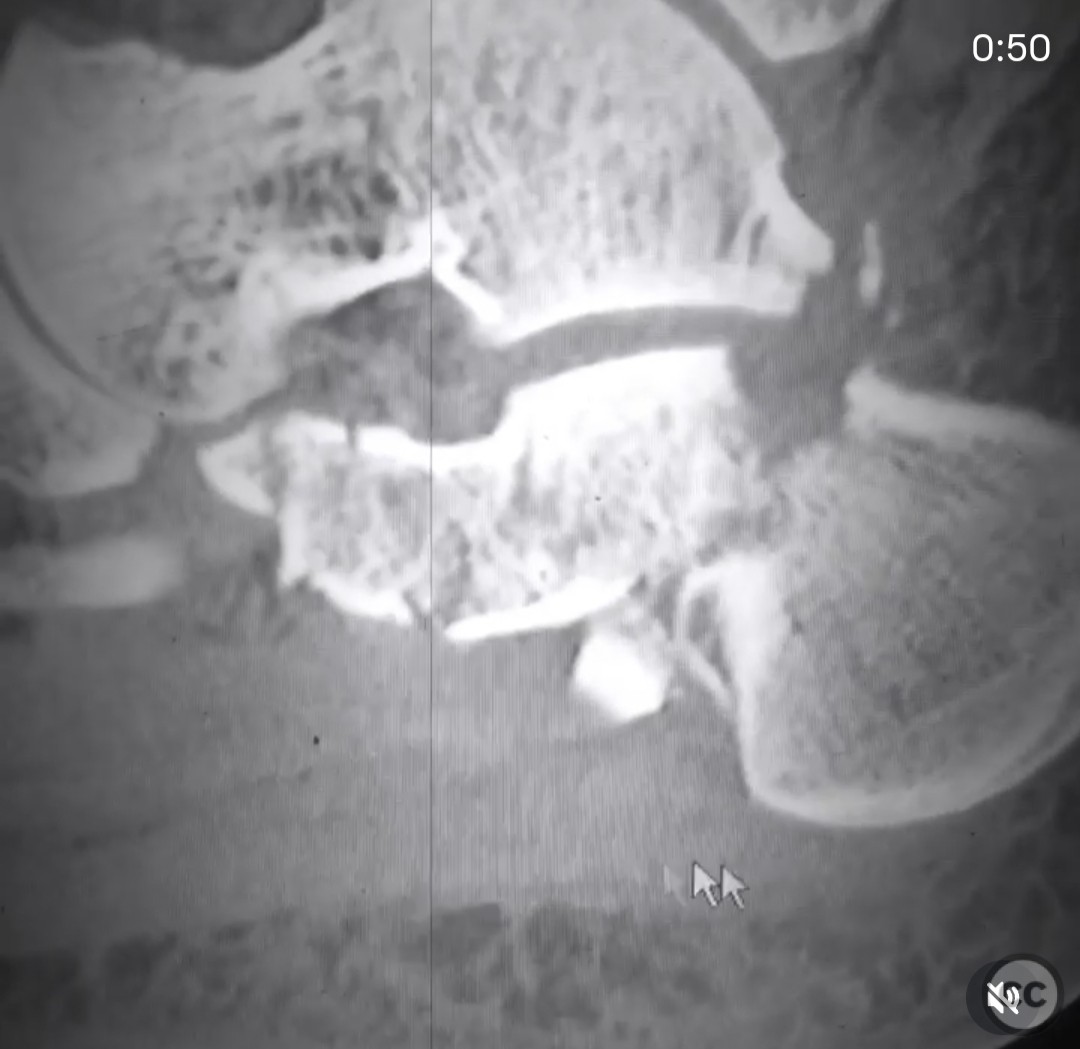
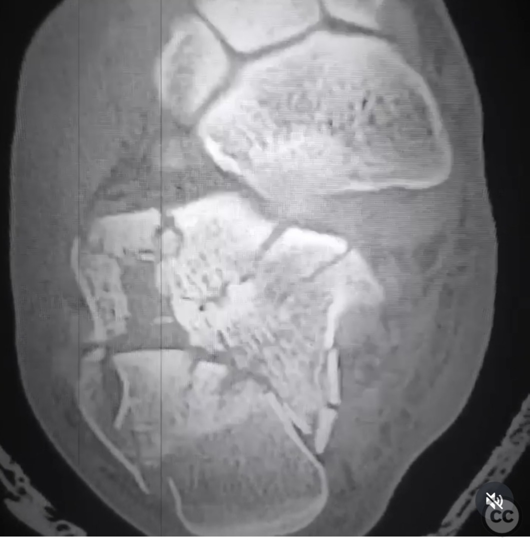
Clinical and radiological findings: A 31-year-old male sustained a closed joint depression type calcaneal fracture classified as Sanders 3ab following a 10-foot fall. The patient is otherwise healthy, a non-smoker, and this was an isolated injury. Initial radiographs and CT scans confirmed the fracture classification. The injury was closed with no significant soft tissue compromise noted at presentation.
Preoperative Plan
Planning remarks: The preoperative plan involved a staged approach. Initially, a closed manipulative reduction with external fixation was performed to stabilize the fracture and improve soft tissue conditions. Definitive open reduction and internal fixation (ORIF) were planned through an extensile lateral approach, scheduled for 10 days post-injury once soft tissue swelling had subsided.
Surgical Discussion
Patient positioning: The patient was positioned in a lateral decubitus position to facilitate access to the lateral aspect of the calcaneus during the surgical procedure.
Anatomical surgical approach: An extensile lateral approach was utilized. A lateral L-shaped incision was made, extending from the posterior aspect of the heel to the sinus tarsi, allowing for full-thickness flap elevation. The lateral wall blowout and free posterior facet fragments were removed, marked, and placed in bacitracin solution. The anterior process was reduced and temporarily fixed with wires. The tuberosity was distracted and displaced posteriorly and medially to facilitate reduction of the anterior process to the constant fragment. The middle facet was reduced first, followed by the posterior facet fragments. The tuberosity was then repositioned anatomically, and the lateral wall was flattened and replaced to confirm reduction.
Operative remarks:The surgeon emphasized the importance of restoring the normal relationship between the anterior process and the constant fragment at the critical angle to ensure room for an anatomical reduction of the posterior facet. The staged approach with initial external fixation facilitated soft tissue recovery and simplified subsequent ORIF by achieving early morphological restoration of the calcaneus.
Postoperative protocol: Postoperatively, the patient was advised to remain non-weight bearing on the affected limb for 6 weeks, followed by gradual weight-bearing as tolerated. Range of motion exercises were initiated early to prevent stiffness, with progressive strengthening exercises introduced as healing permitted.
Follow up: Not specified.
Orthopaedic implants used: External fixator, Kirschner wires, calcaneal plate and screws.
Search for Related Literature

orthopaedic_trauma
- United States , Seattle
- Area of Specialty - General Trauma
- Position - Specialist Consultant

Industry Sponsership
contact us for advertising opportunities
Article viewed 342 times
28 Jul 2025
Add to Bookmarks
Full Citation
Cite this article:
Surname, Initial. (2025). Sanders 3ab Calcaneal Fracture with Staged ORIF.. Journal of Orthopaedic Surgery and Traumatology. Case Report 25635939 Published Online Jul 28 2025.