Pathologic Femoral Neck Fracture in a Young Adult with Stress Fracture Progression.
Score and Comment on this Case
Clinical Details
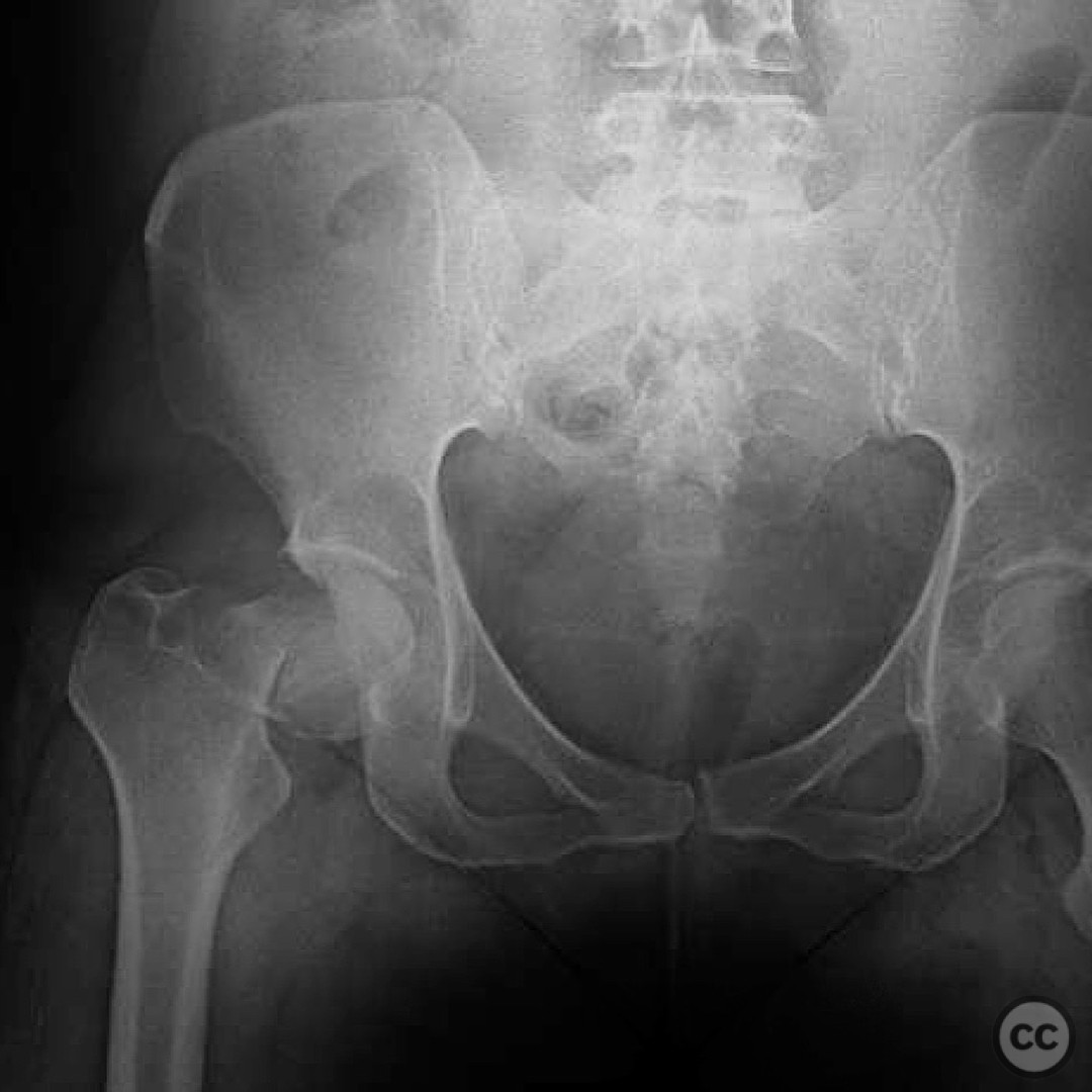
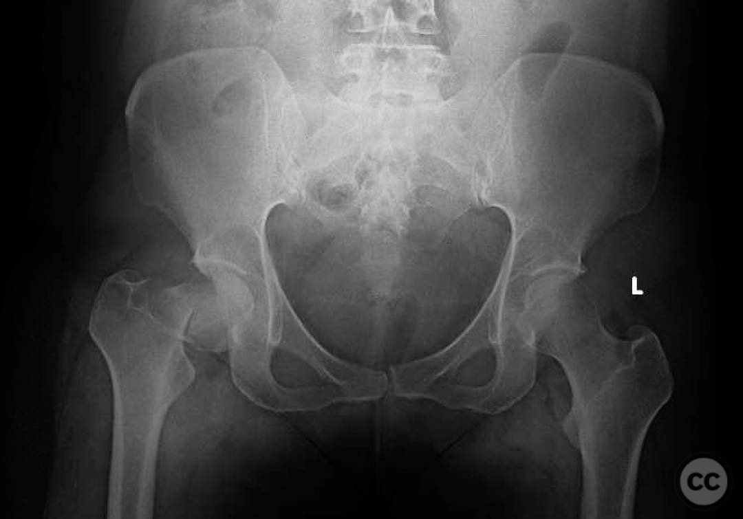
Clinical and radiological findings: A 33-year-old female presented with a sudden onset of hip pain while walking her dog, following a month of antecedent hip pain that began during hiking. The patient is otherwise healthy, with a vegan diet and no significant medical history. Initial radiographs revealed a femoral neck fracture, consistent with a pathologic fracture due to stress fracture progression. No signs of infection, neoplasm, or osteoporosis were noted on imaging and laboratory tests.
Preoperative Plan
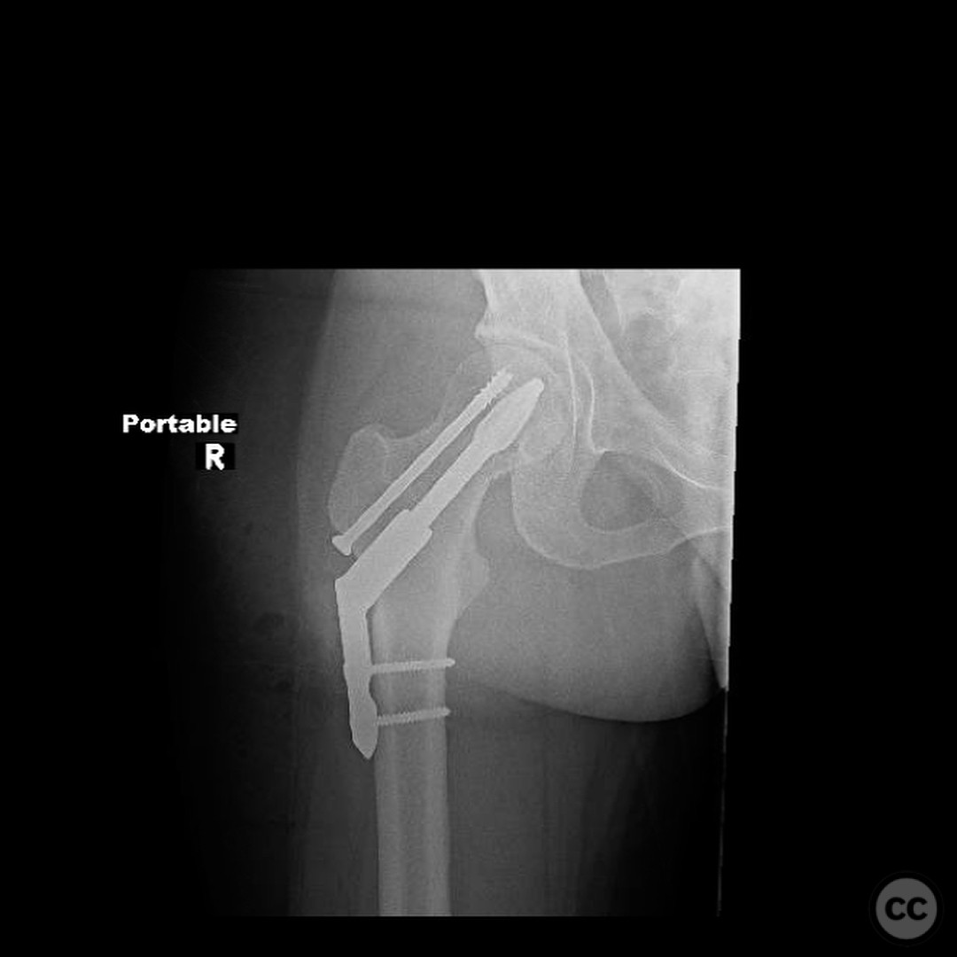
Planning remarks: The preoperative plan involved an open reduction and internal fixation (ORIF) of the femoral neck fracture. The surgical approach was planned through a direct lateral incision to access the proximal femur and achieve anatomical reduction.
Surgical Discussion
Patient positioning: The patient was positioned supine on a fracture table to allow for optimal access and manipulation of the proximal femur during the procedure.
Anatomical surgical approach: A direct lateral approach was utilized, involving an incision over the lateral aspect of the hip. Dissection proceeded through the fascia lata and gluteus medius, with careful subperiosteal elevation to expose the femoral neck and head for reduction and fixation.
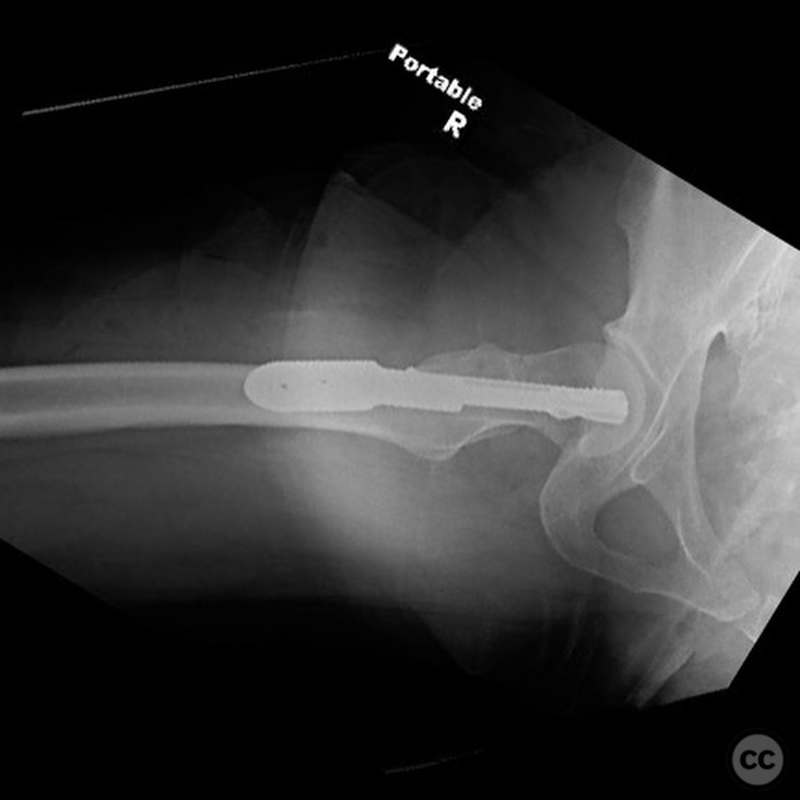
Operative remarks:The surgeon noted that achieving an anatomical reduction was critical, followed by compression fixation to ensure stability. Given the pathologic nature of the fracture, there was an elevated risk of nonunion, although the risk of avascular necrosis was considered low due to the low-energy mechanism and minimal displacement.
Postoperative protocol: Postoperative rehabilitation included protected weight-bearing with crutches for 6 weeks, followed by gradual progression to full weight-bearing as tolerated. Physical therapy focused on range of motion and strengthening exercises.
Follow up: Not specified.
Orthopaedic implants used: Cannulated screws for internal fixation.
Search for Related Literature

orthopaedic_trauma
- United States , Seattle
- Area of Specialty - General Trauma
- Position - Specialist Consultant

Industry Sponsership
contact us for advertising opportunities
Article viewed 293 times
25 Jul 2025
Add to Bookmarks
Full Citation
Cite this article:
Surname, Initial. (2025). Pathologic Femoral Neck Fracture in a Young Adult with Stress Fracture Progression.. Journal of Orthopaedic Surgery and Traumatology. Case Report 2405757 Published Online Jul 25 2025.