Geriatric Supracondylar Distal Humerus Fracture in a Rheumatoid Arthritis Patient with Ipsilateral Shoulder Hemiarthroplasty.
Score and Comment on this Case
Clinical Details
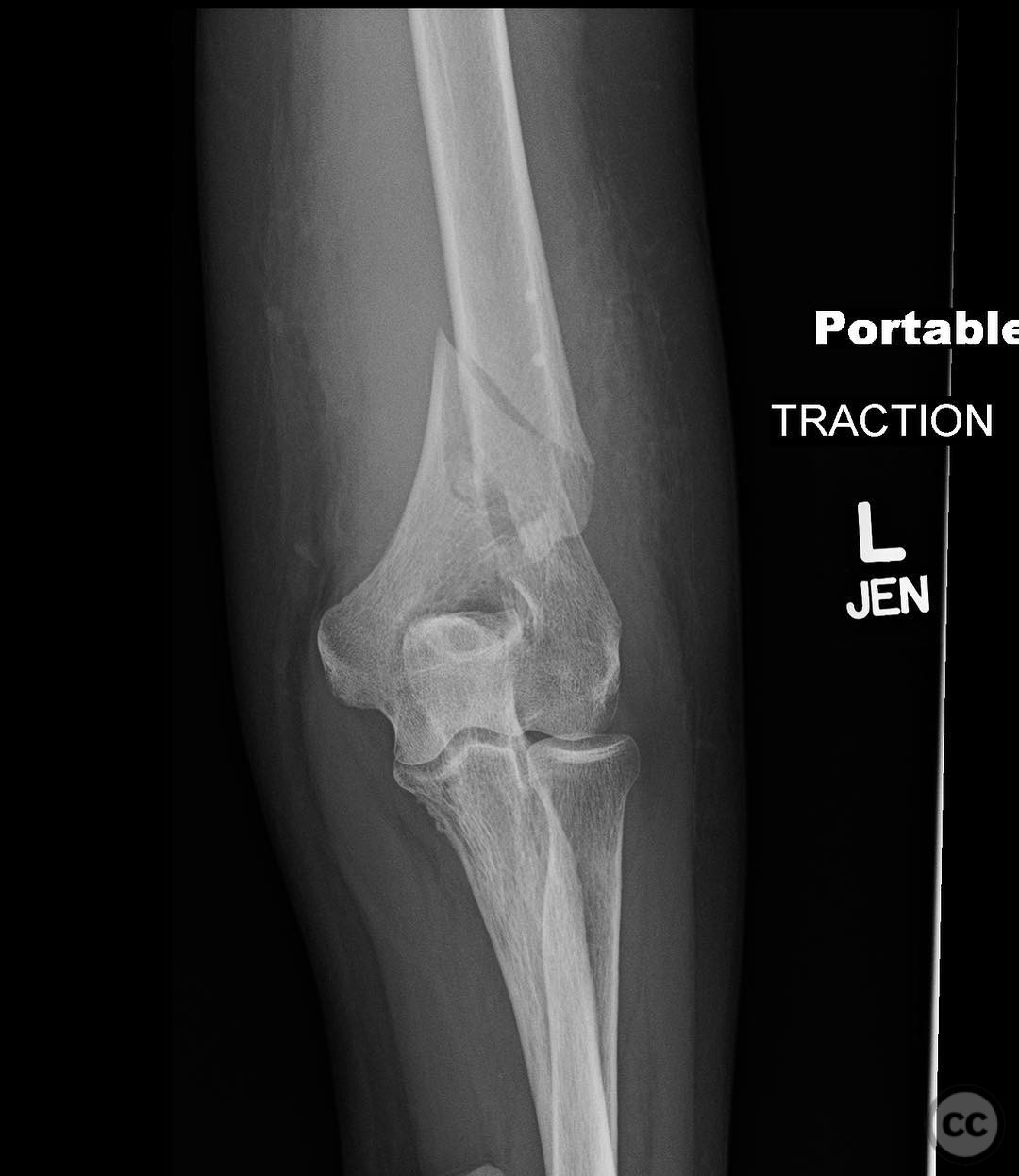
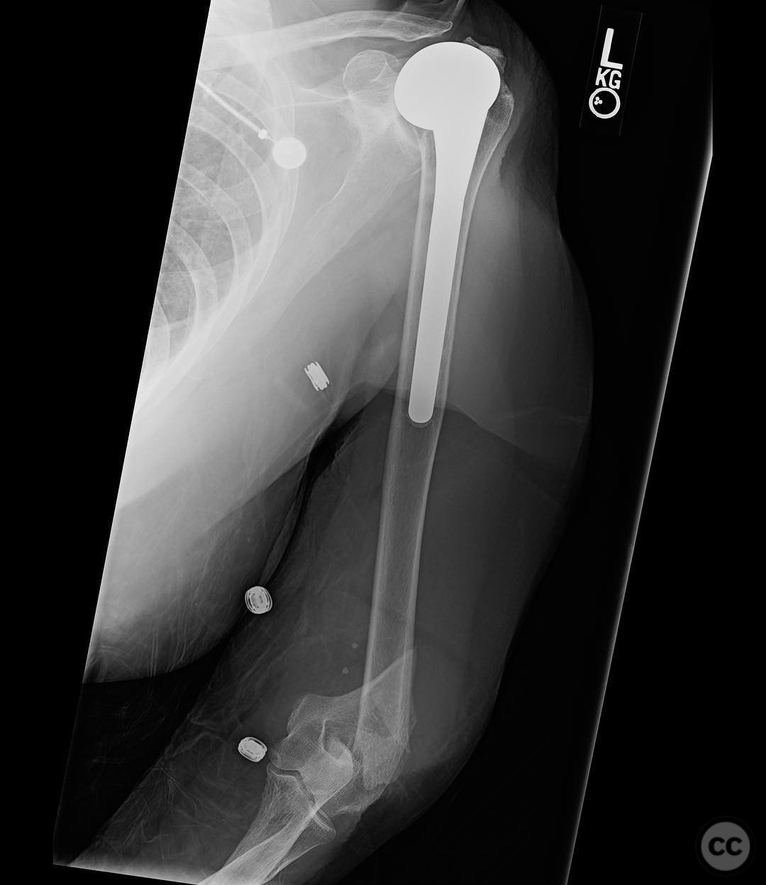
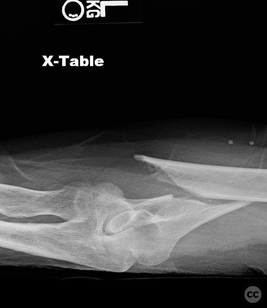
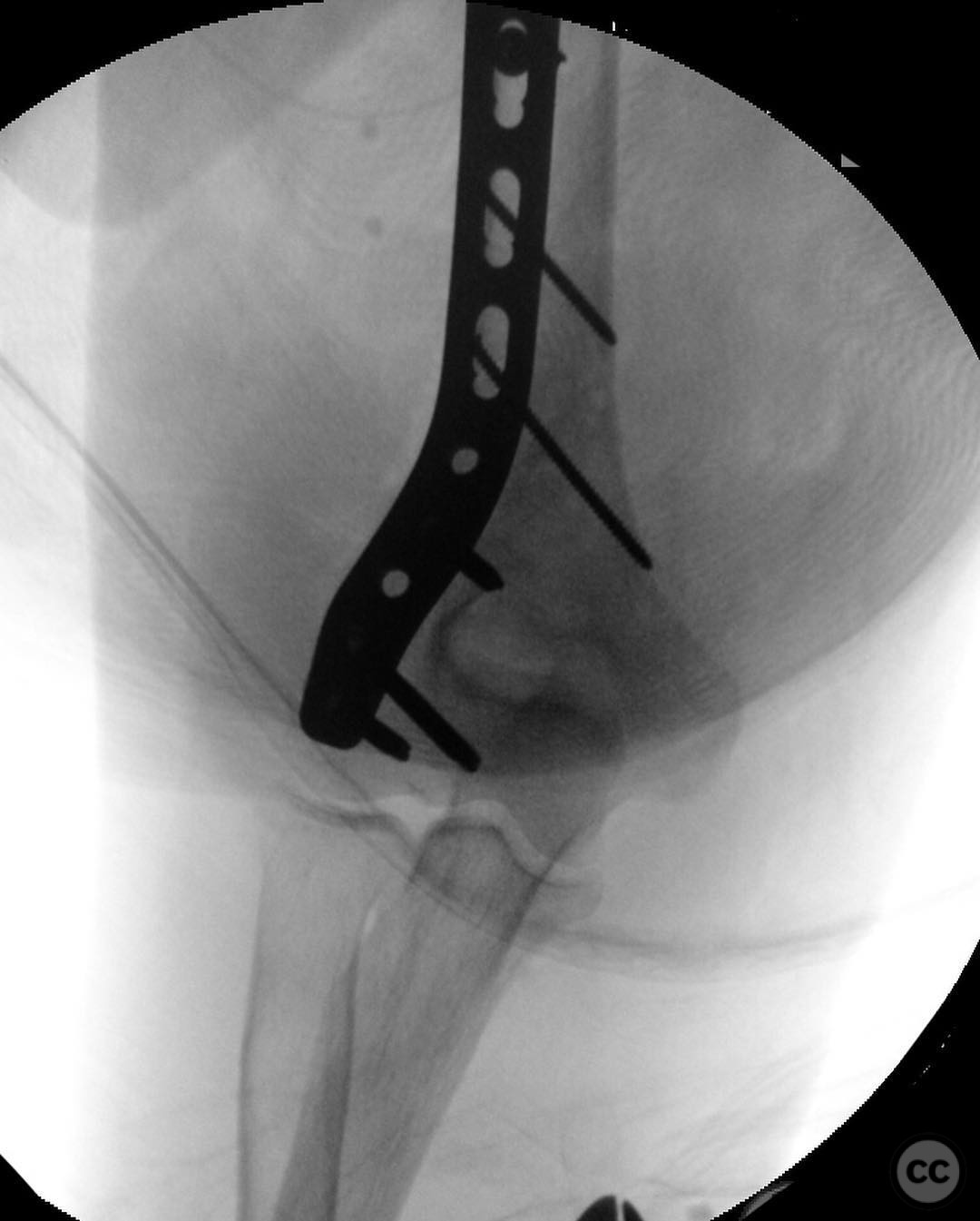
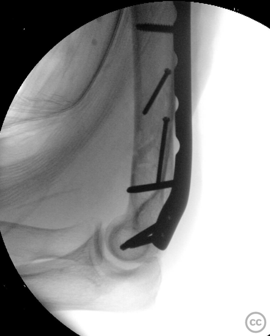
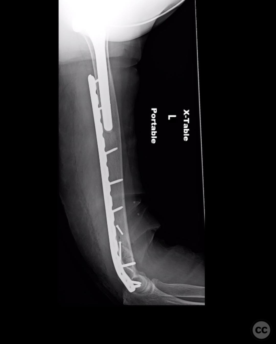
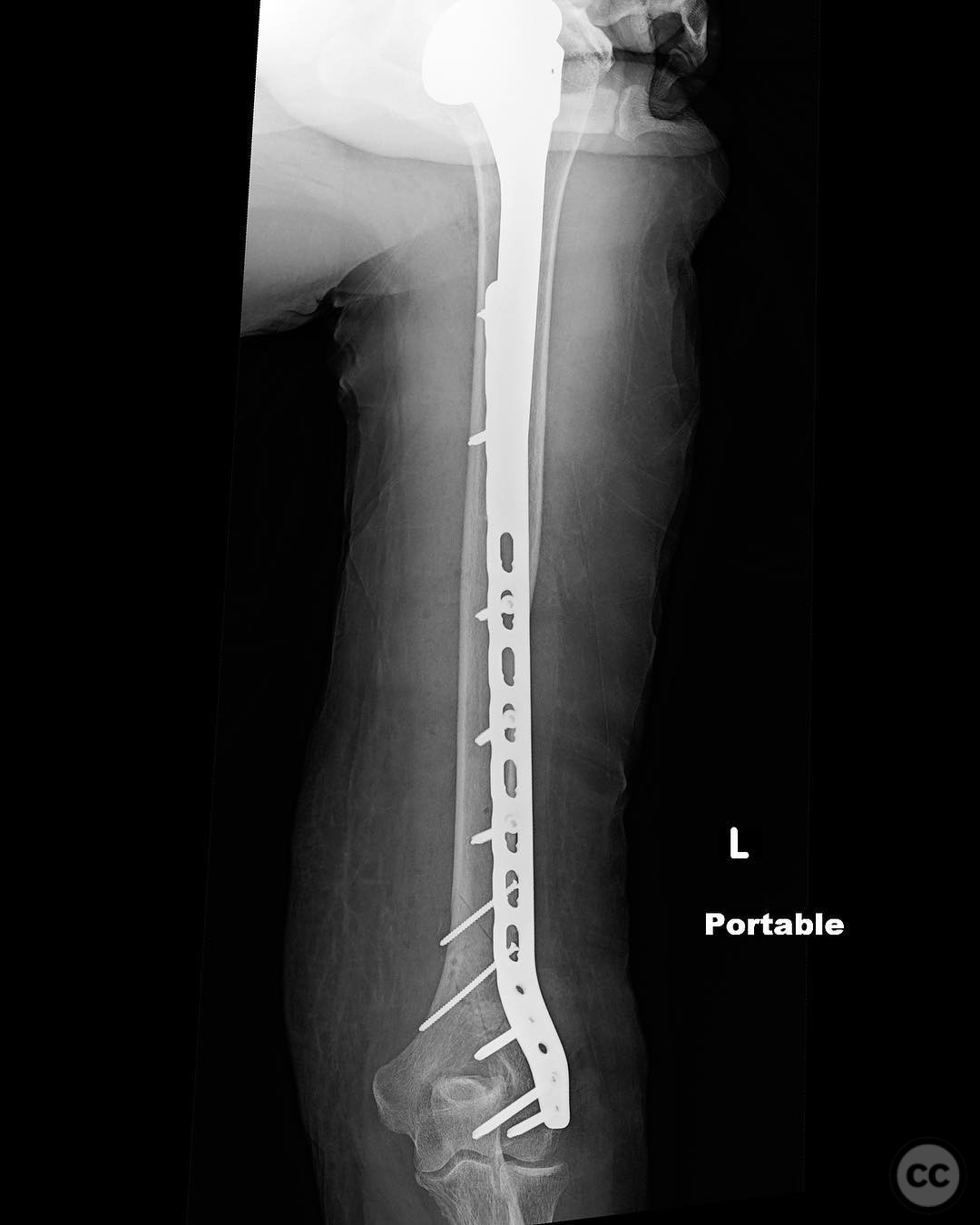
Clinical and radiological findings: A 79-year-old female with a history of rheumatoid arthritis presented following a ground-level fall. The patient was otherwise healthy with no other injuries reported. Radiological assessment revealed a simple supracondylar fracture of the distal humerus. The patient also had an ipsilateral shoulder hemiarthroplasty. The fracture was classified as AO/OTA 13-A2.
Preoperative Plan
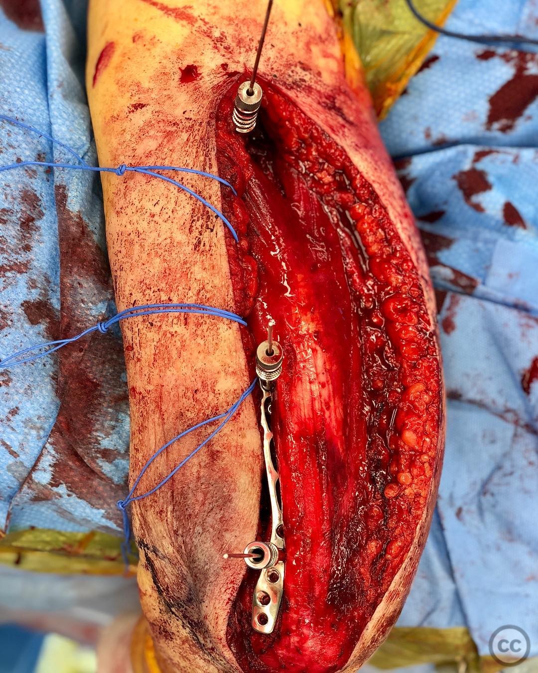
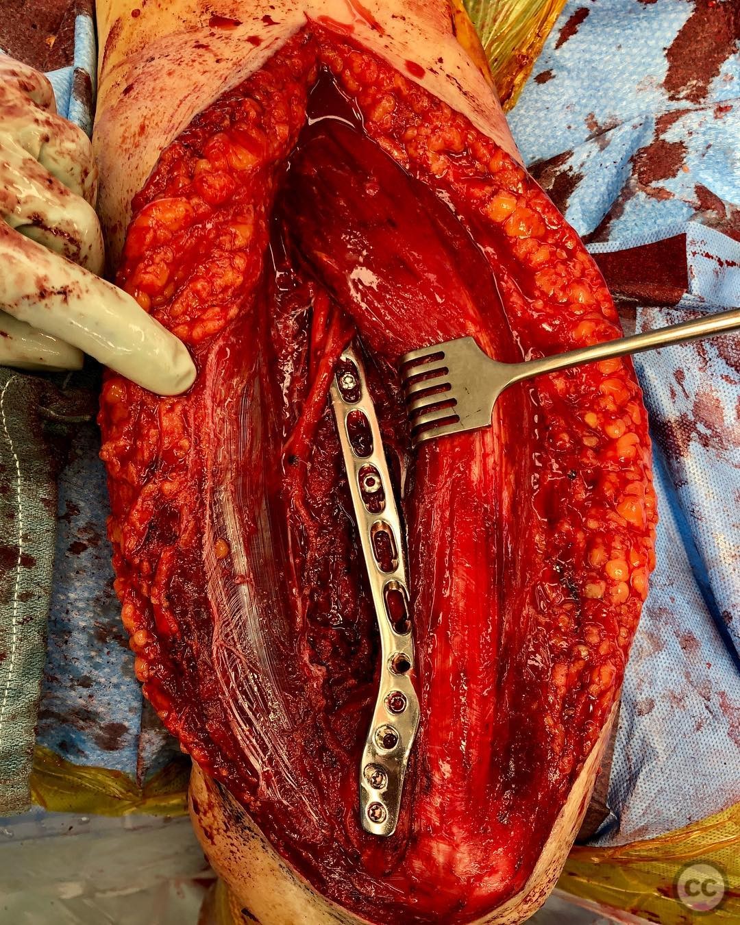
Planning remarks: The preoperative plan involved a direct anatomic reduction using clamps, followed by interfragmentary compression with 2.4mm lag screws by technique, and stabilization with a neutralization plate. Due to the risk of stress risers, a long lateral column plate was chosen to span the fracture and provide overlapping fixation.
Surgical Discussion
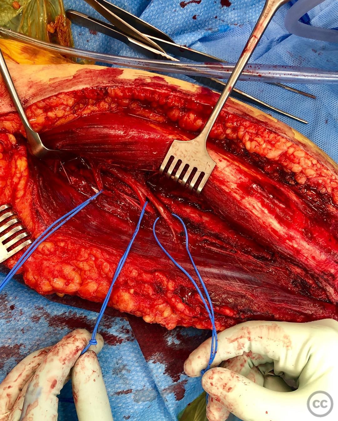
Patient positioning: The patient was positioned in a lateral decubitus position to facilitate access to the posterior aspect of the humerus.
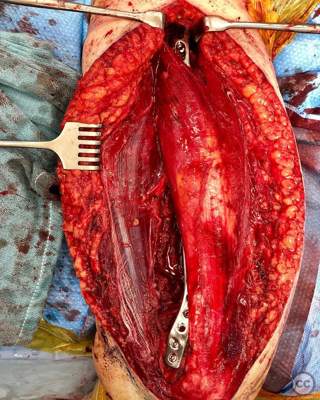
Anatomical surgical approach: A modified Gerwin approach was utilized, which is a triceps-sparing technique. The triceps was elevated laterally until limited by the lateral head. Careful protection of the radial nerve and its branches was ensured. The interval between the long and lateral head of the triceps was developed to access the proximal humeral shaft. Retraction of the deltoid and axillary nerve allowed access up to the neck if necessary.
Operative remarks:The surgeon opted for a long lateral column plate to eliminate the possibility of stress risers, considering the patient's history and potential for future falls. Although a medial column plate was not used, it was noted that it could provide additional stability against varus failure, especially in more complex fracture patterns.
Postoperative protocol: Postoperative rehabilitation included early mobilization with range of motion exercises as tolerated, avoiding weight-bearing activities on the affected limb for an initial period, followed by gradual strengthening exercises.
Follow up: Not specified.
Orthopaedic implants used: Long lateral column plate, 2.4mm lag screws.
Search for Related Literature

orthopaedic_trauma
- United States , Seattle
- Area of Specialty - General Trauma
- Position - Specialist Consultant

Industry Sponsership
contact us for advertising opportunities
Article viewed 272 times
23 Jul 2025
Add to Bookmarks
Full Citation
Cite this article:
Surname, Initial. (2025). Geriatric Supracondylar Distal Humerus Fracture in a Rheumatoid Arthritis Patient with Ipsilateral Shoulder Hemiarthroplasty.. Journal of Orthopaedic Surgery and Traumatology. Case Report 23452892 Published Online Jul 23 2025.