_0.jpg)
_1.jpg)

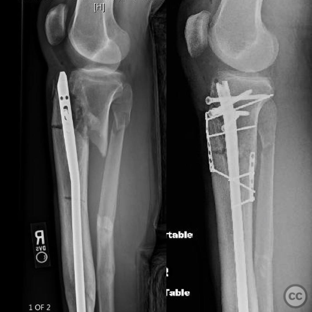
Complex Tibial Fracture with Intramedullary Nail Removal and Reconstruction
Score and Comment on this Case
Clinical Details
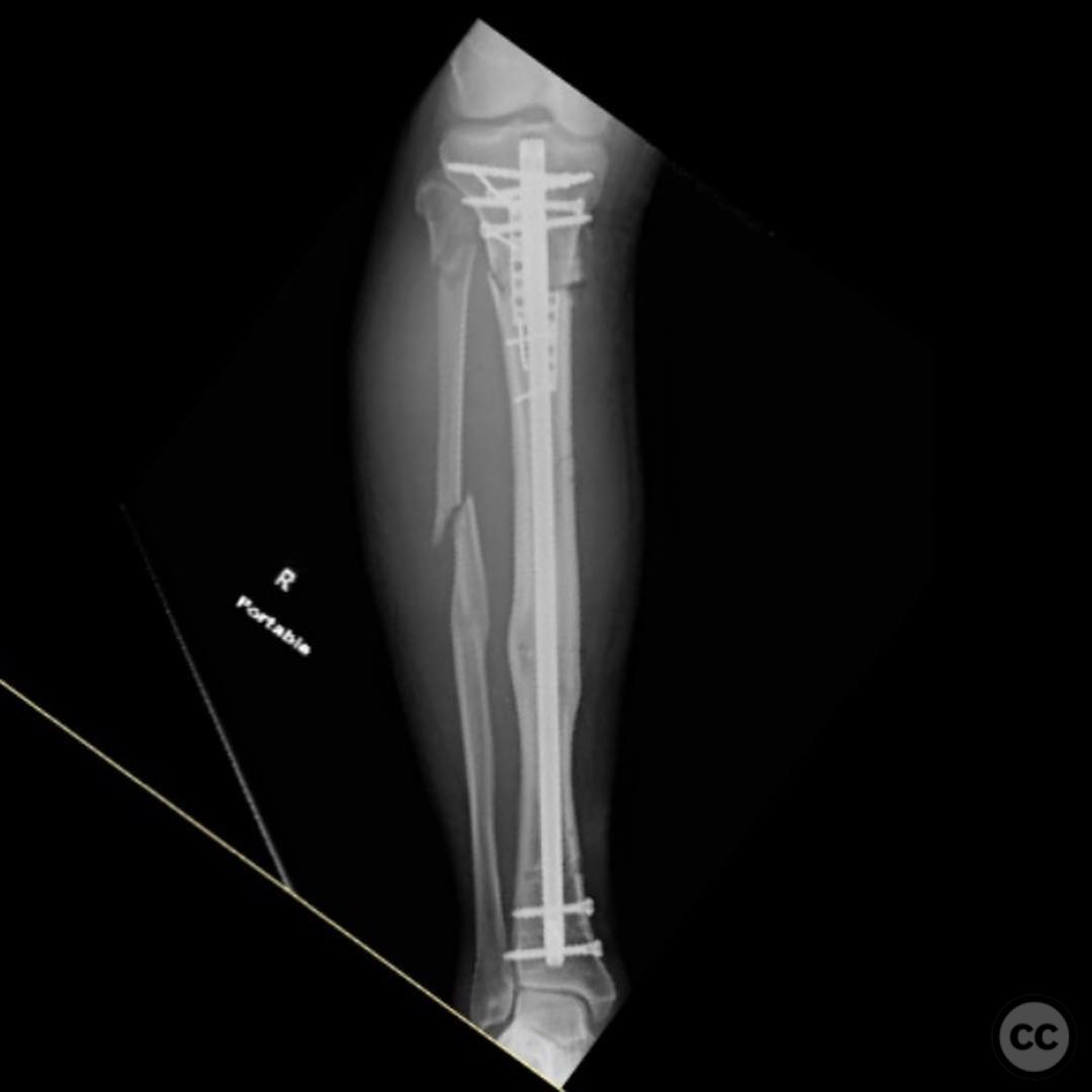
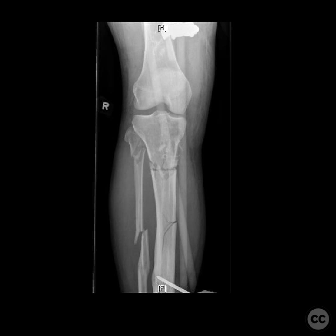
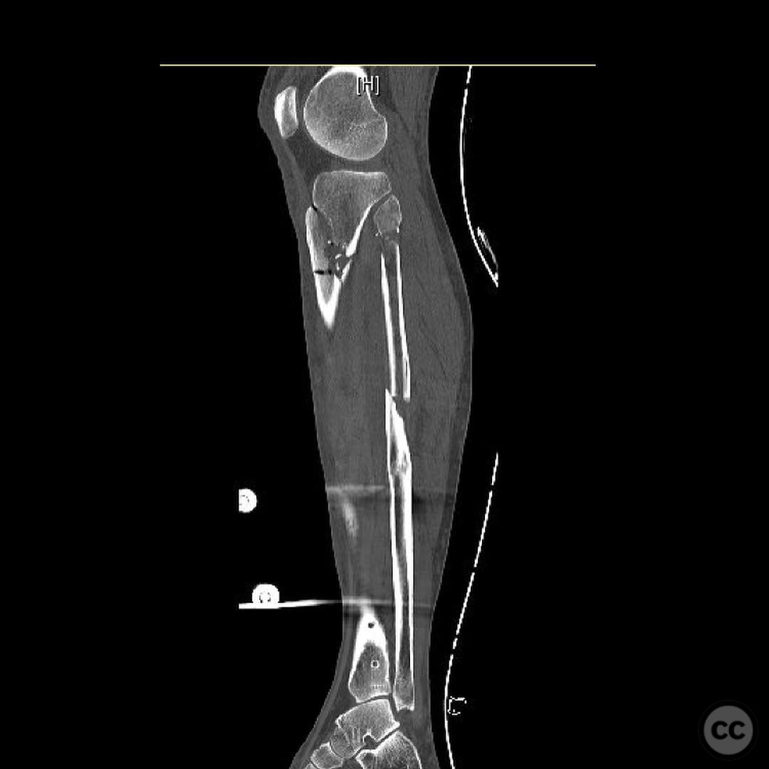
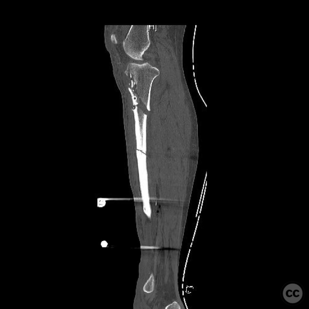
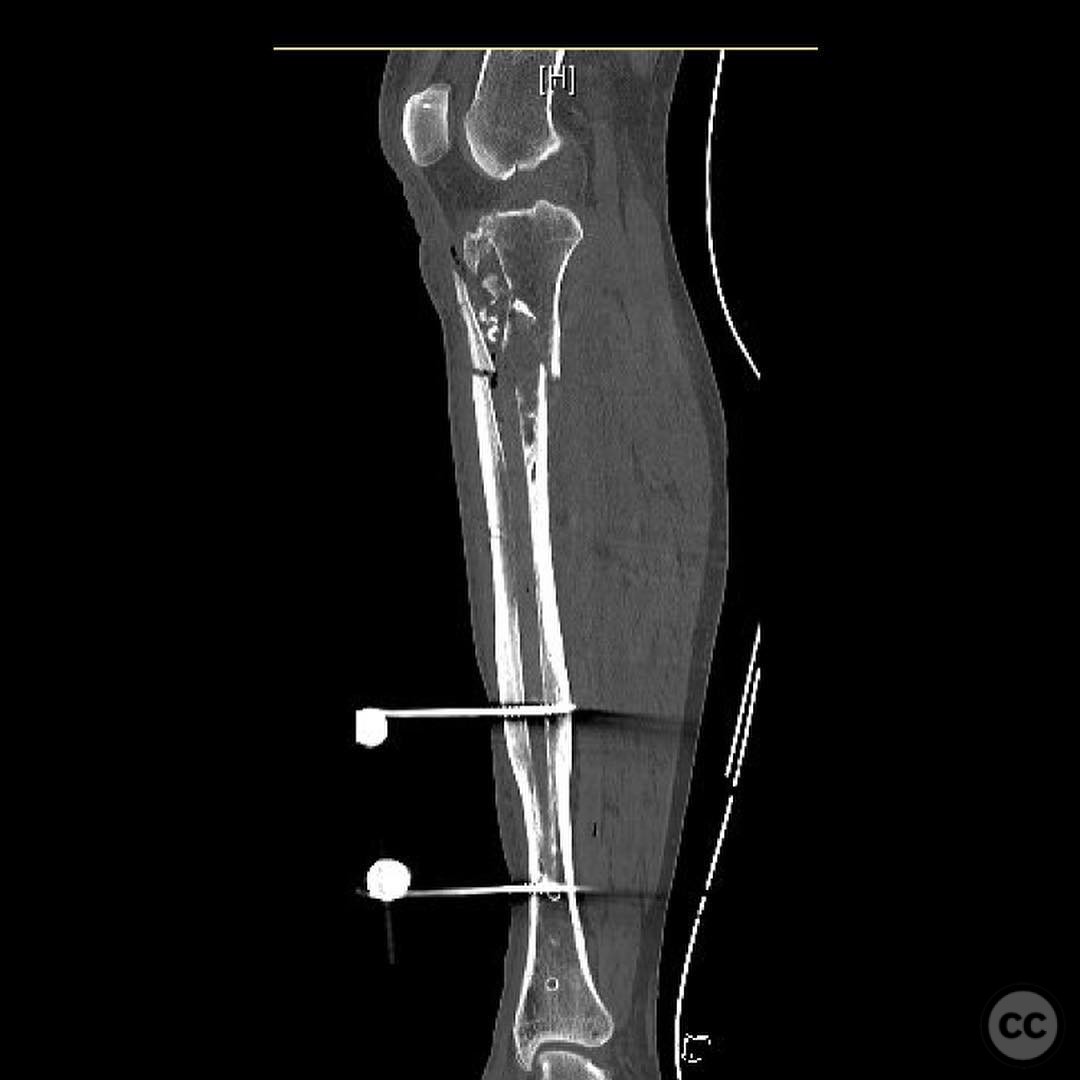
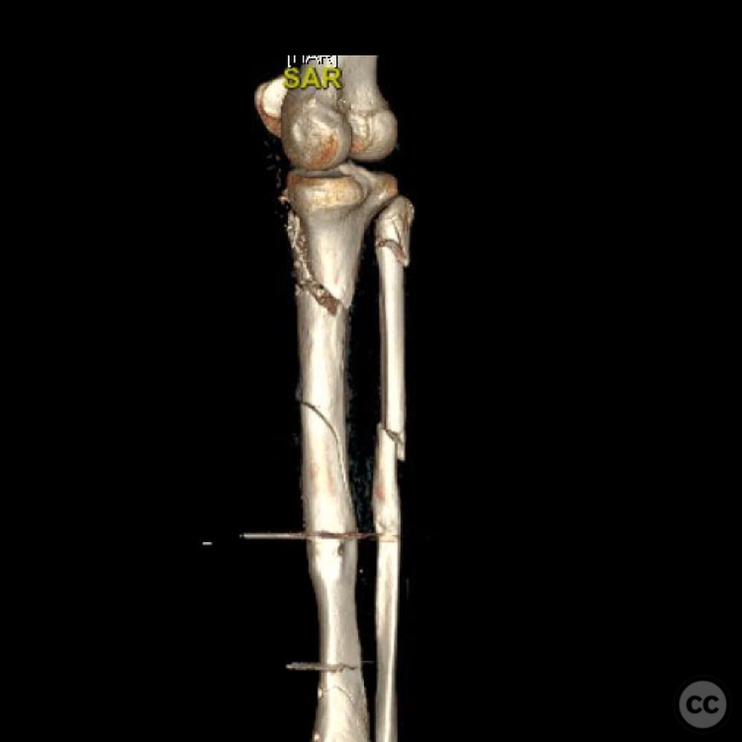
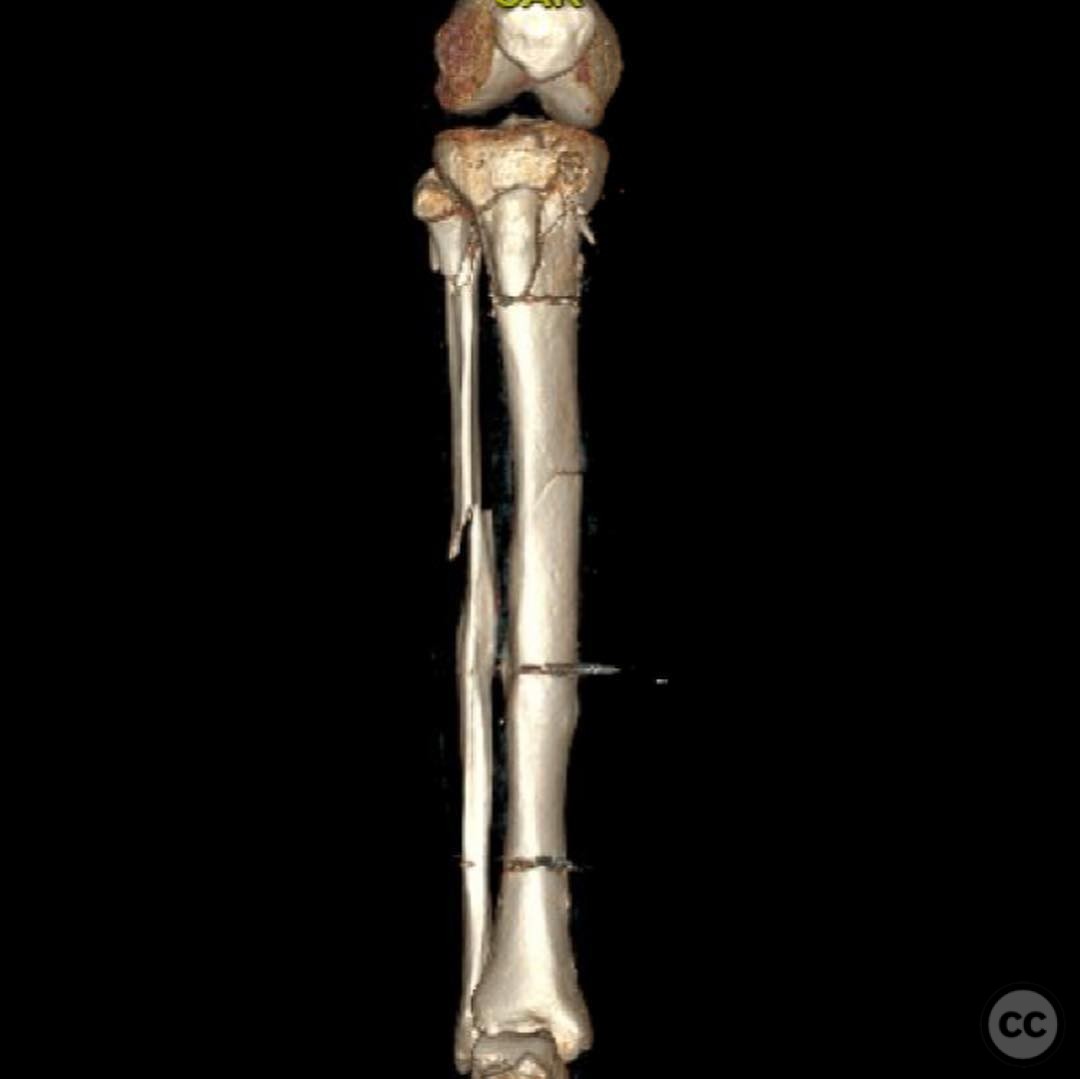
Clinical and radiological findings: A 26-year-old male pedestrian was struck by an automobile, resulting in closed tibial fractures. Initial radiographs indicated a complex fracture pattern with a bent intramedullary nail present. The fracture exhibited three-point bending with a significant proximal fracture and a distal fracture near the distal bolt holes of the nail. A CT scan was performed to assess the distal fracture configuration and to evaluate the proximal fracture pattern and tubercle fragment for potential nailing.
Preoperative Plan
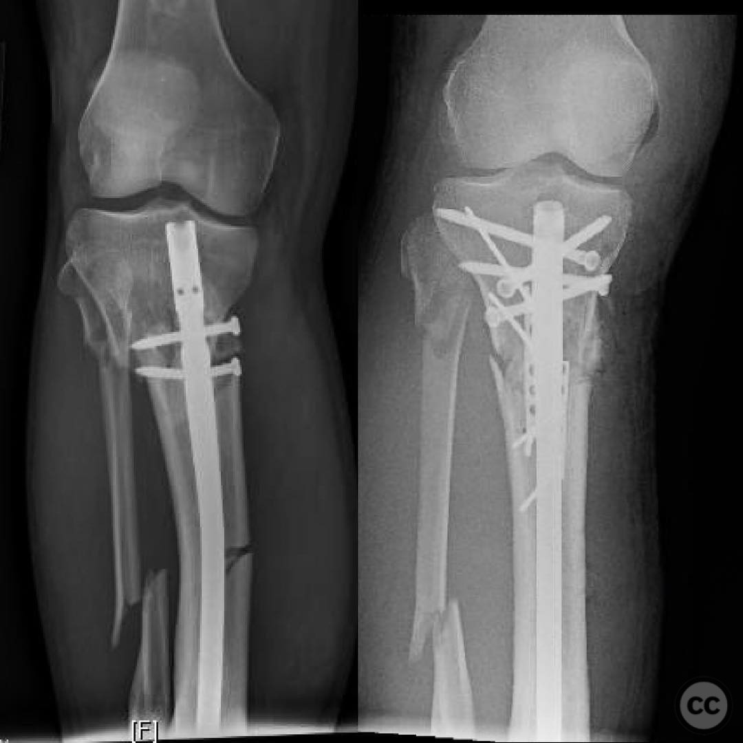
Planning remarks: The preoperative plan involved removal of the existing intramedullary nail, followed by application of an external fixator to restore length, alignment, and rotation. This was followed by obtaining a CT scan to further evaluate the fracture pattern. The surgical plan included open reduction and internal fixation with a buttress plate for the main segment displacement in shear, a tension band plate for the tubercle fragment, and subsequent careful intramedullary nailing.
Surgical Discussion
Patient positioning: Supine position on a radiolucent table to facilitate imaging and access for both nail removal and subsequent fixation procedures.
Anatomical surgical approach: A longitudinal medial approach to the tibia was utilized for removal of the intramedullary nail and application of the external fixator. For open reduction and internal fixation, a direct anterior approach was employed to access the proximal tibial fracture and tubercle fragment.
Operative remarks:The surgeon noted the importance of meticulous planning due to the complexity of the fracture pattern and the presence of the intramedullary nail. Removal of the nail was achieved without complication, allowing for precise application of the external fixator. The open reduction of the main segment required careful alignment to address shear displacement, while placement of the tension band plate on the tubercle fragment required strategic screw placement to avoid interference with subsequent nailing. The final step involved careful intramedullary nailing, ensuring that screws did not obstruct nail placement.
Postoperative protocol: Postoperative rehabilitation included non-weight bearing with crutches for 6 weeks, followed by progressive weight bearing as tolerated. Range of motion exercises were initiated immediately postoperatively to prevent stiffness.
Follow up: Not specified.
Orthopaedic implants used: External fixator, buttress plate, tension band plate, intramedullary nail.
Search for Related Literature

orthopaedic_trauma
- United States , Seattle
- Area of Specialty - General Trauma
- Position - Specialist Consultant

Industry Sponsership
contact us for advertising opportunities
Article viewed 313 times
23 Jul 2025
Add to Bookmarks
Full Citation
Cite this article:
Surname, Initial. (2025). Complex Tibial Fracture with Intramedullary Nail Removal and Reconstruction. Journal of Orthopaedic Surgery and Traumatology. Case Report 23279998 Published Online Jul 23 2025.