. It depends on _3.jpg)
. It depends on _4.jpg)
. It depends on _5.jpg)
. It depends on _7.jpg)
. It depends on _6.jpg)
. It depends on _2.jpg)
. It depends on _9.jpg)
. It depends on _8.jpg)
. It depends on t(.jpg)
Complex Ipsilateral Tibial Plateau and Shaft Fracture Management in a Young Adult Cyclist.
Score and Comment on this Case
Clinical Details
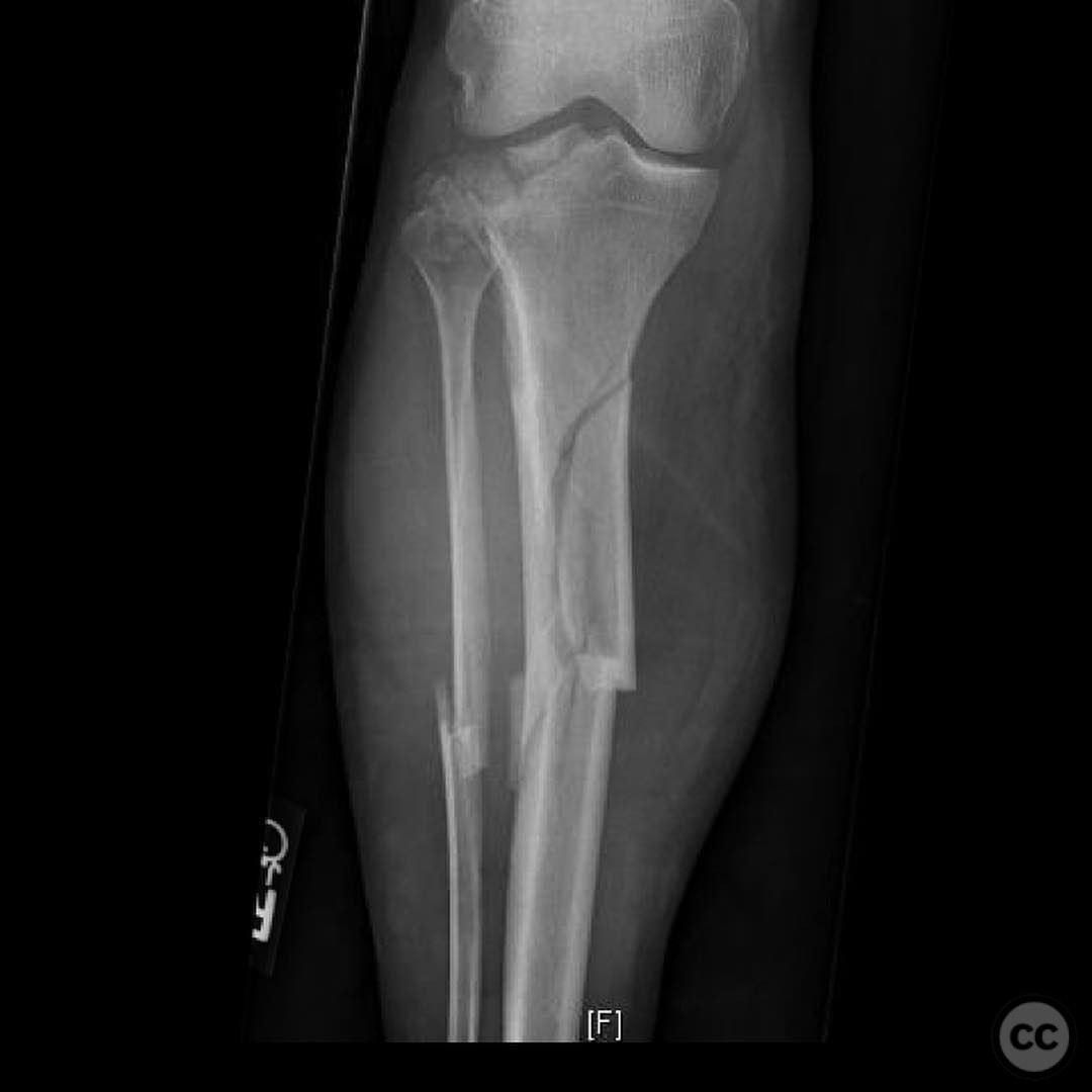
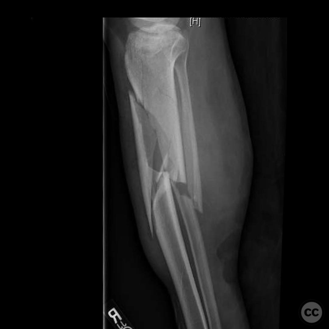
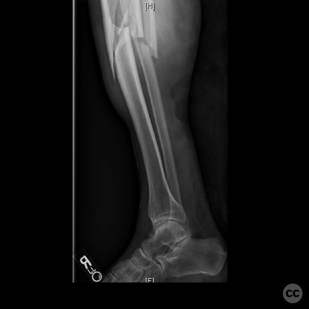
Clinical and radiological findings: A 23-year-old male bicyclist was involved in a collision with a truck bumper, resulting in a closed injury to the right lower extremity. Initial clinical evaluation revealed no signs of compartment syndrome. Radiological assessment, including CT imaging, identified a complex fracture pattern involving both the tibial plateau and the tibial shaft. The tibial plateau fracture was classified as AO/OTA 41-B3, indicating a partial articular fracture with depression and split components. The tibial shaft fracture was classified as AO/OTA 42-A1, a simple spiral fracture.
Preoperative Plan
Planning remarks: The preoperative plan involved addressing the tibial plateau fracture first using a standard lateral approach. The surgical strategy included the use of a varus moment for joint distraction and visualization, with careful placement of implants to avoid interference with subsequent intramedullary nailing of the tibial shaft. A blocking screw was planned to prevent valgus displacement during nailing.
Surgical Discussion
Patient positioning: The patient was positioned supine on the operating table. A leg holder was utilized to facilitate varus manipulation for joint distraction, with the knee slightly flexed to allow optimal access to the lateral aspect of the tibia.
Anatomical surgical approach: A lateral parapatellar approach was employed for the tibial plateau fixation. The incision extended from the lateral aspect of the patella, curving distally along the lateral border of the tibial tuberosity. Subperiosteal dissection was performed to expose the lateral tibial plateau. Joint visualization was enhanced using a medial and lateral dialing distractor system.
Operative remarks:Intraoperatively, careful sequencing was critical due to the complex fracture pattern. The tibial plateau was stabilized first using standard fixation techniques, ensuring that implant placement did not obstruct subsequent nailing of the shaft fracture. A blocking screw was inserted to prevent valgus malalignment during intramedullary nail insertion. An intraoperative flat plate X-ray was obtained prior to locking the nail to confirm accurate alignment, as fluoroscopic assessment alone was deemed insufficient.
Postoperative protocol: Postoperatively, the patient was placed in a hinged knee brace allowing controlled range of motion exercises starting from day one. Toe-touch weight bearing (TTWB) was advised for the first six weeks, progressing to partial weight bearing (PWB) by week seven, and full weight bearing (FWB) by week twelve, contingent on radiographic evidence of healing.
Follow up: Not specified.
Orthopaedic implants used: Lateral tibial plateau locking plate, intramedullary nail, blocking screw.
Search for Related Literature

orthopaedic_trauma
- United States , Seattle
- Area of Specialty - General Trauma
- Position - Specialist Consultant

Industry Sponsership
contact us for advertising opportunities
Article viewed 295 times
23 Jul 2025
Add to Bookmarks
Full Citation
Cite this article:
Surname, Initial. (2025). Complex Ipsilateral Tibial Plateau and Shaft Fracture Management in a Young Adult Cyclist.. Journal of Orthopaedic Surgery and Traumatology. Case Report 2325883 Published Online Jul 23 2025.