Pseudoarthrosis of Proximal Metaphyseal Tibia with Suprapatellar Tibial Nail Fixation
Score and Comment on this Case
Clinical Details
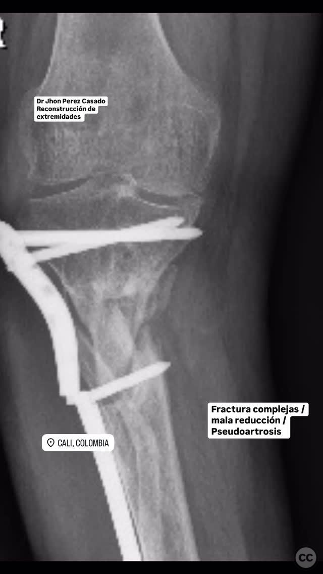
Clinical and radiological findings: An 80-year-old patient presented with a complex fracture of the proximal metaphyseal tibia, characterized by poor reduction and subsequent pseudoarthrosis. Radiological evaluation confirmed the presence of a non-union at the fracture site. The AO/OTA classification for this fracture is likely 41-A3, indicating a proximal tibial fracture with involvement of the metaphysis.
Preoperative Plan
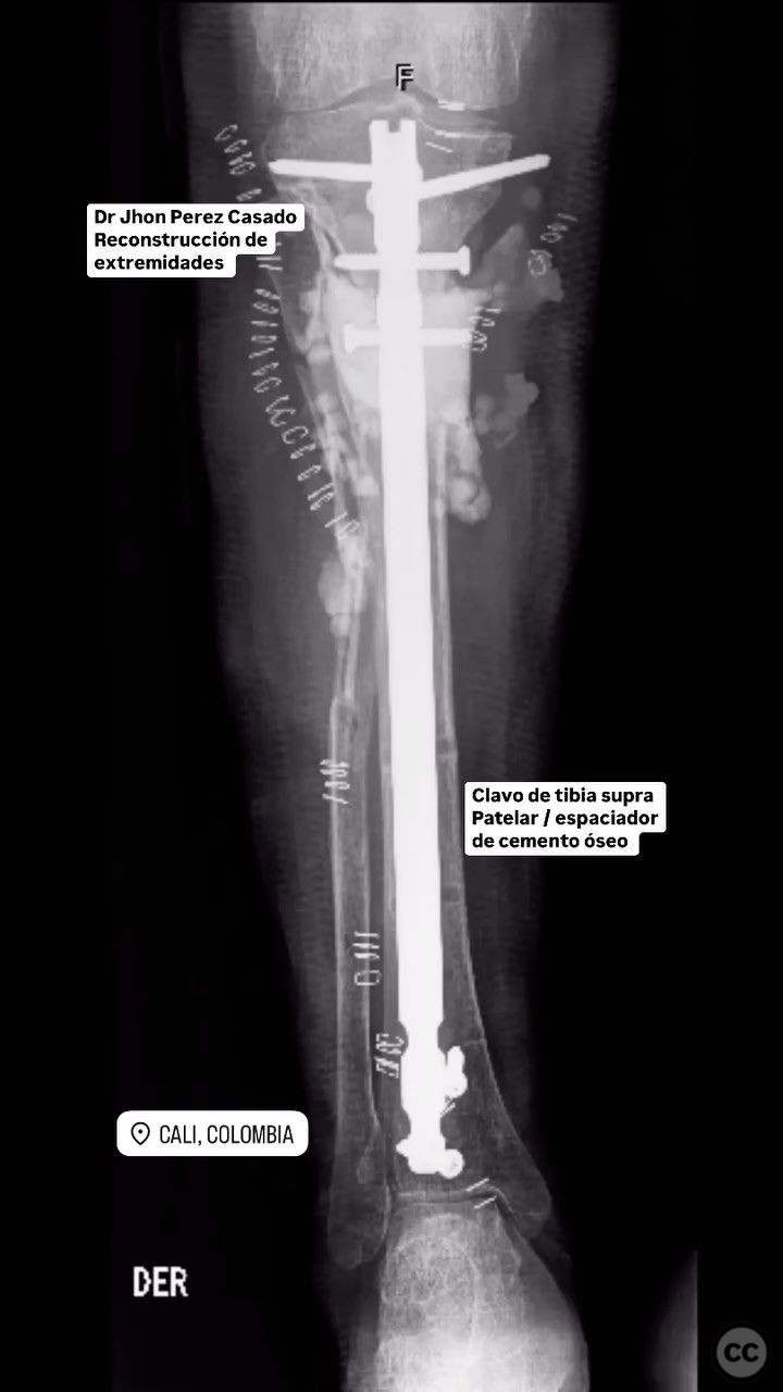
Planning remarks: The preoperative plan included removal of existing material, resection of the pseudoarthrosis, and stabilization using a suprapatellar tibial nail. Posterior bone substitute and a cement spacer were planned to address bone loss and provide structural support.
Surgical Discussion
Patient positioning: The patient was positioned supine on the operating table. A radiolucent leg holder was utilized to facilitate access to the proximal tibia and allow for intraoperative imaging.
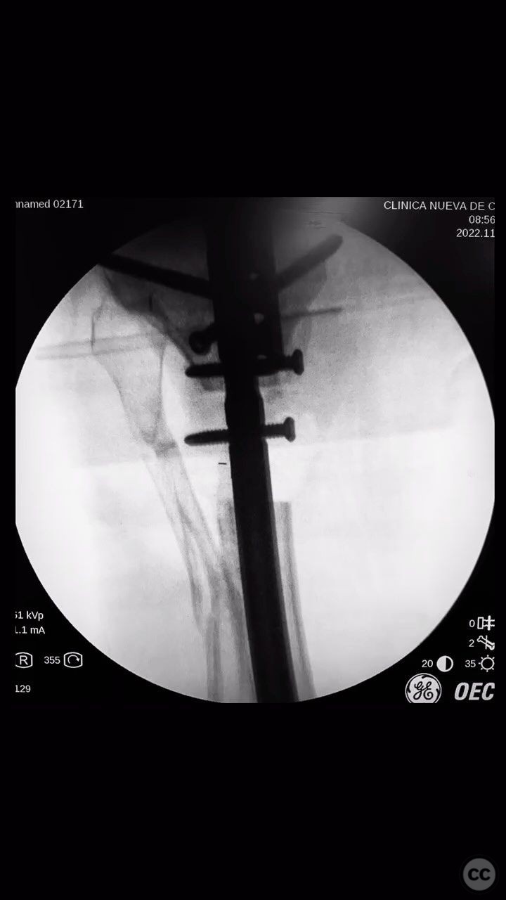
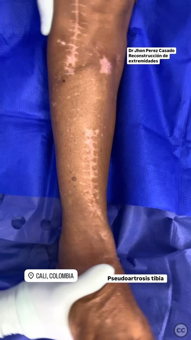
Anatomical surgical approach: A longitudinal incision was made over the patellar tendon, extending proximally to allow access to the knee joint. The suprapatellar approach was utilized, involving an arthrotomy through the quadriceps tendon to gain entry into the knee joint and access the proximal tibia for nail insertion.
Operative remarks:The surgeon noted that the pseudoarthrosis required meticulous resection to achieve viable bone ends. The existing hardware was removed, and a suprapatellar tibial nail was inserted to stabilize the fracture. Posterior bone substitute was applied to fill any voids, and a cement spacer was used to provide additional support and promote healing.
Postoperative protocol: Postoperatively, the patient was advised to maintain non-weight bearing status for 6 weeks, followed by gradual progression to partial weight bearing as tolerated. Physical therapy focused on range of motion exercises for the knee and strengthening of the quadriceps and hamstring muscles.
Follow up: Not specified.
Orthopaedic implants used: Suprapatellar tibial nail, posterior bone substitute, cement spacer.
Search for Related Literature

Jhon Perez Casado
- Colombia , Cali - Valle del Cauca
- Area of Specialty - Lower Limb
- Position - Specialist Consultant

Industry Sponsership
contact us for advertising opportunities
Article viewed 279 times
23 Jul 2025
Add to Bookmarks
Full Citation
Cite this article:
Perez Casado, J.J.. (2025). Pseudoarthrosis of Proximal Metaphyseal Tibia with Suprapatellar Tibial Nail Fixation. Journal of Orthopaedic Surgery and Traumatology. Case Report 22722312 Published Online Jul 23 2025.