Complex Distal Femur Fracture with Osteomyelitis and Hardware Loosening
Score and Comment on this Case
Clinical Details
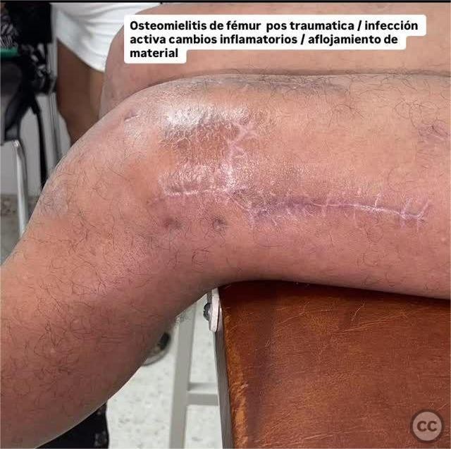
Clinical and radiological findings: The patient presented with a complex distal femur fracture following a traffic accident, complicated by active infection, hardware loosening, and osteomyelitis. Initial radiological assessment revealed instability of the lateral plates and evidence of infection around the fracture site.
Preoperative Plan
Planning remarks: The preoperative plan involved the removal of unstable lateral plates, radical debridement of infected soft tissue and bone, and placement of a provisional cement spacer. Cultures were to be obtained to guide antibiotic therapy, and broad-spectrum antibiotics were initiated.
Surgical Discussion
Patient positioning: The patient was positioned supine on the operating table to facilitate access to the lateral aspect of the distal femur.
Anatomical surgical approach: A lateral approach to the distal femur was utilized. The incision was made over the previous surgical scar, extending proximally and distally as needed to expose the hardware. Subperiosteal dissection was performed to access the lateral plates, which were removed. Radical debridement of infected tissue was carried out, followed by placement of a provisional cement spacer.
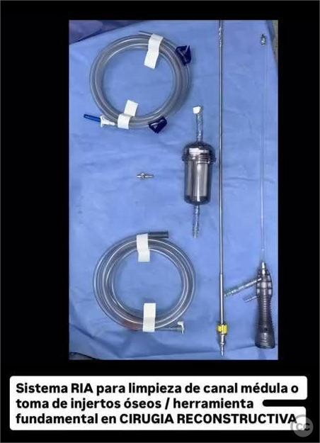
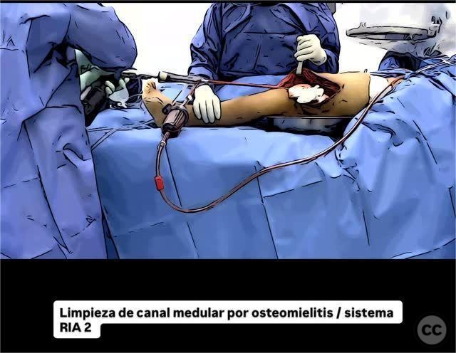
Operative remarks:The surgeon noted significant purulence and necrotic tissue at the site of infection. Thorough irrigation and debridement were performed. The intramedullary canal was cleaned using the RIA system 2. A temporary spacer with bone cement was placed to maintain limb length and alignment while addressing the infection.
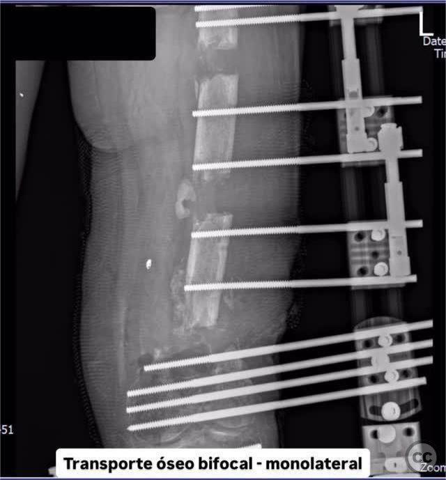
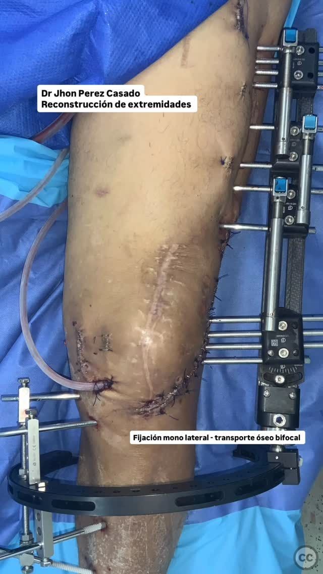
Postoperative protocol: Postoperatively, the patient was managed with a monolateral external fixation device for bifocal bone transport. Tricalcium phosphate was used to fill the bone defect. Antibiotic therapy was adjusted based on culture results.
Follow up: Not specified.
Orthopaedic implants used: Provisional cement spacer, monolateral external fixation system, tricalcium phosphate.
Search for Related Literature

Jhon Perez Casado
- Colombia , Cali - Valle del Cauca
- Area of Specialty - Lower Limb
- Position - Specialist Consultant

Industry Sponsership
contact us for advertising opportunities
Article viewed 259 times
19 Jul 2025
Add to Bookmarks
Full Citation
Cite this article:
Perez Casado, J.J.. (2025). Complex Distal Femur Fracture with Osteomyelitis and Hardware Loosening. Journal of Orthopaedic Surgery and Traumatology. Case Report 22710544 Published Online Jul 19 2025.