Low Energy Femoral Stress Fracture in a Young Runner
Score and Comment on this Case
Clinical Details
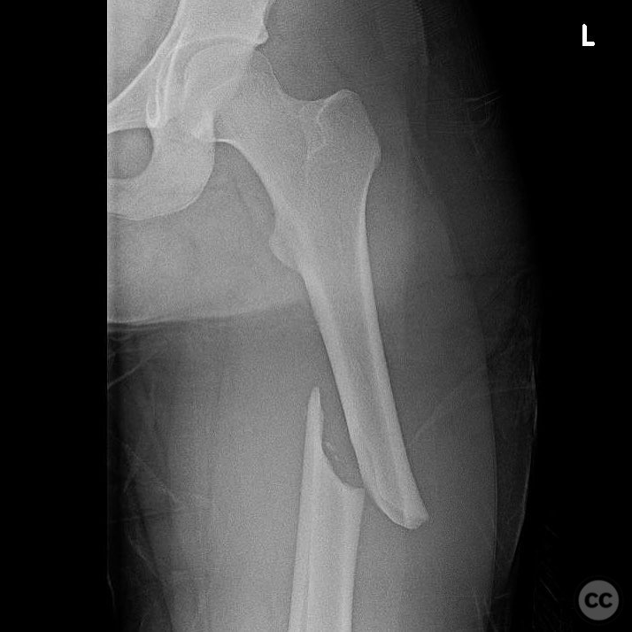
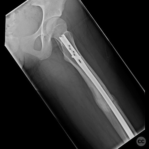
Clinical and radiological findings: A young female recreational runner presented with a low energy femoral fracture. She had no significant past medical or surgical history, maintained a balanced diet, and was not on any medications. There was no preceding or contralateral thigh pain. CT imaging revealed very small intracortical cysts without any soft tissue component. Nutritional and metabolic laboratory results were normal. Initial working diagnosis was a completed stress fracture.
Preoperative Plan
Planning remarks: The preoperative plan included an open biopsy to obtain a specimen for diagnosis, given the unusual presentation and absence of risk factors. The biopsy was performed to rule out any neoplastic processes before proceeding with definitive treatment.
Surgical Discussion
Patient positioning: Supine positioning on the operating table was utilized for both the biopsy and subsequent intramedullary nailing procedure.
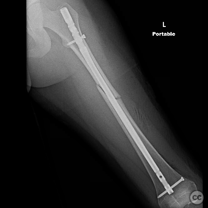
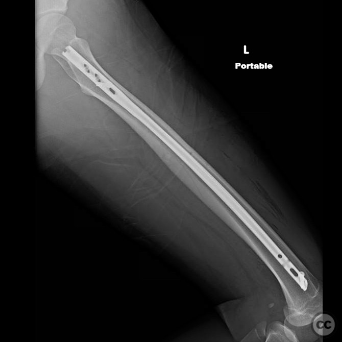
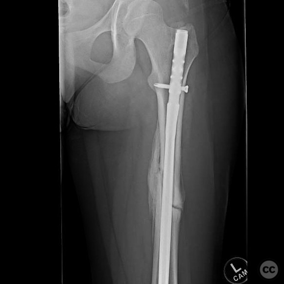
Anatomical surgical approach: A lateral approach to the femur was employed for the open biopsy, ensuring adequate exposure for specimen collection. Following confirmation of an adequate specimen, an antegrade intramedullary nailing was performed through a standard piriformis fossa entry point.
Operative remarks:The surgeon noted the importance of considering a pathologic fracture in low energy femoral fractures in young patients and emphasized the necessity of thorough workup before definitive treatment. The open biopsy was crucial in ruling out neoplastic causes, allowing for appropriate management with intramedullary nailing.
Postoperative protocol: Postoperative rehabilitation included protected weight-bearing with crutches for 6 weeks, followed by gradual progression to full weight-bearing as tolerated. Range of motion exercises were initiated immediately postoperatively to maintain joint mobility.
Follow up: Not specified.
Orthopaedic implants used: Antegrade intramedullary nail.
Search for Related Literature

orthopaedic_trauma
- United States , Seattle
- Area of Specialty - General Trauma
- Position - Specialist Consultant

Industry Sponsership
contact us for advertising opportunities
Article viewed 289 times
28 Jul 2025
Add to Bookmarks
Full Citation
Cite this article:
Surname, Initial. (2025). Low Energy Femoral Stress Fracture in a Young Runner. Journal of Orthopaedic Surgery and Traumatology. Case Report 2211833 Published Online Jul 28 2025.