High-energy polytrauma with open femoral shaft fracture and associated knee instability.
Score and Comment on this Case
Clinical Details
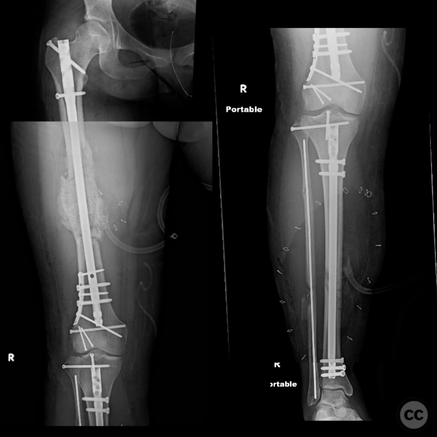
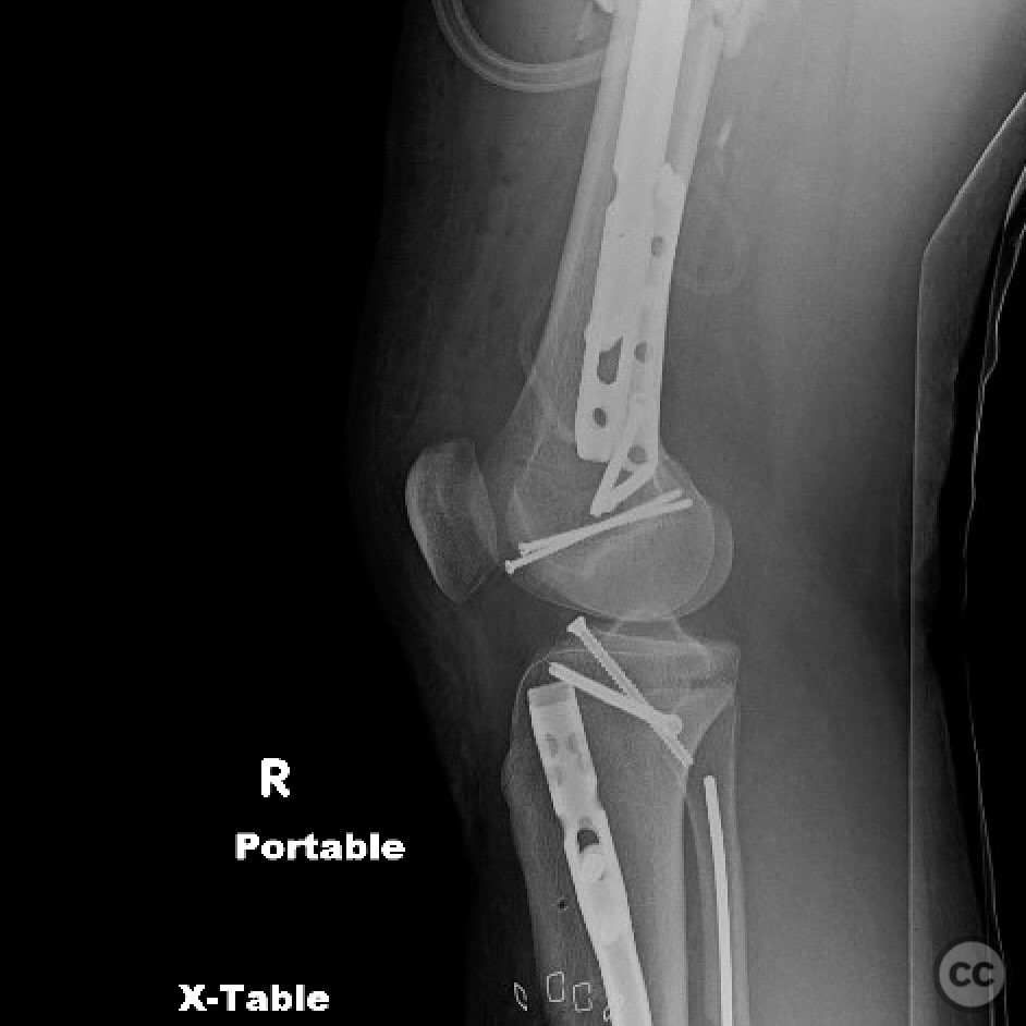
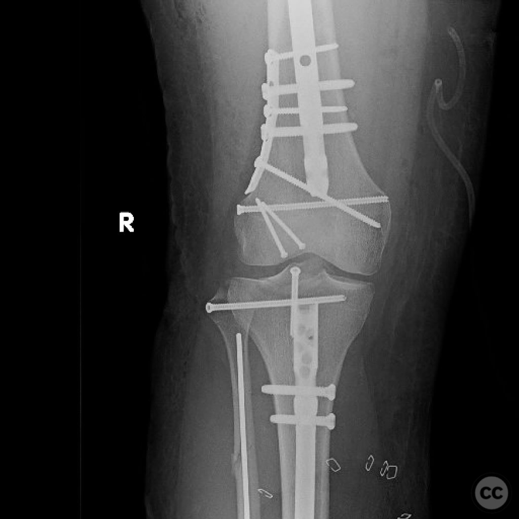
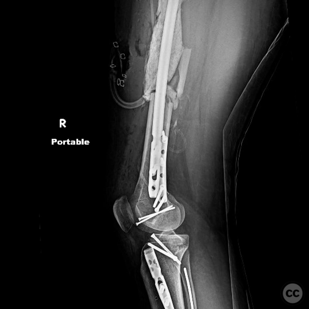
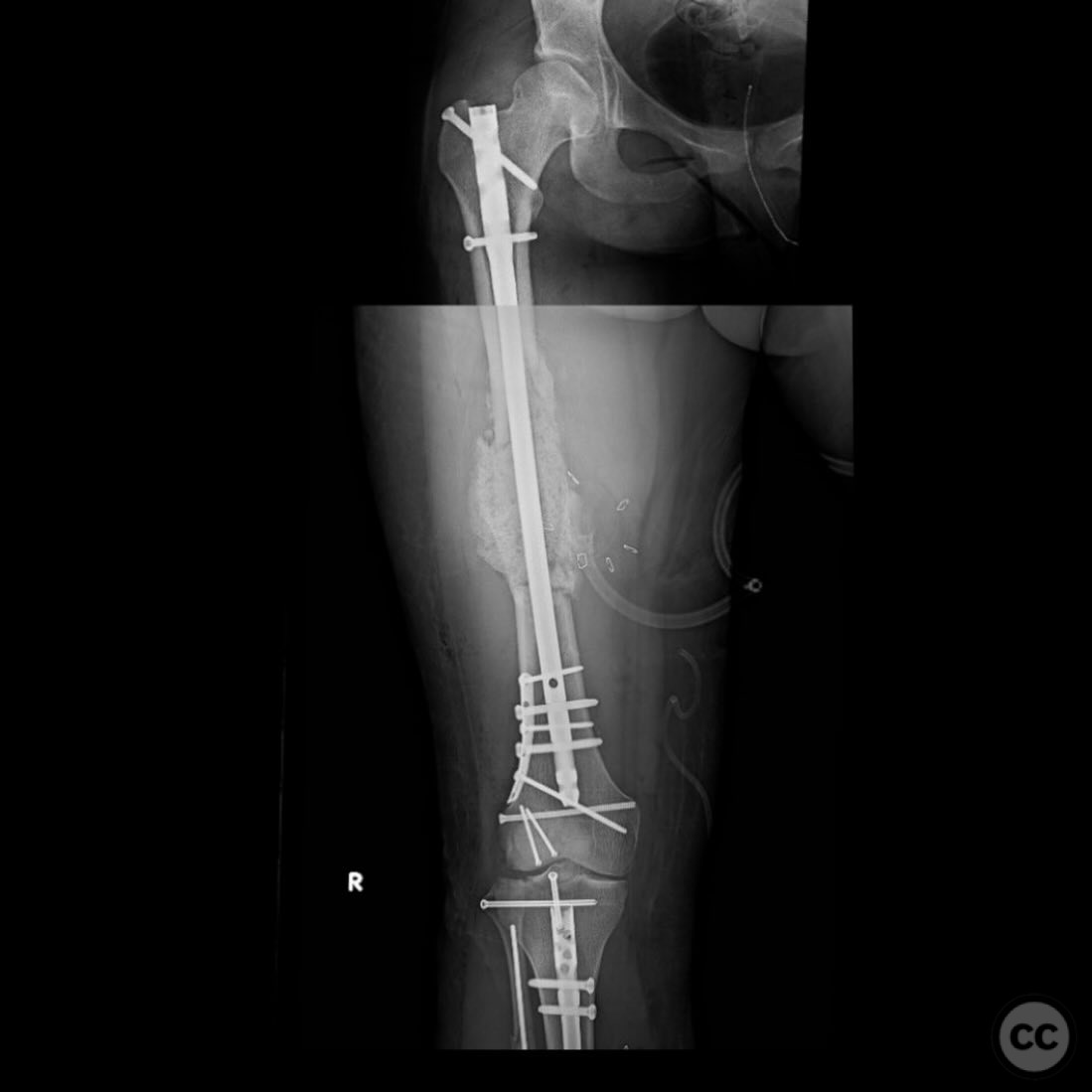
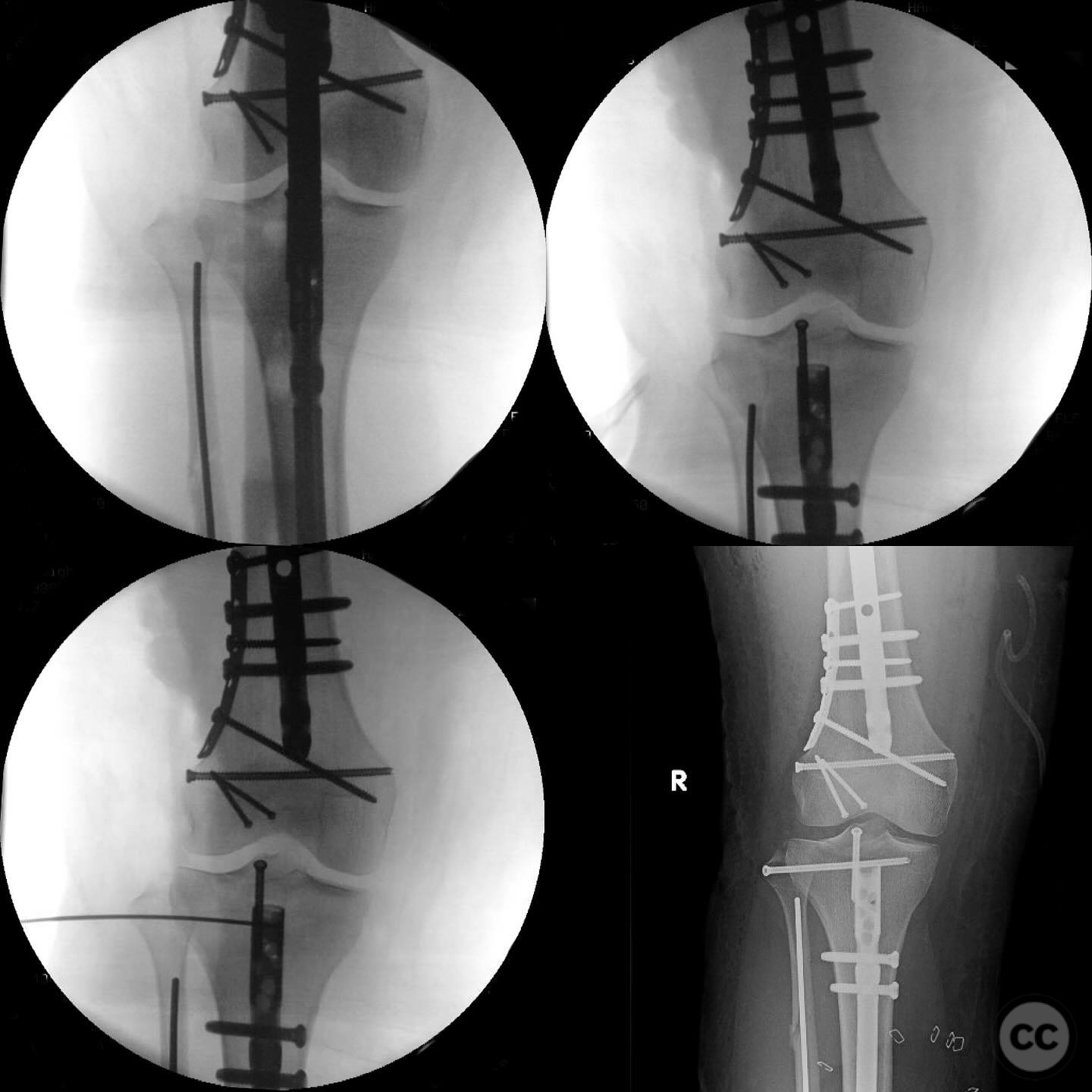
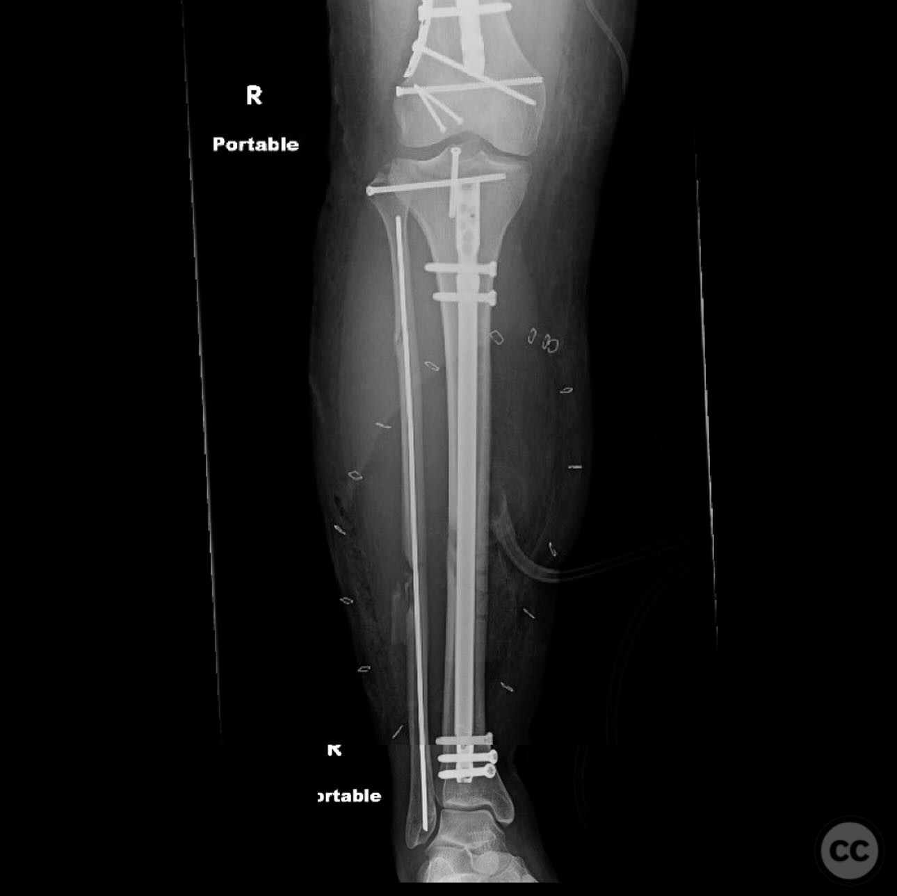
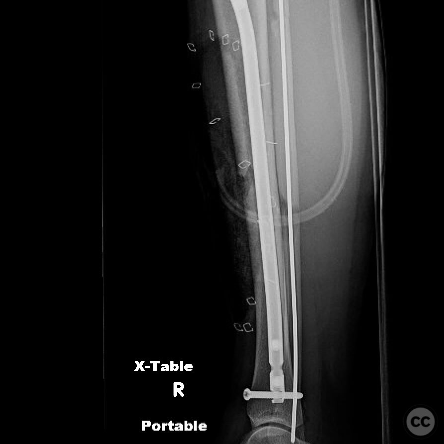
Clinical and radiological findings: A 23-year-old female involved in a high-speed motorcycle collision presented as a polytrauma case. Initial assessment revealed no major head or abdominal injuries. The patient was well resuscitated with a lactate level of 3 upon arrival, and ongoing resuscitation was in progress. Bilateral pneumothoraces were managed with chest tubes. The patient sustained an open diaphyseal femur fracture with several direct anterior irregular wounds, classified as AO/OTA 32-C3. The distal femur fracture was closed. The diaphyseal tibia fracture was closed but complicated by a massive Morel-Lavallée lesion involving the leg. There was no vascular injury, and compartment pressures were soft. A segmental fibula fracture with proximal interosseous membrane disruption and proximal tibiofibular joint dislocation was noted, along with a displaced tibial spine fracture causing knee instability.
Preoperative Plan
Planning remarks: The preoperative plan included antegrade intramedullary nailing of the femur to allow for potential exchange nailing if necessary. The femoral length and version were to be measured using fluoroscopy on the operating table, referencing the contralateral femur. Management of the critical size defect in the femur was planned using intramedullary transport (IMT) as it is suitable for the defect size and location and is less burdensome for the patient.
Surgical Discussion
Patient positioning: The patient was positioned supine on a radiolucent table to facilitate fluoroscopic imaging and surgical access.
Anatomical surgical approach: A standard antegrade approach to the femur was utilized, involving a longitudinal incision over the greater trochanter. The entry point for the intramedullary nail was established at the piriformis fossa. The tibial fracture was addressed through a separate anteromedial incision, allowing access to the proximal tibiofibular joint and tibial spine.
Operative remarks:The surgical team noted the complexity of the tibial injury, emphasizing the need to address subtle abnormalities indicative of severe injury due to the high-energy mechanism. The segmental fibula fracture, proximal interosseous membrane disruption, and proximal tibiofibular joint dislocation were stabilized to provide a stable knee joint and skeletal structure, essential for supporting the compromised soft tissue envelope.
Postoperative protocol: Postoperative rehabilitation included non-weight bearing on the affected limb initially, with gradual progression to partial weight bearing as tolerated. Range of motion exercises for the knee were initiated early to prevent stiffness.
Follow up: Not specified.
Orthopaedic implants used: Intramedullary nail for femur, autologous bone grafting materials.
Search for Related Literature

orthopaedic_trauma
- United States , Seattle
- Area of Specialty - General Trauma
- Position - Specialist Consultant

Industry Sponsership
contact us for advertising opportunities
Article viewed 307 times
08 Jul 2025
Add to Bookmarks
Full Citation
Cite this article:
Surname, Initial. (2025). High-energy polytrauma with open femoral shaft fracture and associated knee instability.. Journal of Orthopaedic Surgery and Traumatology. Case Report 21986298 Published Online Jul 08 2025.