
Pelvic Ring Injury with Unstable LC3 Pattern Managed with Progressive EUA.
Score and Comment on this Case
Clinical Details
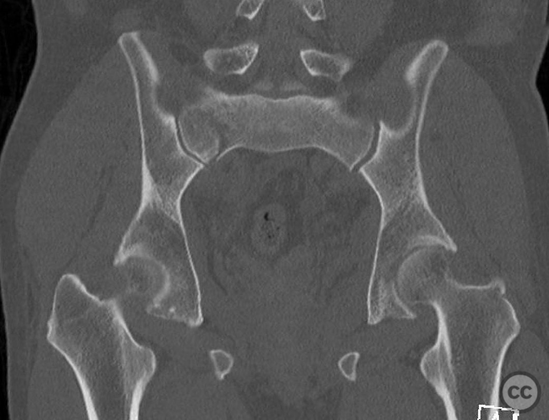
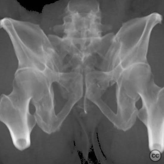
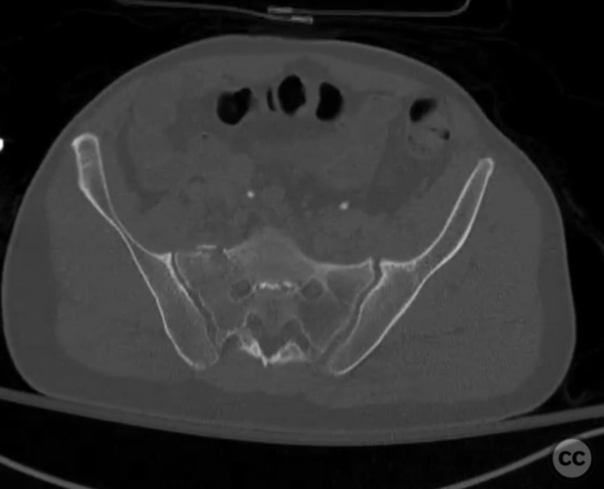
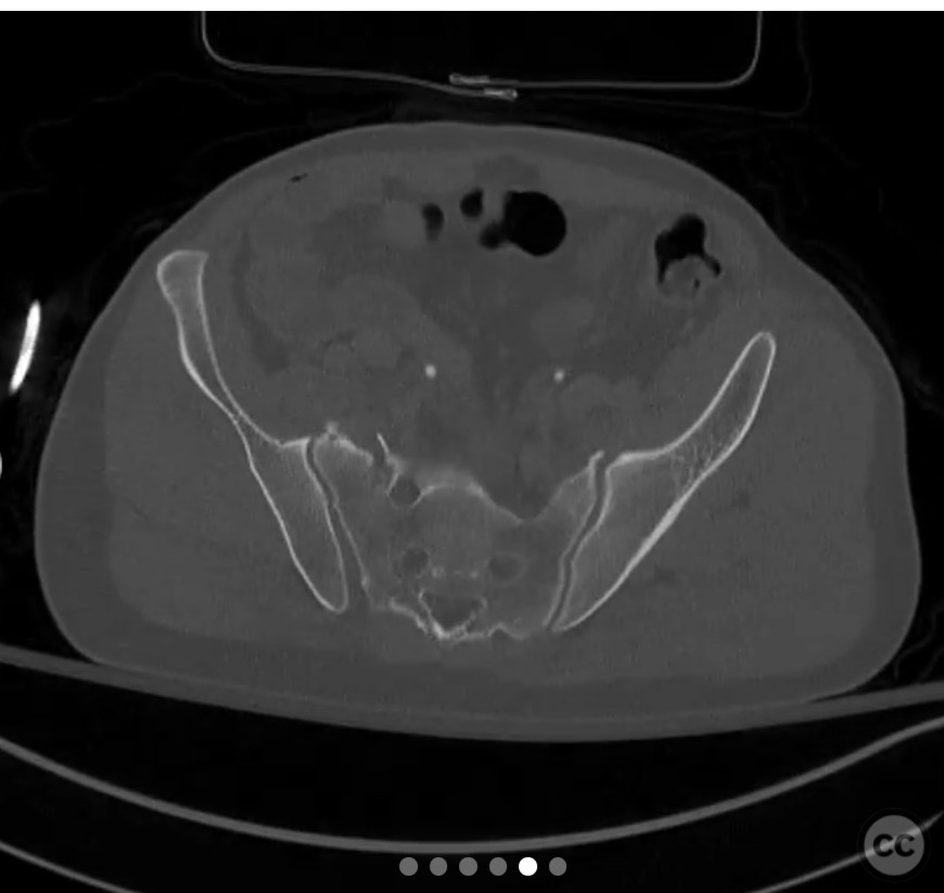
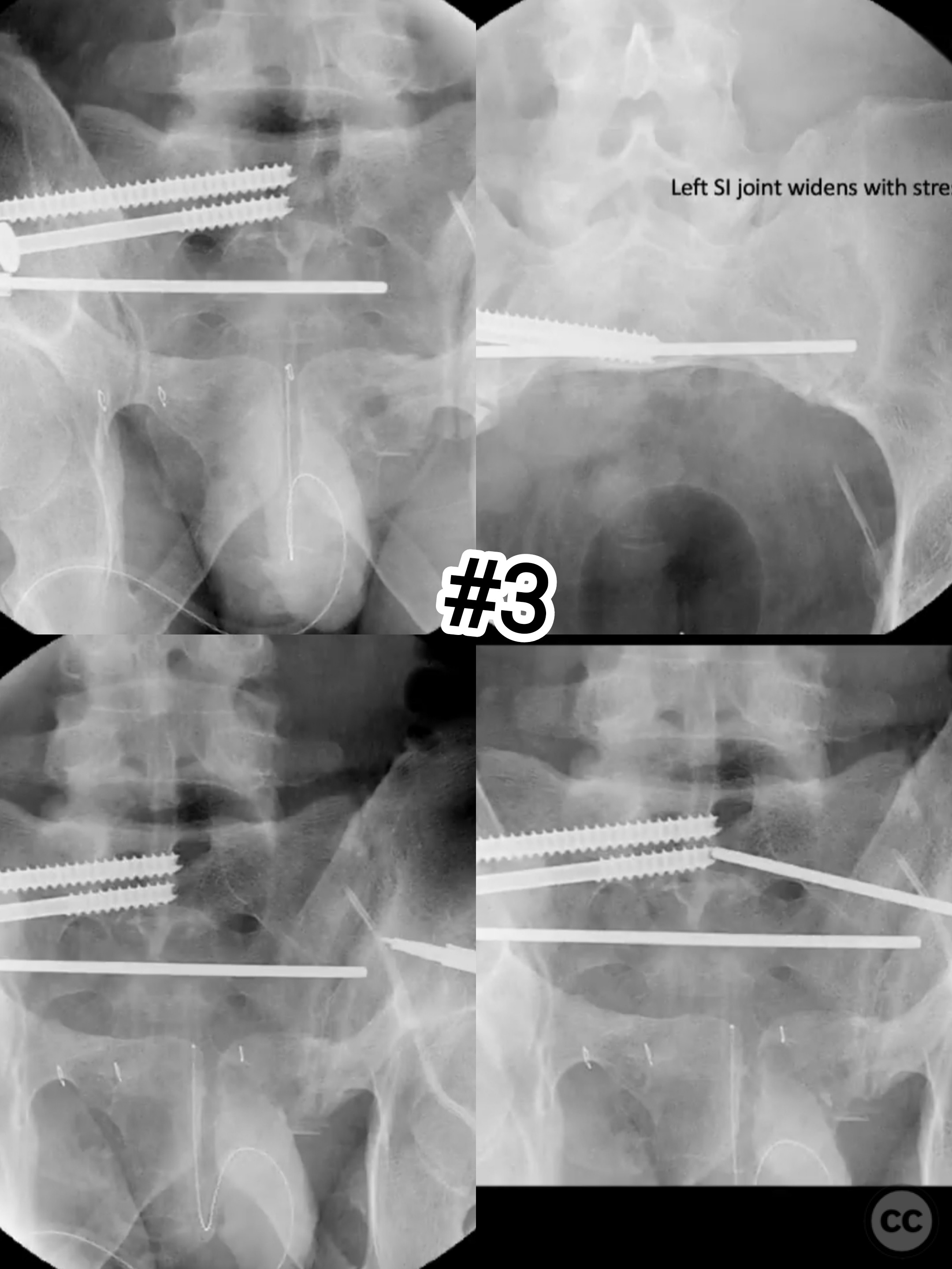
Clinical and radiological findings: A 43-year-old male involved in a motor vehicle collision presented with a high-energy mechanism of injury, having fallen from a height of two stories. Initial pelvic X-rays appeared unimpressive, showing normal ring morphology. However, CT imaging revealed subtle widening of the left sacroiliac (SI) joint, suggestive of an unstable pelvic ring injury. Despite initial impressions of an LC1 injury, further scrutiny identified this as an LC3 pattern, characterized by instability not immediately apparent on standard imaging due to potential masking by CT gantry angles.
Preoperative Plan
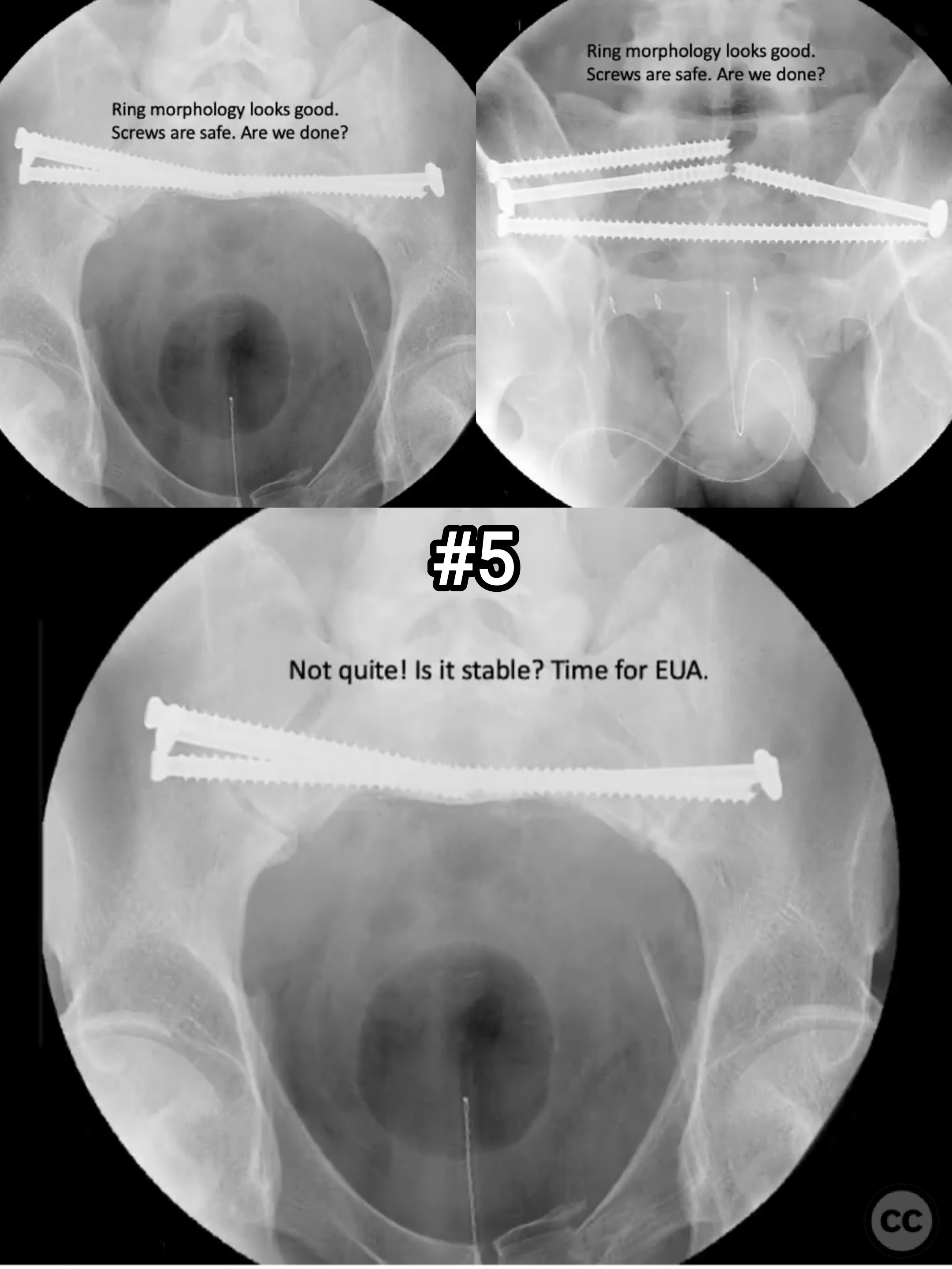
Planning remarks: The preoperative plan involved a progressive examination under anesthesia (EUA) to assess the degree of instability in the pelvic ring. Initial stabilization was planned for the right hemipelvis using screws to provide a stable base for further stress testing. Subsequent stress testing of the left SI joint was anticipated to reveal instability, necessitating stabilization with a screw. Anterior ring stability was to be assessed through lateral compression testing.
Surgical Discussion
Patient positioning: The patient was positioned supine on a radiolucent operating table to facilitate fluoroscopic imaging and access to both anterior and posterior aspects of the pelvis.
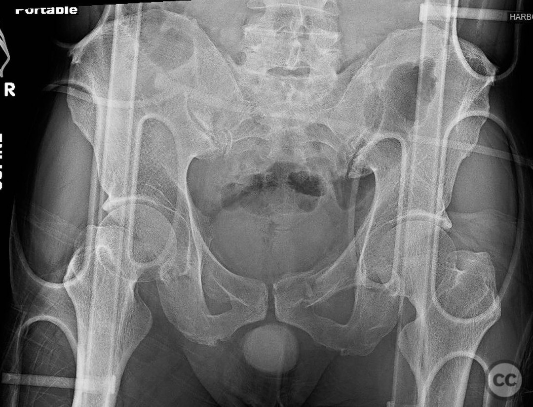
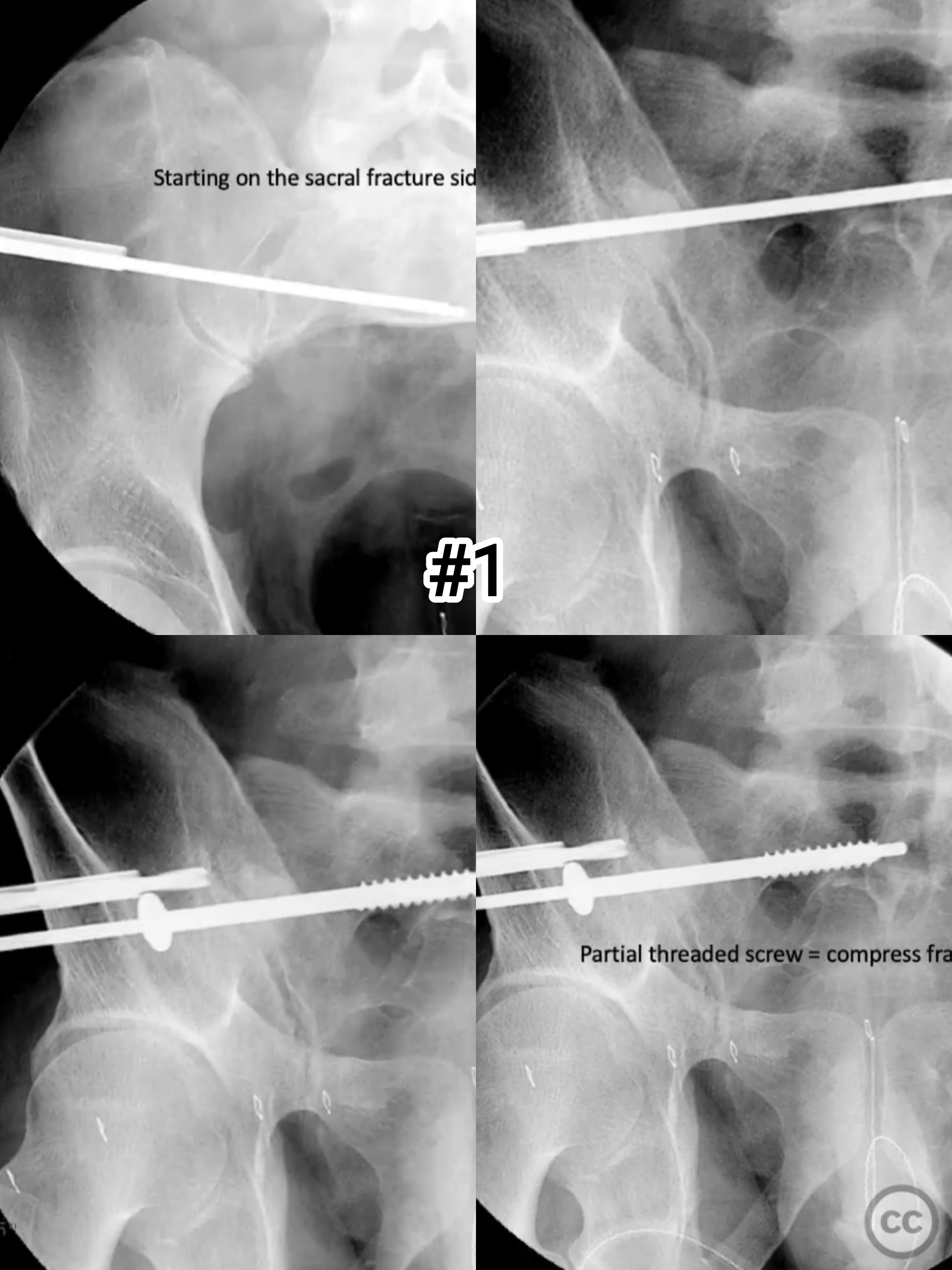
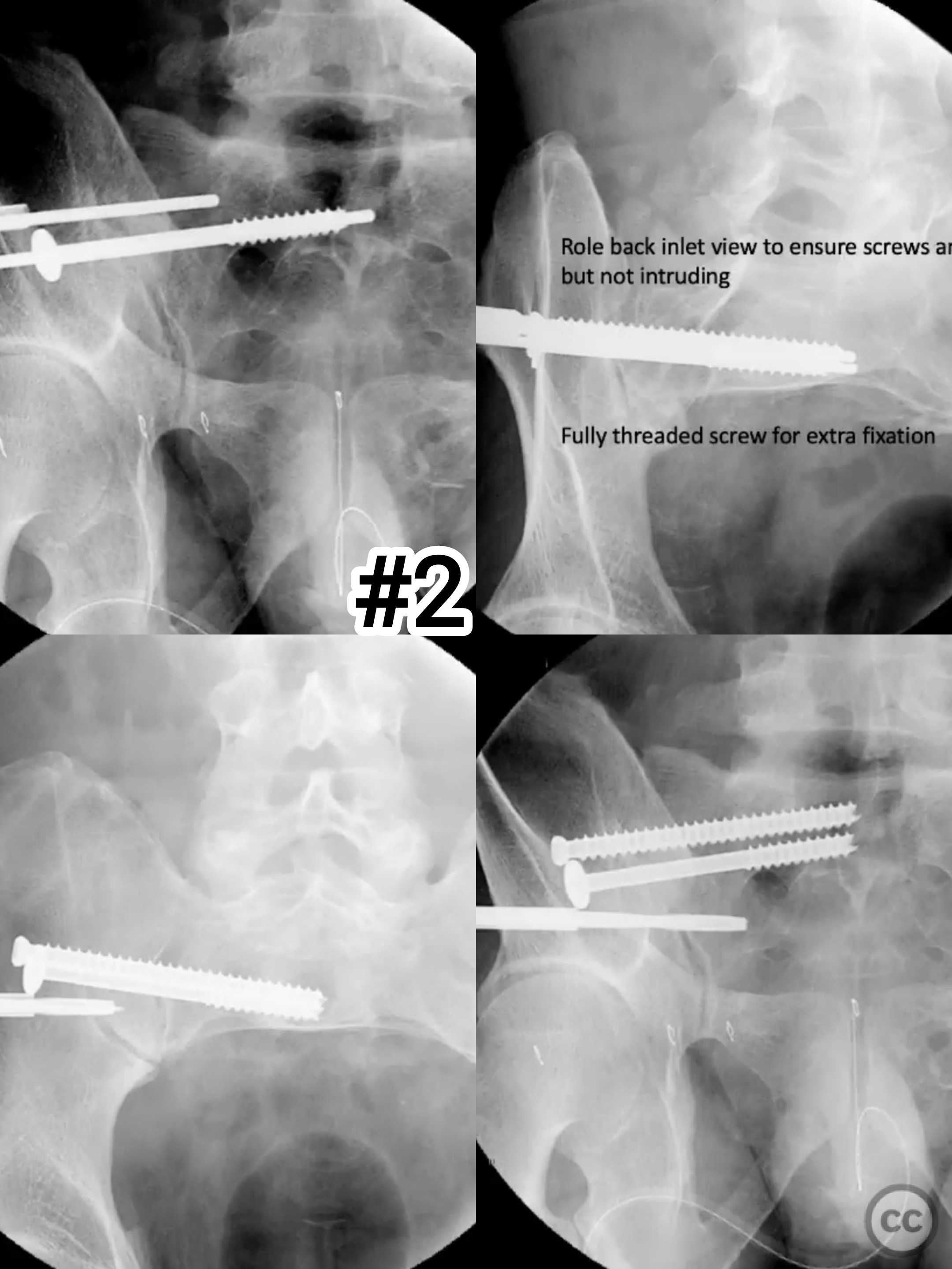
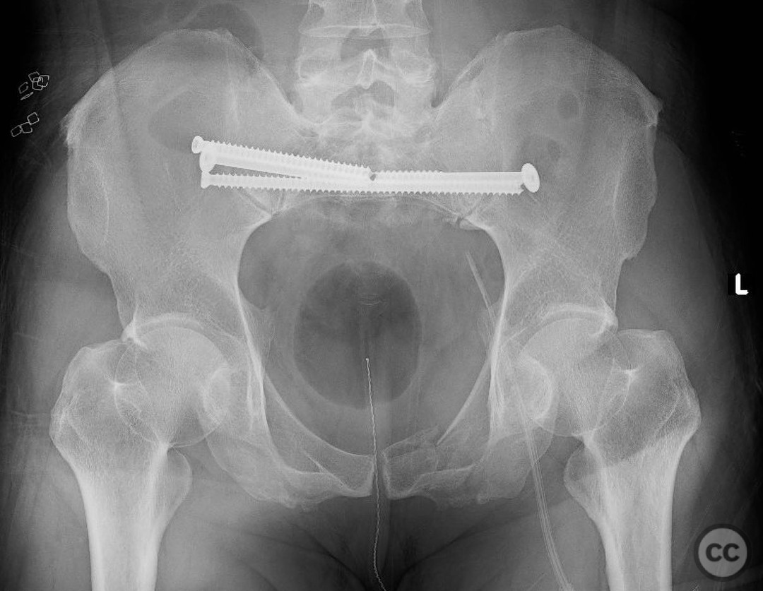
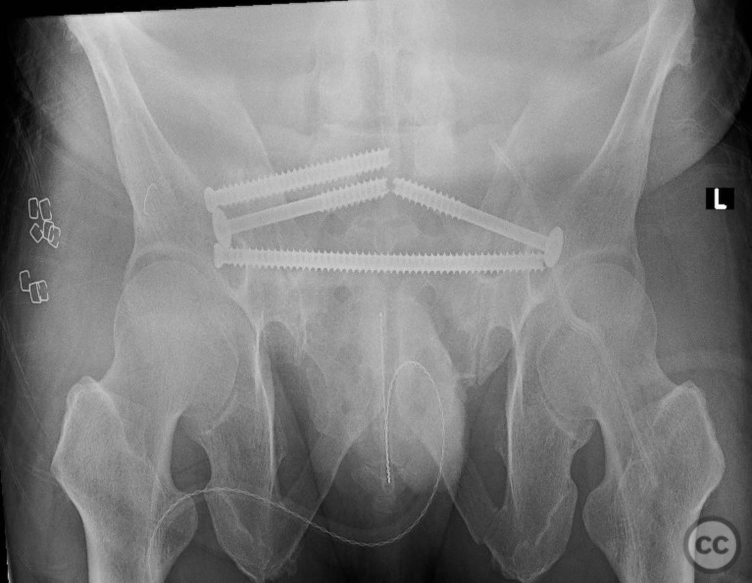
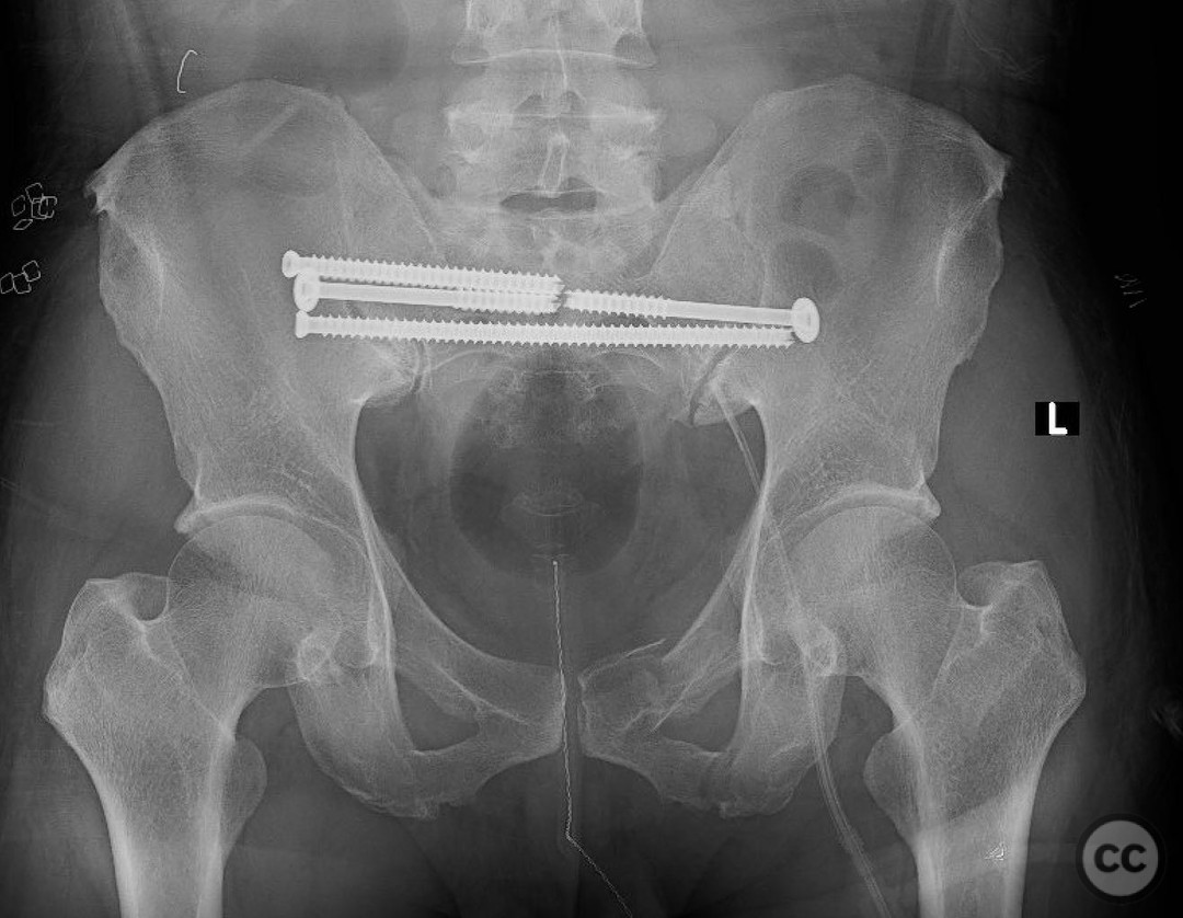
Anatomical surgical approach: A posterior approach was utilized for stabilization of the pelvic ring. Percutaneous screw fixation was performed on the right hemipelvis through small incisions overlying the posterior superior iliac spine and directed towards the sacroiliac joint. The left SI joint was similarly accessed for screw placement following stress testing.
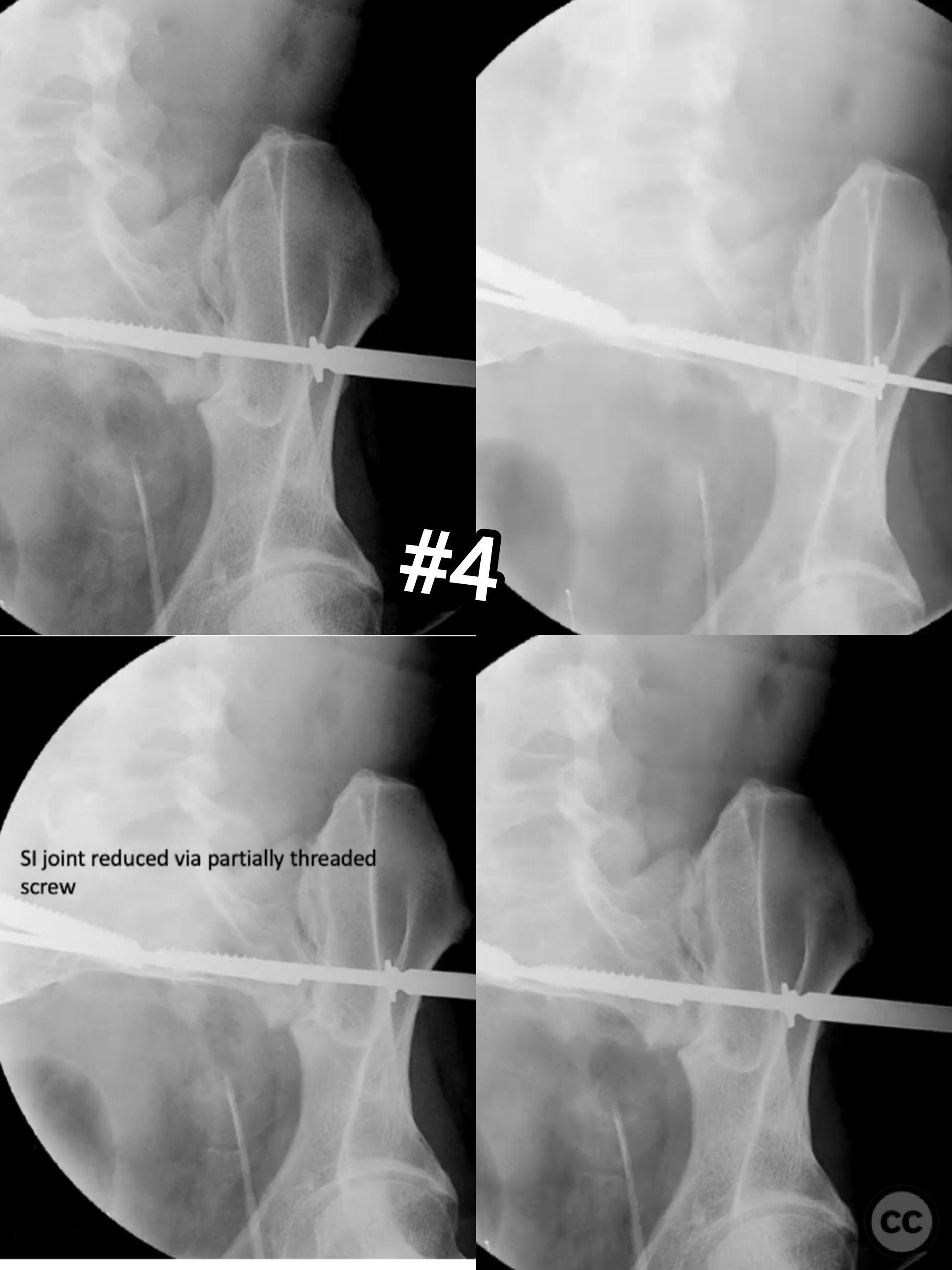
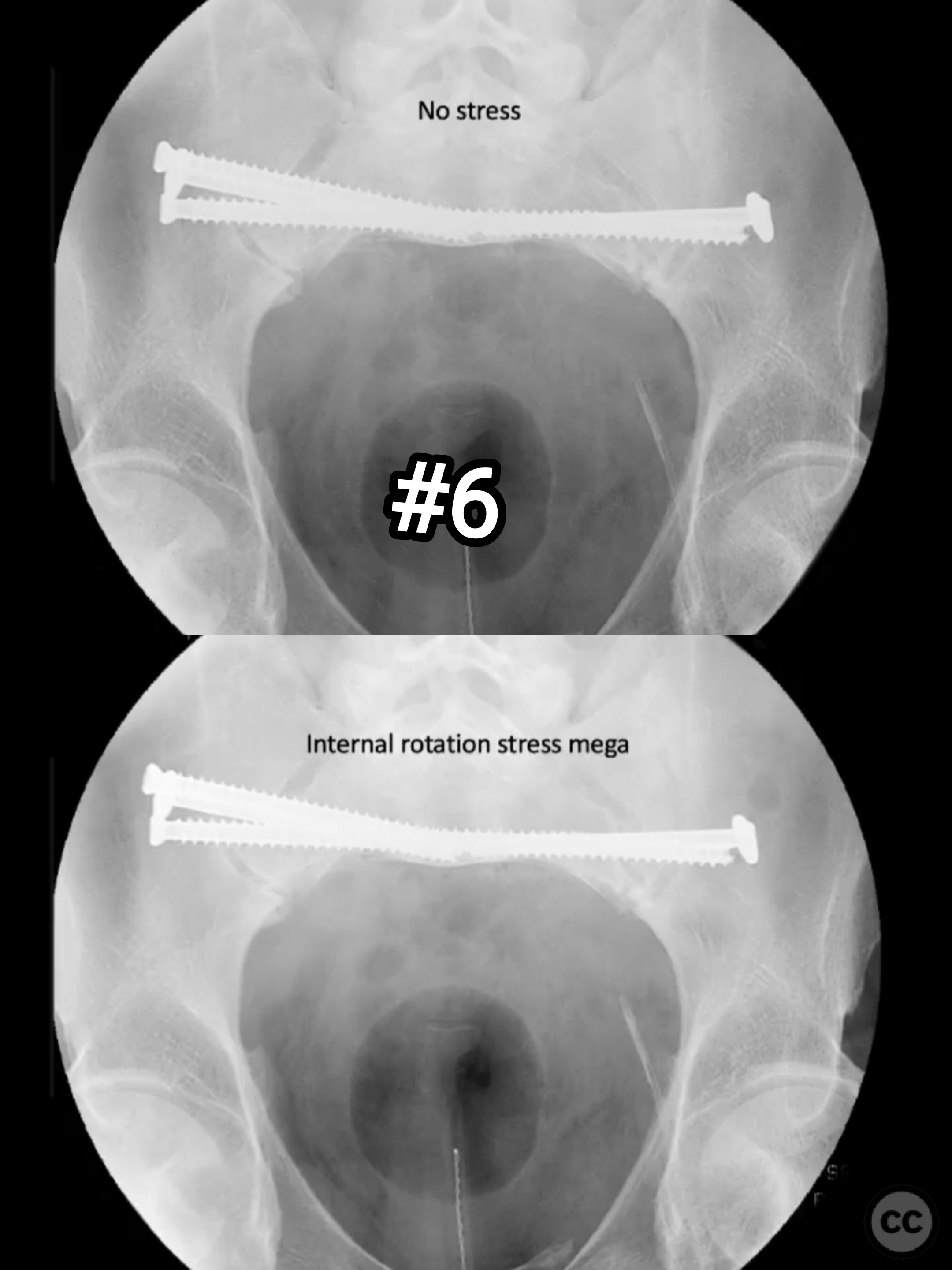
Operative remarks:Intraoperative findings confirmed significant instability of the left SI joint upon stress testing after stabilization of the right side. A screw was placed across the left SI joint to achieve stabilization. Further examination under anesthesia demonstrated no motion or deformity in the anterior ring upon lateral compression, obviating the need for anterior fixation. The concept of a progressive EUA was pivotal in guiding the selective fixation strategy, minimizing unnecessary surgical intervention and associated risks.
Postoperative protocol: Postoperatively, the patient was managed with partial weight-bearing restrictions on the affected side for six weeks, progressing to full weight-bearing as tolerated thereafter. Physical therapy focused on maintaining range of motion and strengthening exercises commenced immediately.
Follow up: Not specified.
Orthopaedic implants used: Percutaneous sacroiliac screws.
Search for Related Literature

orthopaedic_trauma
- United States , Seattle
- Area of Specialty - General Trauma
- Position - Specialist Consultant

Industry Sponsership
contact us for advertising opportunities
Article viewed 239 times
12 Jul 2025
Add to Bookmarks
Full Citation
Cite this article:
Surname, Initial. (2025). Pelvic Ring Injury with Unstable LC3 Pattern Managed with Progressive EUA.. Journal of Orthopaedic Surgery and Traumatology. Case Report 21451147 Published Online Jul 12 2025.