
Pilon Nonunion Repair with Deformity Correction
Score and Comment on this Case
Clinical Details
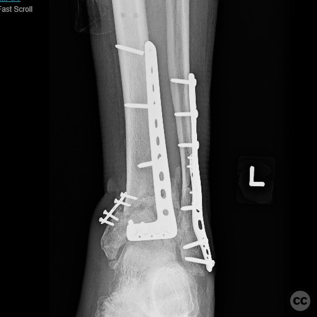
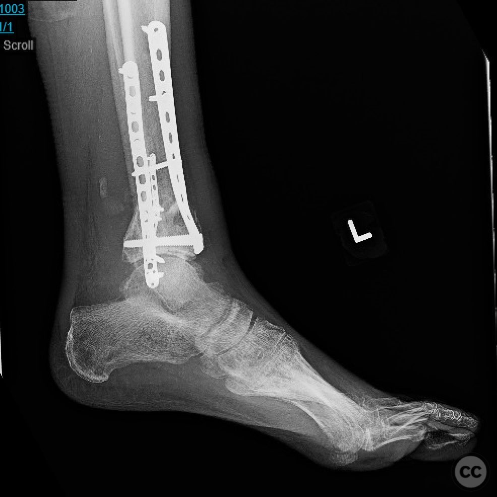
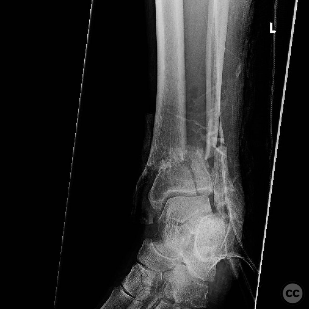
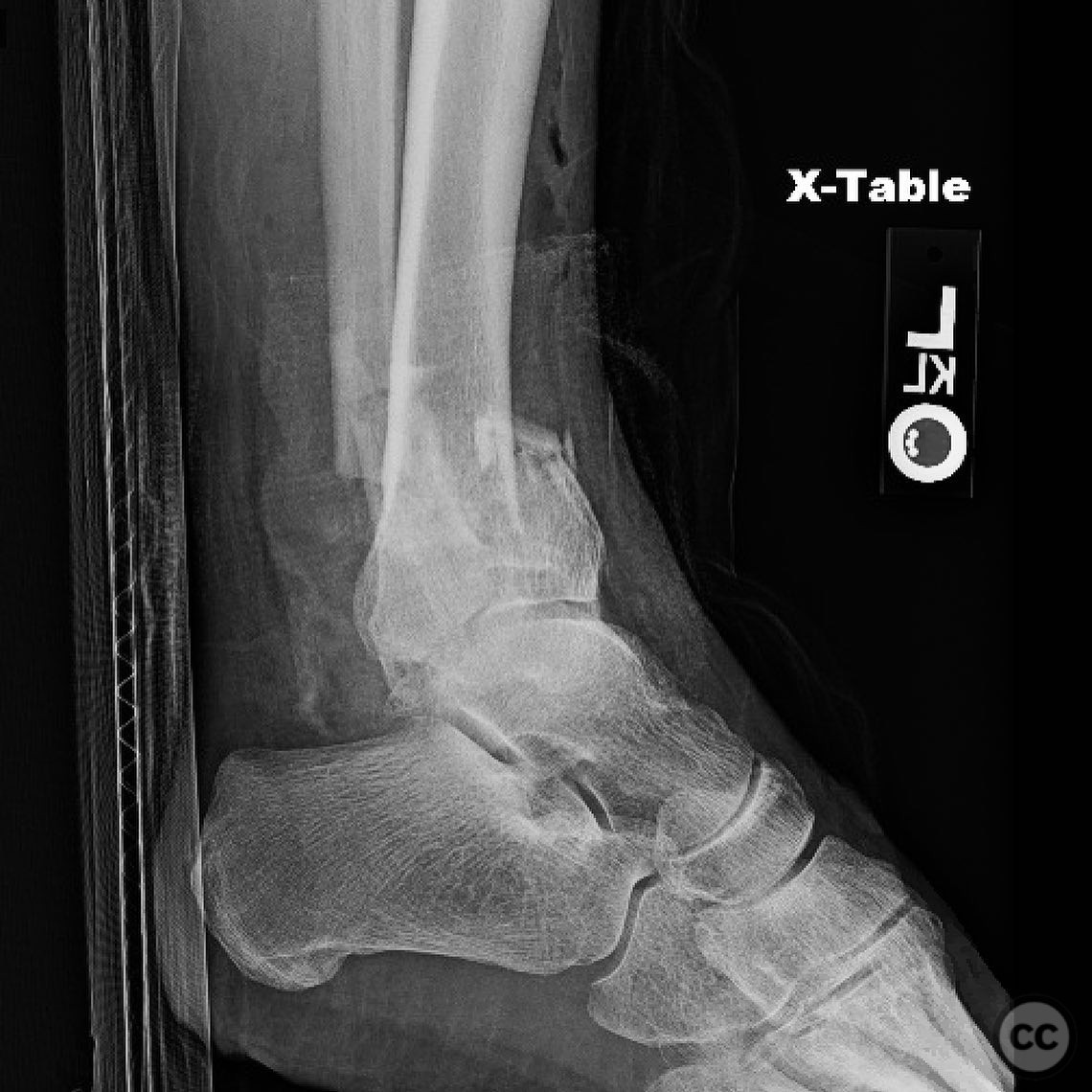
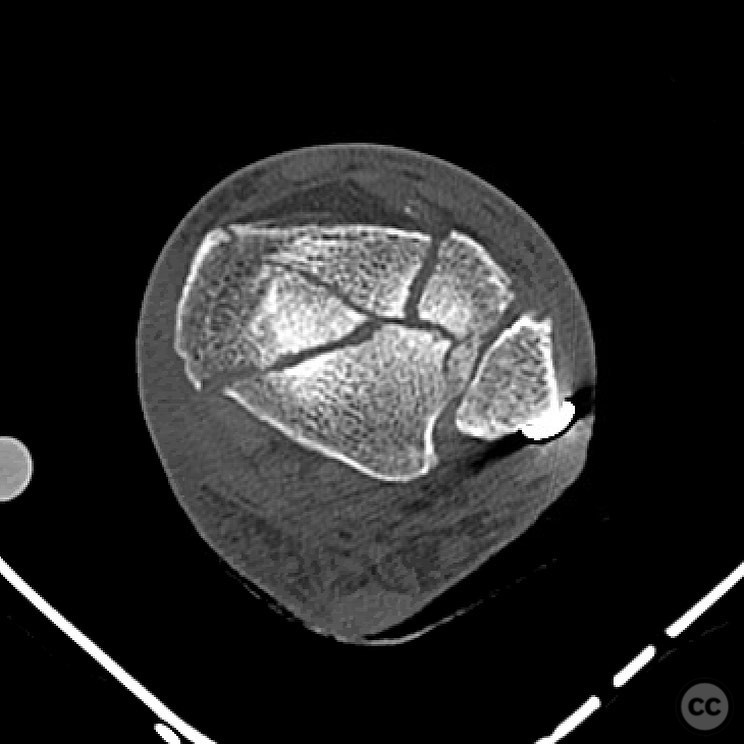
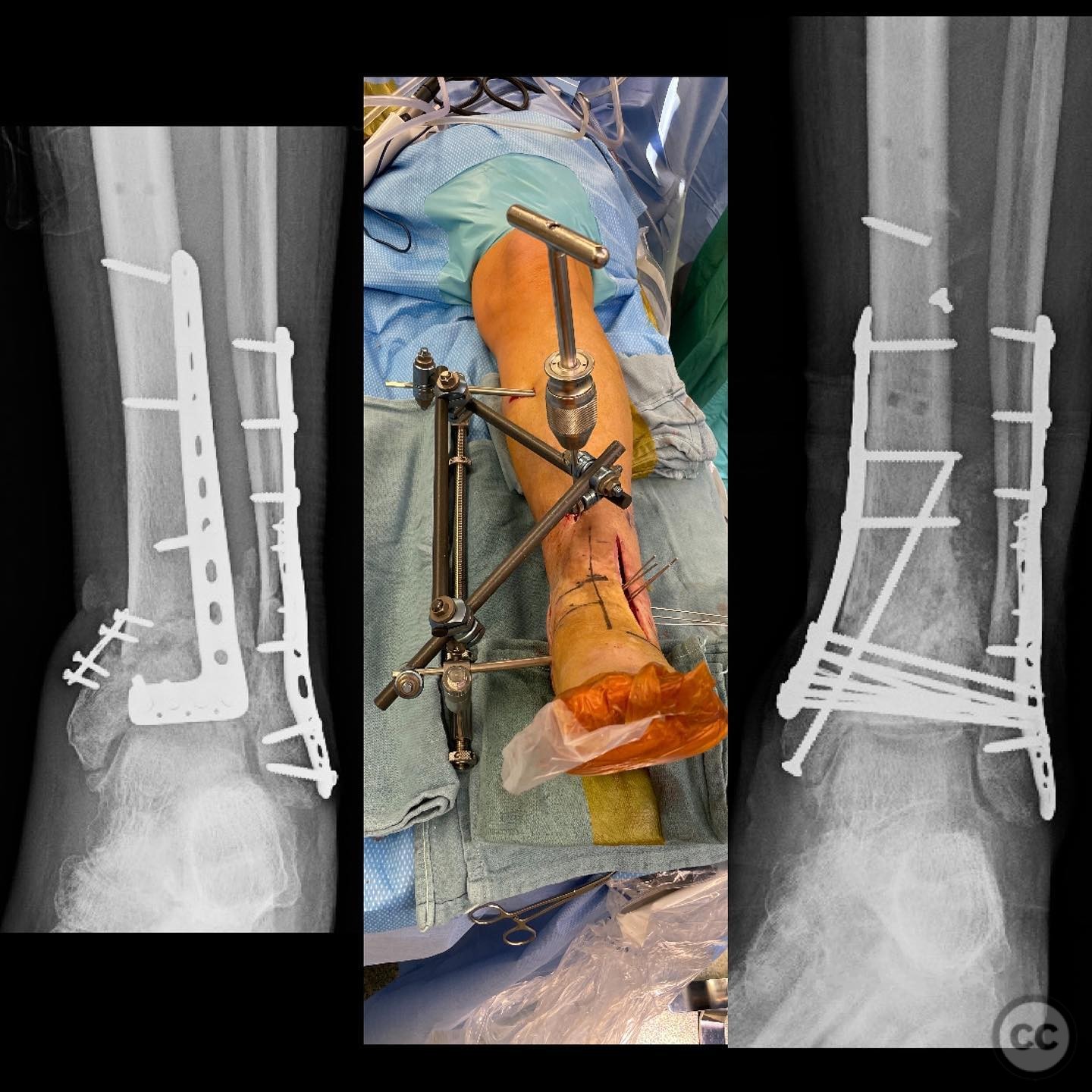
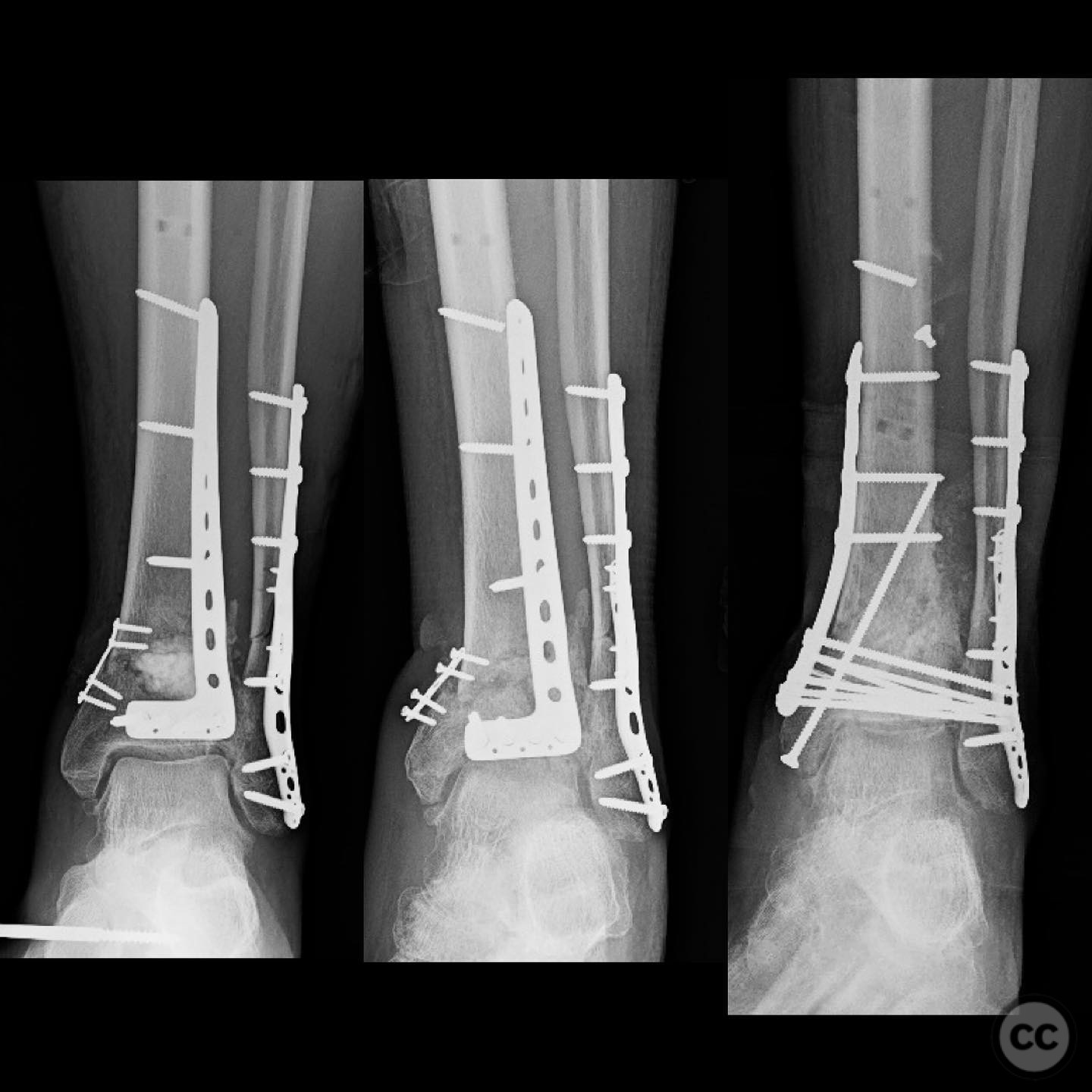
Clinical and radiological findings: The patient is a male with a history of valgus failure and medial open pilon fracture, initially fixed via an anterolateral approach. Due to a diagnosis of lung cancer and subsequent chemotherapy, definitive management was delayed, resulting in a nonunion. The primary deformity is in the coronal plane (varus). Radiological assessment revealed a nonunion with varus deformity and posterior translation/recurvatum. Laboratory markers for infection were unreliable due to recent chemotherapy, necessitating a biopsy to rule out infection, which returned negative cultures.
Preoperative Plan
Planning remarks: The preoperative plan involved correction of the varus deformity using indirect means with a medial distractor. A single-stage repair was planned following confirmation of the absence of infection. The plan included structural support with femoral head allograft and biological augmentation using synthetic biologics with lineage-directed stem cells.
Surgical Discussion
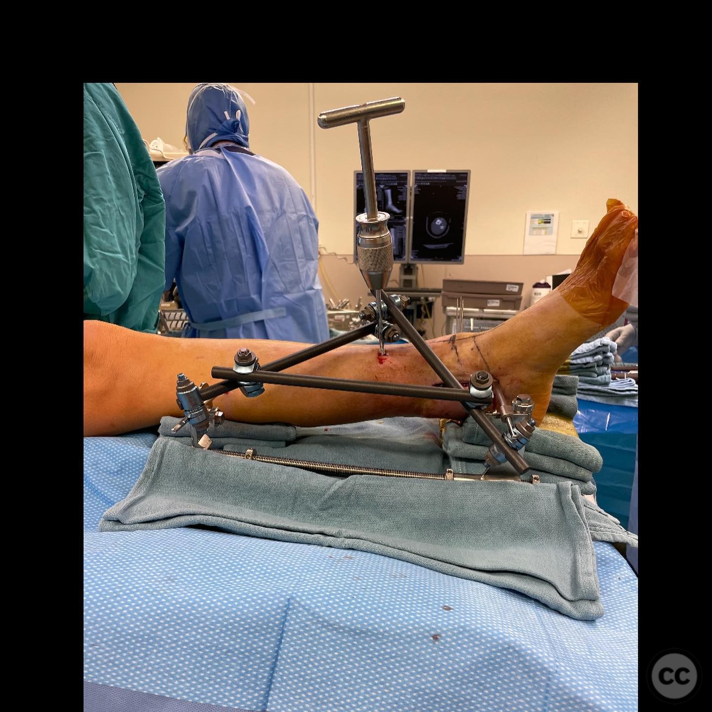
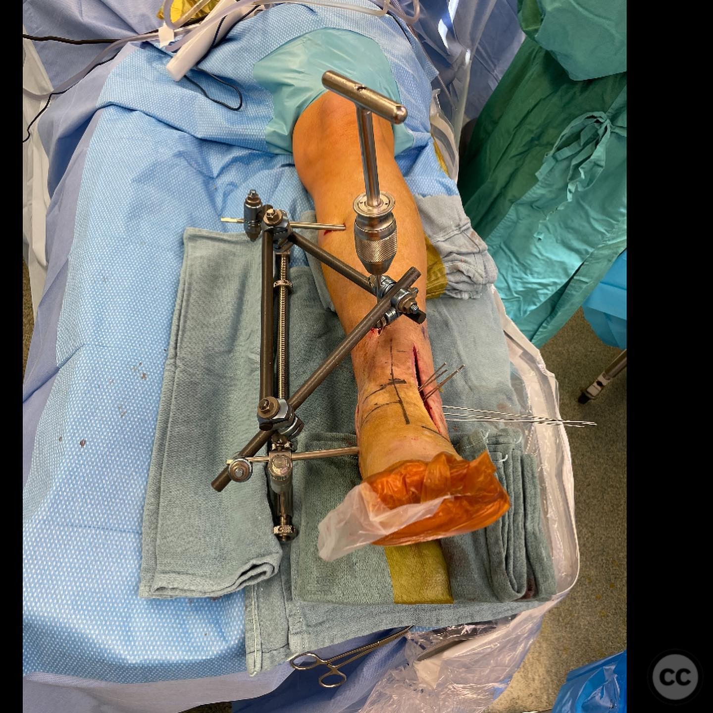
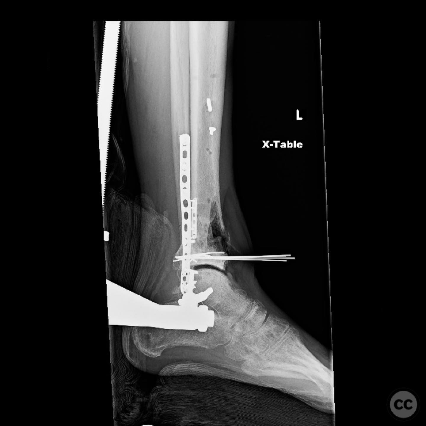
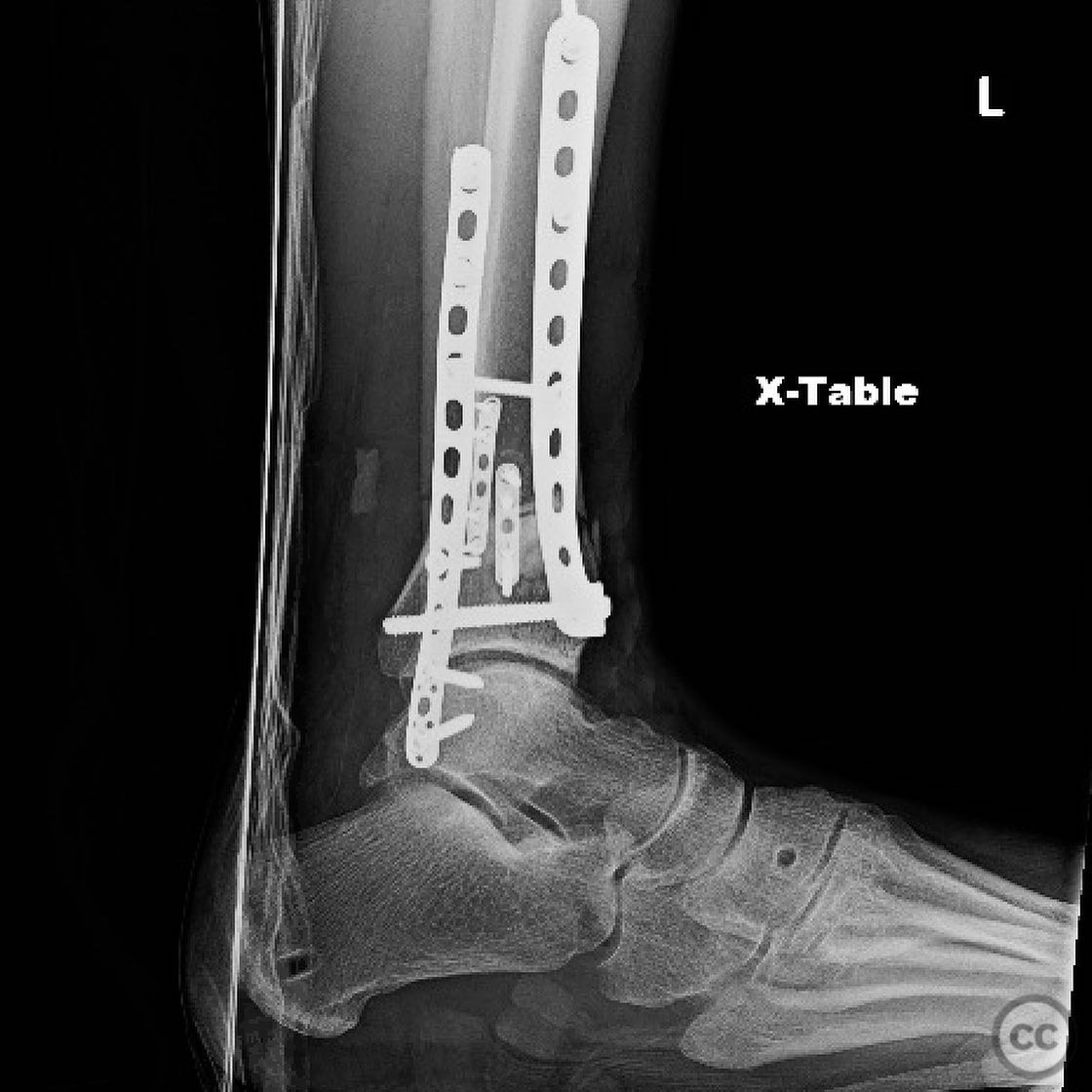
Patient positioning: The patient was positioned supine with a bump placed under the heel to facilitate posterior translation of the tibia during correction.
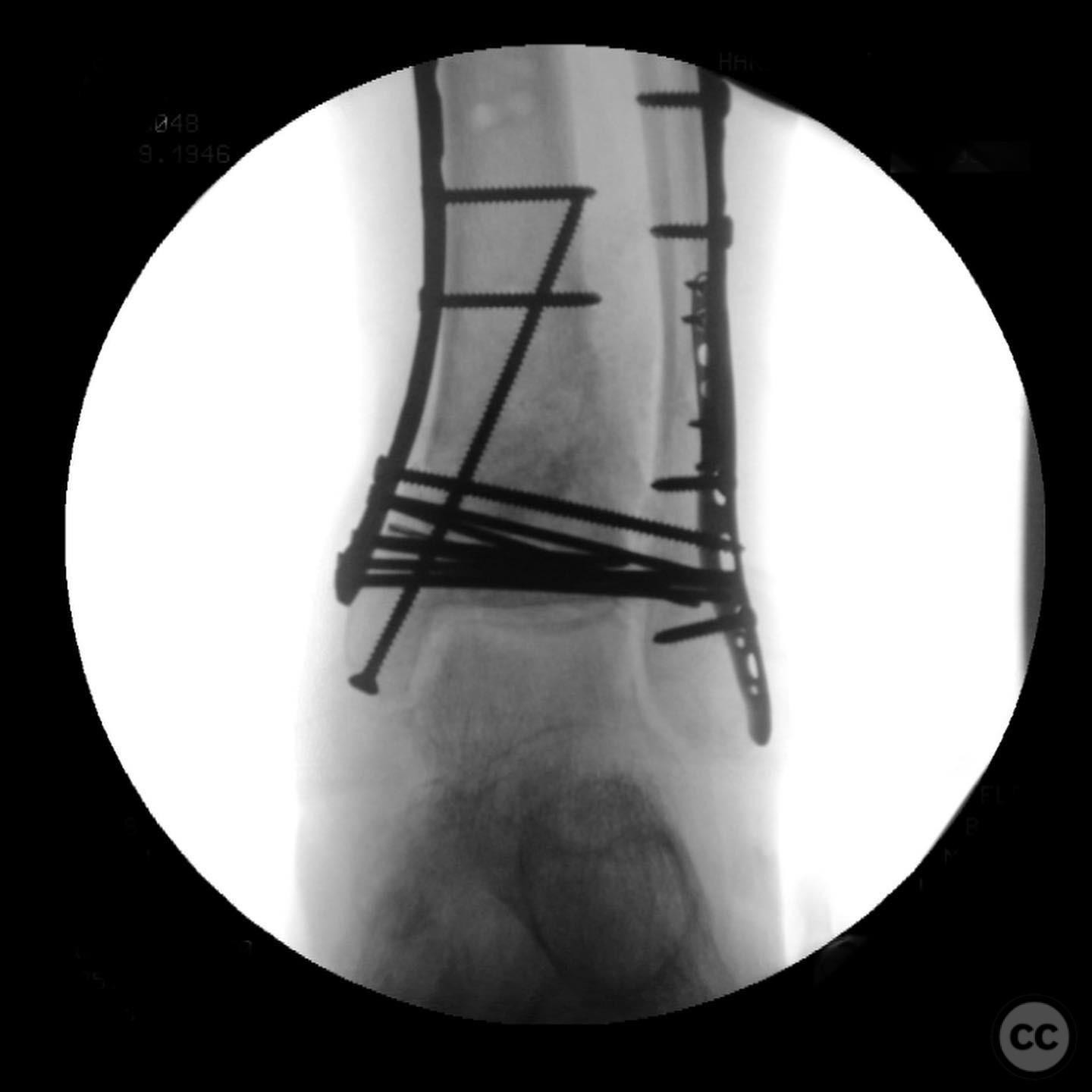
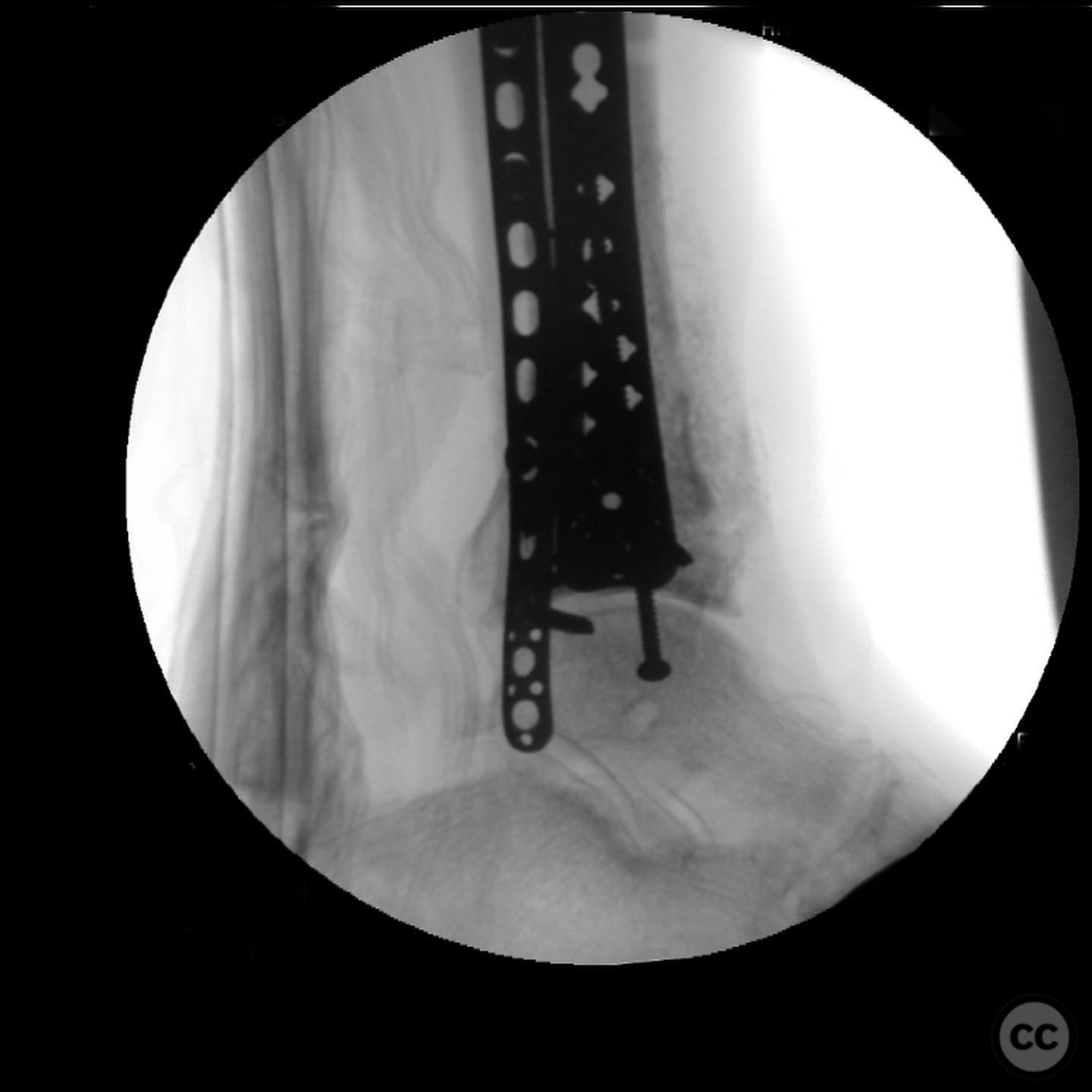
Anatomical surgical approach: A medial approach was utilized for the surgical procedure. The varus deformity was corrected using a medial distractor, with a Schanz pin connected to the distractor for multiplanar indirect reduction. The void at the nonunion site was filled with femoral head allograft for structural support, and synthetic biologics were used for biological augmentation.
Operative remarks:The surgeon noted that the primary deformity in the coronal plane was corrected via indirect means using a medial distractor. Posterior translation/recurvatum was addressed intraoperatively with a "push pin" technique connected to the distractor. The medial plate was applied to address the varus deformity, with screws engaging quad cortical purchase into the fibula. The importance of medial column support in C-type pilon nonunions was emphasized, as these often fail in varus due to fibular healing.
Postoperative protocol: Postoperative rehabilitation protocol was not specified.
Follow up: Not specified.
Orthopaedic implants used: Medial plate, femoral head allograft, synthetic biologics with lineage-directed stem cells, Schanz pin, distractor system.
Search for Related Literature

orthopaedic_trauma
- United States , Seattle
- Area of Specialty - General Trauma
- Position - Specialist Consultant

Industry Sponsership
contact us for advertising opportunities
Article viewed 263 times
13 Jul 2025
Add to Bookmarks
Full Citation
Cite this article:
Surname, Initial. (2025). Pilon Nonunion Repair with Deformity Correction. Journal of Orthopaedic Surgery and Traumatology. Case Report 17372487 Published Online Jul 13 2025.