Complex Combined Pelvic Ring and Acetabular Fracture Management in a Young Male
Score and Comment on this Case
Clinical Details
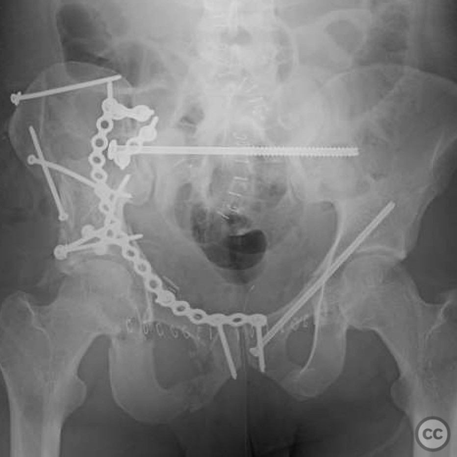
Clinical and radiological findings: A 27-year-old male presented with a severe combined pelvic ring and acetabular fracture following a high-energy trauma. Initial radiographs and CT imaging revealed a complex fracture pattern involving both the pelvic ring and acetabulum, indicative of a high-impact injury. The fracture was classified as AO/OTA 62-C3, suggesting a complete disruption of the pelvic ring with associated acetabular involvement.
Preoperative Plan
Planning remarks: The preoperative plan involved a dual approach to address both the pelvic ring and acetabular components of the fracture. An anterior approach via an ilioinguinal incision was planned for pelvic ring stabilization, while a separate Kocher-Langenbeck approach was considered for acetabular reconstruction.
Surgical Discussion
Patient positioning: The patient was positioned supine on a radiolucent table to facilitate anterior access, with the potential for repositioning to lateral decubitus if necessary for posterior acetabular access.
Anatomical surgical approach: The surgical approach commenced with an ilioinguinal incision, providing access to the anterior column of the pelvis. Dissection proceeded through the external oblique aponeurosis, with careful retraction of the neurovascular structures to expose the fracture site. For the acetabular component, a Kocher-Langenbeck approach was utilized, involving an incision along the posterior aspect of the hip, with detachment of the gluteus maximus and exposure of the posterior column.
Operative remarks:The operative procedure was challenging due to the complexity and comminution of the fracture fragments. Meticulous reduction techniques were employed, utilizing a combination of direct and indirect reduction methods. Temporary fixation with K-wires facilitated stabilization during definitive fixation. The use of intraoperative fluoroscopy ensured accurate alignment and fixation of both pelvic and acetabular components.
Postoperative protocol: Postoperatively, the patient was placed on a non-weight-bearing protocol for 8 weeks, with gradual progression to partial weight-bearing as tolerated. Early mobilization with physiotherapy focused on maintaining range of motion and preventing joint stiffness.
Follow up: Not specified.
Orthopaedic implants used: Pelvic reconstruction plate, acetabular reconstruction plate, K-wires for temporary fixation.
Search for Related Literature

orthopaedic_trauma
- United States , Seattle
- Area of Specialty - General Trauma
- Position - Specialist Consultant

Industry Sponsership
contact us for advertising opportunities
Article viewed 306 times
25 Jul 2025
Add to Bookmarks
Full Citation
Cite this article:
Surname, Initial. (2025). Complex Combined Pelvic Ring and Acetabular Fracture Management in a Young Male. Journal of Orthopaedic Surgery and Traumatology. Case Report 16135690 Published Online Jul 25 2025.